
Stenostygnus Simon, 1879
|
publication ID |
https://doi.org/ 10.11646/zootaxa.4984.1.15 |
|
publication LSID |
lsid:zoobank.org:pub:71CB5F50-A5B0-4C2C-B499-FD73F7FE666C |
|
DOI |
https://doi.org/10.5281/zenodo.4926889 |
|
persistent identifier |
https://treatment.plazi.org/id/2D0A576D-FF9E-3120-A895-B59DFAEEF852 |
|
treatment provided by |
Plazi |
|
scientific name |
Stenostygnus Simon, 1879 |
| status |
|
Stenostygnus Simon, 1879 View in CoL
Stenostygnus Simon 1879: 224 View in CoL . Roewer 1913: 163. 1923: 460. Mello-Leitão 1923: 133. 1932: 418. Sørensen in Henriksen 1932: 284. Kästner 1937: 389. Pinto-da-Rocha 1995: 194. Kury 2003: 36
Type species. Stenostygnus pusio Simon, 1879 View in CoL (by monotypy).
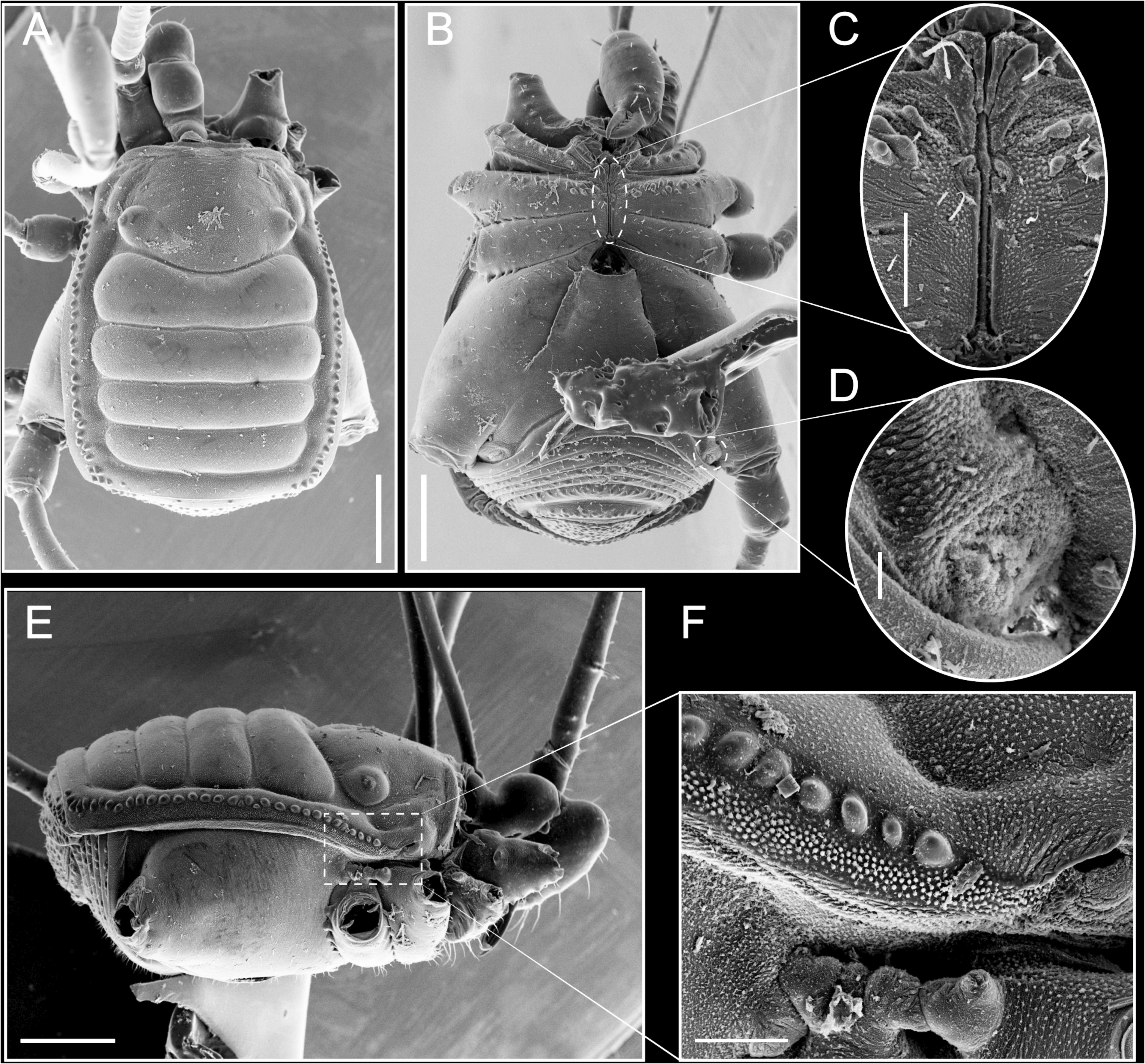
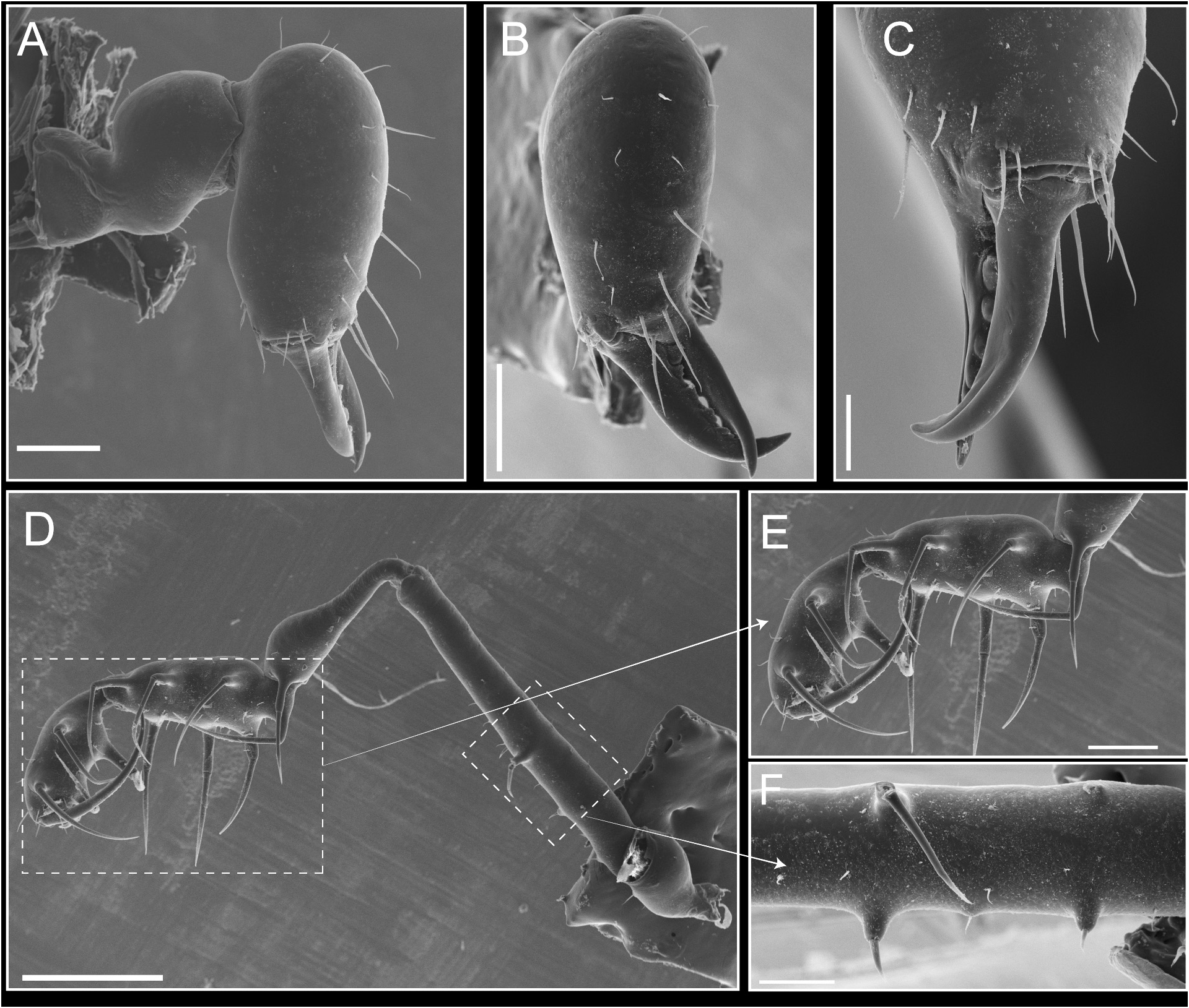
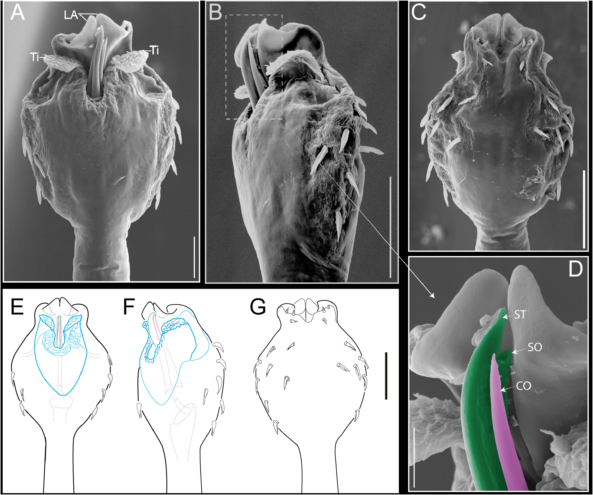
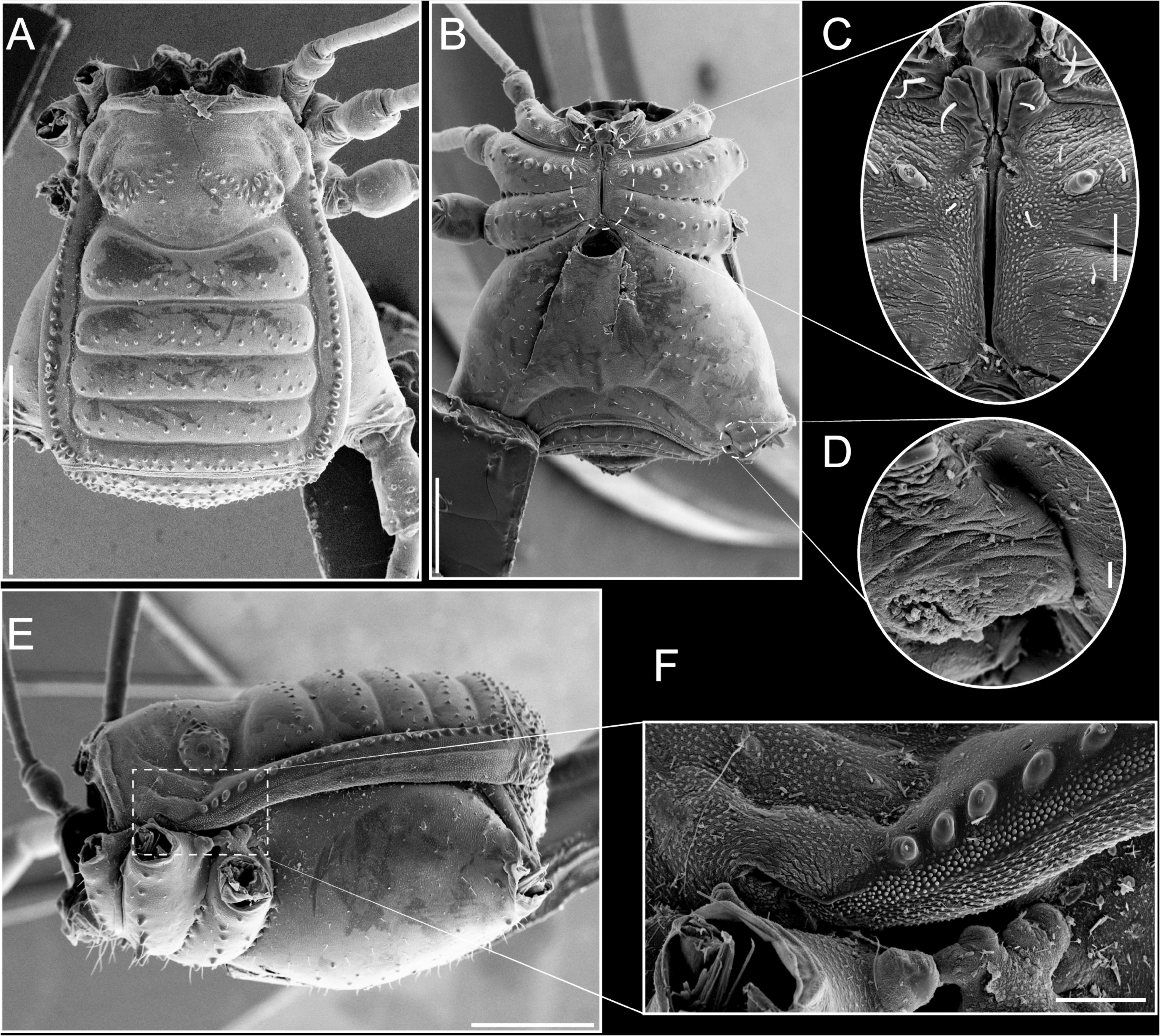
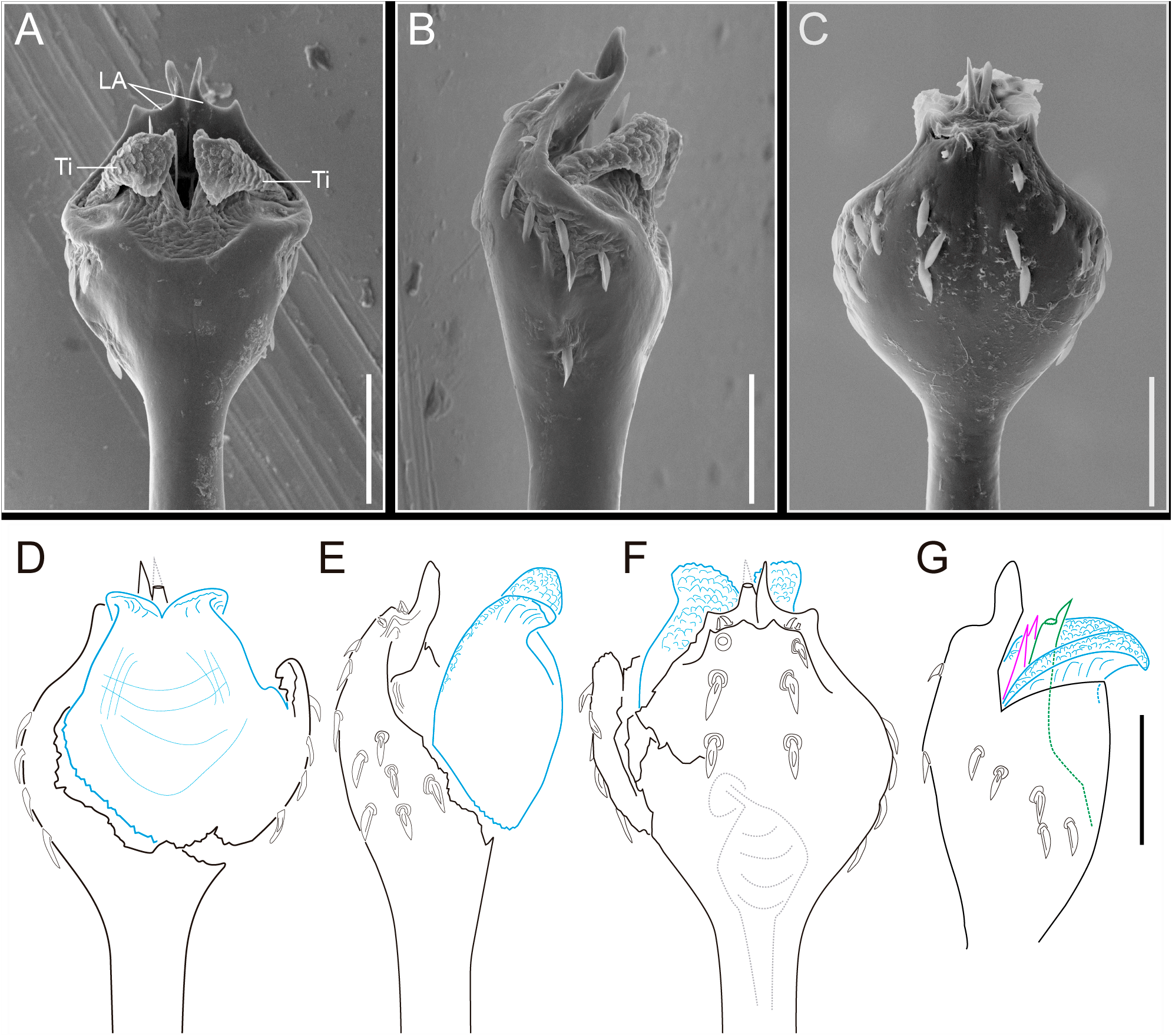
Emended diagnosis. Area I without a median longitudinal groove ( Fig. 2 A View FIGURES 2 ). Mesotergal areas and free tergites unarmed, only with scarce small granules or smooth ( Figs 2 A View FIGURES 2 ; 8 A View FIGURES 8 ). Male (α) chelicera not strongly swollen (only slightly bigger than in females) ( Fig. 6 C View FIGURES 6 vs A). Patella of the pedipalp with or without meso-distal spine ( Fig. 3 D View FIGURES 3 ). Males without metatarsus III enlarged. Males with armature on patella IV or tibia IV ( Figs 6 D View FIGURES 6 ; 12 D, F View FIGURES 12 ). Tarsal counts: 6–8(3): 9–15(3–4): 6: 6. Pars distalis of penis ending in a lamina apicalis medially divided ( Figs 5 A, C View FIGURES 5 ; 11 A, C View FIGURES 11 Pinto-da-Rocha 1995, figs 5; 7). Presence of two titillators with their apical edges dorsally projected, the internal surface of titillators is covered by small digitiform projections ( Figs 5 A View FIGURES 5 ; 11 A View FIGURES 11 ; Pinto-da-Rocha 1995, fig. 5). Stenostygnus View in CoL can be distinguished from the other Stenostygninae View in CoL genera by the absence of the male sexually dimorphic characteristics of swollen chelicerae and metatarsus III enlarged. Also, by the presence of a pars distalis with a lamina apicalis medially divided and two titillators with their apical edges dorsally twisted and covered by digitiform projections on the internal surface. In addition, Stenostygnus View in CoL (as well as Bidoma Šilhavý, 1973 View in CoL , and Decuella Avram, 1977 View in CoL ) also differs by the absence of a median longitudinal groove in area I. Stenostygnus View in CoL can be distinguished from Bidoma View in CoL by the absence of a tubercle on the eye mounds connected with the opposite tubercle located on the anterior margin of area I ( Figs 2 A View FIGURES 2 vs Šilhavý 1973, figs 71, 74). Differentially, Stenostygnus View in CoL and Manahunca Šilhavý, 1973 View in CoL have the mesotergal areas and free tergites without spiniform apophyses. However, Stenostygnus View in CoL can be easily differentiated from Manahunca View in CoL by lacking tubercles, being armed only with scarce small granules or smooth.
Species included. Stenostygnus huberi View in CoL spec. nov., Stenostygnus martensi View in CoL spec. nov., Stenostygnus pusio View in CoL .


No known copyright restrictions apply. See Agosti, D., Egloff, W., 2009. Taxonomic information exchange and copyright: the Plazi approach. BMC Research Notes 2009, 2:53 for further explanation.
|
Kingdom |
|
|
Phylum |
|
|
Class |
|
|
Order |
|
|
Family |
Stenostygnus Simon, 1879
| Mamani, Claudia Vanesa, Porto, Willians, Iglesias, Patricia P. & González, Abel Pérez- 2021 |
Stenostygnus
| Kury, A. B. 2003: 36 |
| Pinto-da-Rocha, R. 1995: 194 |
| Kastner, A. 1937: 389 |
| Mello-Leitao, C. F. de 1932: 418 |
| Roewer 1913: 163 . 1923 |
| Roewer, C. F. 1923: 460 |
| Mello-Leitao, C. F. de 1923: 133 |
| Simon, E. 1879: 224 |