
Pseudophilomedes ferulanus Kornicker 1958
|
publication ID |
https://doi.org/ 10.11646/zootaxa.1565.1.1 |
|
publication LSID |
lsid:zoobank.org:pub:A2CDD9CB-CA5E-418B-A471-9EEFDC5CCF16 |
|
DOI |
https://doi.org/10.5281/zenodo.5096311 |
|
persistent identifier |
https://treatment.plazi.org/id/2A5087FF-3E60-FC54-3A91-FB71FAE36B1C |
|
treatment provided by |
Felipe |
|
scientific name |
Pseudophilomedes ferulanus Kornicker 1958 |
| status |
|
Pseudophilomedes ferulanus Kornicker 1958 View in CoL
Figs. 46 View FIGURE 46 , 47 View FIGURE 47
Pseudophilomedes ferulana Kornicker 1958: 235 View in CoL , figs. 46: 1a,b, 2a,b: 56A–D.
Pseudophilomedes ferulanus View in CoL .—Kornicker 1989: 8.
Pseudophilomedes ferulana View in CoL .— Darby 1965: 64–70, Fig. 10 View FIGURE 10 , pls. 11,12.
Not Pseudophilomedes ferulanus View in CoL .— Kornicker 1967: 8, figs. 1–6, pl. 1.—1969: 119, figs. 3, 4, pl. 11.—1977: 792.— 1984: 33, figs. 14–16.
Holotype. USNM 113287 View Materials , adult female on slides.
Type locality. Bimini, Bahamas, Great Bahama Bank.
Material. Great Bahama Bank, Exuma Cays, Great Exuma, Stocking Island (Bottomly’s Blue Hole), Sta 99-065: USNM 10214561 View Materials , 1 specimen on slide and in alcohol .
Distribution. See Kornicker (1984: 33). Herein, Great Bahama Bank, Stocking Island (Bottomly’s Blue Hole).
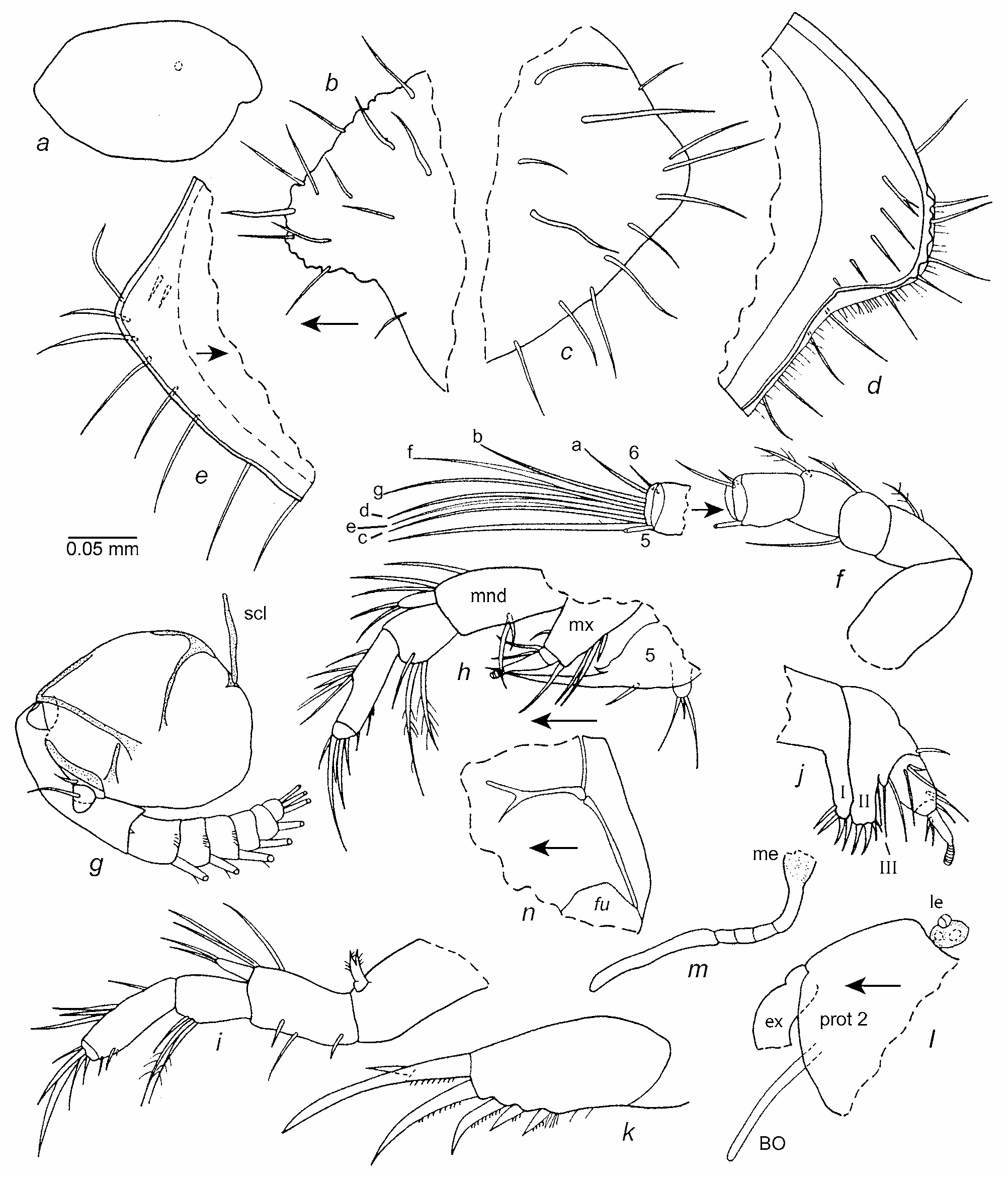
Description of instar II (sex unknown) ( Figs. 46 View FIGURE 46 , 47 View FIGURE 47 ). Carapace elongate with projecting caudal process ( Fig. 46 a,c,e View FIGURE 46 ); incisure forming obtuse-angle ( Fig. 46 a,b,d View FIGURE 46 ).
Ornamentation ( Fig. 46 b–e View FIGURE 46 ): Surface with widely distributed long and medium length bristles; long bristles with broad triangular basal part. Outer edge of rostrum with small nodes ( Fig. 46 b,d View FIGURE 46 ). Ribs, punctations, and reticulations absent on USNM 1021461.
Infold: Rostral infold broad with 4 long bristles forming row, and 1 minute bristle near ventral end of rostrum ( Fig. 46 d View FIGURE 46 ). Infold of caudal process broad, with 2 indistinct bristles ( Fig. 46 e View FIGURE 46 ). Narrow infold along anteroventral, ventral and posterior margins of valve. Anteroventral infold without bristles or parallel striations.
Selvage: Narrow fringed lamellar prolongation along anterior and ventral margins of valve. Prolongation not divided at incisure.
Central adductor muscle attachments: Indistinct on USNM 10214561.
Carapace size (length, height in mm): USNM 10214561, 0.69, 0.41.
First antenna ( Fig. 46 f View FIGURE 46 ): Segment 2 with 1 dorsal bristle. Segment 3 with 2 bristles (1 ventral, 1 dorsal). Segment 4 with 1 dorsal bristle. Long ventral bristle of segment 5 with small proximal filament. Segment 6 with small medial bristle near dorsal margin. Segment 7 with 3 bristles: a-bristle about 3 times length of bristle of 6th segment; b-bristle about twice length of a-bristle; c-bristle long, bare. Segment 8 with 4 long bare bristles.
Second antenna ( Fig. 46 g,l View FIGURE 46 ): Protopodite bare; proximal connecting sclerite fairly linear. Endopod with 2 segments, each with 1 bristle. Exopod: Right limb of USNM 10214561 aberrant in having only 7 segments ( Fig. 46 g View FIGURE 46 ); left limb with 9 segments; terminal segment of left limb with 2 bristle, of right limb with 3 bristles. Most bristles with natatory hairs, some also with slender proximal ventral spines; 1st segment with minute terminal medial spine-like bristle; some segments with distal spines forming row.
Mandible ( Fig. 46 h,i View FIGURE 46 ): Coxa endite bifurcate, spinous. Basis: dorsal margin with 1 distal and 2 terminal bristles; medial surface with 1 or 2 short bristles near ventral margin; ventral margin with 1 bristle near midlength. Exopod elongate with 2 terminal bristles. Endopod: 1st segment with 3 terminal ventral bristles; 2nd segment with 4 or 5 bristles on or near dorsal margin and 2 short distal bristles on ventral margin; 3rd segment with 3 slender bristles and 2 claw-like bristles.
Maxilla ( Fig. 46 j View FIGURE 46 ): Endite I with 2 terminal bristles and 2 terminal pectinate claws; endite II with 1 proximal bristle, 2 terminal bristles and 2 terminal claws; endite III with 1 long terminal bristle. Basis with 1 short dorsal terminal bristle and 1 long ventral terminal bristle. Exopod with 3 bristles (2 long, 1 short); base of exopod bristles on 1st endopod segment close to distal margin of basis. Endopod: 1st segment with distal dorsal bristle; 2nd segment with 3 short bristles and long terminal finger-like process with annulated narrower distal part.
Fifth limb ( Fig. 47 a–e View FIGURE 47 ): Epipod with 28 spinous bristles. Exopodite represented by 2 short bristles. Coxa with 2 endites: endite I with 2 short bristles; endite II with tooth with 3 prongs and 3 bristles. Basis with 2 endites: endite I anterior with tooth with 2 prongs (distal tooth with small marginal prong on inner edge); endite II posterior with 1 long posterior proximal bristle and long slender tooth; inner edge of tooth with 2 teeth (proxima1 with 3 prongs, distal single). Exopod represented by two bristles. Endopod: segment 1 represented by single bristle on posterior surface; segment 2 elongate with 4 bristles (1 short proximal, 3 terminal (stout terminal bristle with stout proximal marginal spines)).
Sixth limb ( Fig. 47 f View FIGURE 47 ): Bilobed with marginal spines and 1 long spinous bristle on smaller lobe.
Seventh limb: Absent.
Furca ( Fig. 46 k View FIGURE 46 ): Each lamella with 5 claws decreasing in length and width posteriorly along lamella. A stout spine adjacent to base of posterior edge of claw 5; additional slender spines along lamella following claw 5. Anterior of right lamella with slender spines near base of claw 1. Claw 1 with stout medially tooth proximal to midlength. All claws with proximal teeth along posterior edge.
Bellonci Organ ( Fig. 46 l,m View FIGURE 46 ): Elongate with rounded tip bearing few spines, and sutures in proximal part.
Eyes: Medial eye only weekly pigmented and small ( Fig. 46 m View FIGURE 46 ). Lateral eye small, pigmented, and with 3 ommatidia ( Fig. 46 l View FIGURE 46 ).
Y-Sclerite ( Figs. 46 n View FIGURE 46 , 47 g View FIGURE 47 ): Branching distally.
Discussion. The second instar of a species of Pseudophilomedes has not been described previously. The specimen from Bottomly’s Blue Hole provided the opportunity to describe instar II of P. ferulanus . The morphology of the first antenna and sixth limb of the species is consistent with members of the Myodocopina in that the fourth segment of the first antenna bears only a dorsal bristle, and the sixth limb bears only one bristle (see Hiruta 1983: 673). The bristle of the sixth limb of instar II of P. ferulanus is unusually long.


No known copyright restrictions apply. See Agosti, D., Egloff, W., 2009. Taxonomic information exchange and copyright: the Plazi approach. BMC Research Notes 2009, 2:53 for further explanation.
|
Kingdom |
|
|
Phylum |
|
|
Class |
|
|
Order |
|
|
Family |
|
|
Genus |
Pseudophilomedes ferulanus Kornicker 1958
| Kornicker, Louis S., Iliffe, Thomas M. & Harrison-Nelson, Elizabeth 2007 |
Pseudophilomedes ferulana
| Darby, D. 1965: 64 |
Pseudophilomedes ferulana
| Kornicker, L. S. 1958: 235 |