
Haplaxius cotinga Bahder & Bartlett, 2022
|
publication ID |
https://doi.org/10.11646/zootaxa.5209.2.6 |
|
publication LSID |
lsid:zoobank.org:pub:92846CE0-28F7-4387-8103-F030217B3A03 |
|
DOI |
https://doi.org/10.5281/zenodo.7331167 |
|
persistent identifier |
https://treatment.plazi.org/id/267E87AB-FF99-FFDA-FF7F-FB49FE0FFE16 |
|
treatment provided by |
Plazi |
|
scientific name |
Haplaxius cotinga Bahder & Bartlett |
| status |
sp. nov. |
Haplaxius cotinga Bahder & Bartlett sp. n.
( Figures 2–9 View FIGURE 2 View FIGURE 3 View FIGURE 4 View FIGURE 5 View FIGURE 6 View FIGURE 7 View FIGURE 8 View FIGURE 9 )
Type locality. La Cotinga Biological Station ( 8.621825, -83.478819), Puntarenas province, Costa Rica ( Fig. 1 View FIGURE 1 ) GoogleMaps .
Diagnosis. A pale species (light green in life ( Fig. 2 View FIGURE 2 ), yellowish when preserved ( Fig. 3 View FIGURE 3 ), with whitish unmarked face and large reddish marking on abdominal terga. Male terminalia bearing gonostyli with sclerotized subapical dorsal ridge. Phallobase complex, bearing large, sinuous ventral projection with hooked apex and a large, elongate dorsal process. Anal segment in lateral view stout and of moderate length, ventral margin with large median process at midlength, apex elongate, strongly curved ventrad.
Description. Color. In life, body pale green, fading to yellow (in ethanol), nearly white on posterior 2/3 of vertex disc and genae excluding temple (yellow), ocelli (yellow with reddish highlights), and antennae (yellow); legs mostly white along with portions of pleuron and paranota; mesonotum paler on posterior margin and between lateral carinae. Forewings transparent, broadly but weakly infuscate except pale along leading margin (humeral region to pterostigma). Abdomen with dorsal portion of terga reddish (approximately from 2 nd- 5 th apparent abdominal tergum).
Morphology. Body length (including wings), males: 2.99 mm ( n = 1), females 2.99–3.02 mm ( n = 5). Head. In dorsal view ( Fig. 4A View FIGURE 4 ), slightly anteriorly projecting, anterior margin convex, posterior margin concave with median convexity (giving appearance of pair of concave dentations; Fig. 4A View FIGURE 4 ), lateral carinae foliate, disc convex, vertex length at midline about twice width at widest point (at posterior margin); widest basally, narrowing slightly at distal margin ( Fig. 4A View FIGURE 4 ). In lateral view ( Fig. 4B View FIGURE 4 ), head anteriorly rounded (slightly compressed), somewhat projected at level of ventral margin of compound eye ( Fig. 4B View FIGURE 4 ). Face (in frontal view— Fig. 4C View FIGURE 4 ) with frons plus clypeus collectively ovate, lateral carinae foliate, median carina distinct, forked between compound eyes, frons distinctly expanding from between compound eyes to below level of antennae, constricting slightly at frontoclypeal suture; frontoclypeal suture straight; median ocellus small, translucent. Clypeus roughly triangular with median carina weak. Antennae bulbous, scape narrow, ring-like; pedicle spheroid, bearing many irregularly placed sensory plaques, flagellum elongate and bristle-like with a bulbous base. Lateral ocelli distinct, anterior to antennae beneath compound eye near leading margin.
Thorax. In dorsal view, pronotum narrow, convex at anterior margin, concave at posterior margin ( Fig. 5A View FIGURE 5 ), median carina indistinct, lateral carinae extending to ventral margin at 2/3 length to ventrolateral margin. Mesonotum tricarinate, median carina evident, reaching posterior margin, lateral carinae subparallel for most of length, diverging posteriorly ( Fig. 5A View FIGURE 5 ). Spinulation of hind leg 6-6-6.
Forewing. Forewing veins with setal pits ( Fig. 6 View FIGURE 6 ), apex of clavus past wing midlength (fusion of PCu and A 1 in basal third of wing), composite vein Pcu+A1 reaching wing margin well before claval apex; fork of R (forming C1 cell) near wing midlength just distad of CuA fork; forewing branching pattern: RA 1-branched, RP 3-branched, MP 5-branched, CuA 2-branched with CuA connected with CuP by icu crossvein. A small cell, spurious, present along the distal margin of C5 at level of claval apex (absent in 3/ 5 females).
Terminalia. Pygofer, in lateral view, irregularly triangular, narrowest at dorsal margin, widest at ventral margin, anterior margin sinuate, posterior margin convex ( Fig. 7A View FIGURE 7 , 8A View FIGURE 8 ). Medioventral process (in lateral view) scooplike, in ventral view, spade-like to sagittate, apex acutely pointed ( Fig. 7B View FIGURE 7 , 8B View FIGURE 8 ). Gonostyli in lateral view strongly expanded distally, sinuate ventrally, dorsally strongly curved to bulbous apex ( Fig. 7A View FIGURE 7 , 8A View FIGURE 8 ), dorsoapical margin with two sclerotized dorsal projections, one arising approximately at 2/3 length and second at apex, lightly sclerotized between ( Fig. 7A View FIGURE 7 , 8A View FIGURE 8 ); in ventral view, inner and outer margins irregularly sinuate, narrowest basally, expanding approximately to twice the width of basal half, apically truncate, inner margins with irregular median serrulations in distal half ( Fig. 7B View FIGURE 7 , 8B View FIGURE 8 ). Aedeagus approximately straight (in lateral view, Figs. 9A View FIGURE 9 , 10A View FIGURE 10 ) for most of length, then angled downward to ventral lobe (A1) followed by dorsal inflection (A2). Phallotheca incompletely enclosing aedeagus, mostly membranous with sclerotized portions, bearing large projection traversing aedeagus dorsum to form large dorsal process (P1), ventral sclerotization with large apical strongly curved process (P2) ( Figs. 7 View FIGURE 7 & 8 View FIGURE 8 ). Anal segment in lateral view stout (not ‘stalklike’, viz. Kramer 1979) moderate length, ventral margin with large median process at midlength, apex elongate, strongly curved ventrad ( Figs. 5A View FIGURE 5 , 6A View FIGURE 6 ); in dorsal view broad with rounded apex ( Figs. 5C View FIGURE 5 , 6C View FIGURE 6 ). Anal column tubular, elongate.
Plant associations. Unknown, collected at light trap. Habitat was an abandoned pasture/grazing land being reforested with hardwood trees.
Distribution. Costa Rica ( Puntarenas), Osa Peninsula.
Etymology. The specific name given is in reference to La Cotinga Biological Station where the specimens were collected.
Material examined. Holotype male “ Costa Rica, Puntarenas / La Cotinga Biological Station / 7.VII.2021 / Light trap near dorms / Coll.: B.W.Bahder // Holotype / Haplaxius cotinga ♂ ” ( FLREC) ; Paratypes, La Cotinga Biological Station [ 7.VII.2021] ( 1 male, 1 female — FSCA, 1 female — FLREC) .
Sequence Data. For the COI gene, a 646 bp product was generated (GenBank Accession No. ON763279 View Materials ), for 18S, a 1,403 bp product was generated (GenBank Accession No. ON758370 View Materials ) and for the H3 gene, a 347 bp product was generated (GenBank Accession No. ON755134 View Materials ). Based on the phylogenetic analyses performed for the 18S and H3 gene, there is strong bootstrap support for the monophyly of Haplaxius , 97 and 94 respectively ( Fig. 9 View FIGURE 9 ). Haplaxius cotinga sp. n. resolves within this clade for both 18S and H3 but separate from all other Haplaxius species analyzed. The placement of Haplaxius cotinga sp. n. is further supported in the consensus tree based on all three loci ( Fig. 9D View FIGURE 9 ), where it resolves with weak support (49) adjacent to H. pictifrons . For the COI, there is weak bootstrap support at all branches (<80) except in the instances of closely related taxa ( Fig. 9A View FIGURE 9 ). The pairwise comparison based on the 18S gene shows that the average level of variation among species within a genus is 0.77% (SE±0.1) for Haplaxius , 0.37% (SE±0.1) for Oecleus , 1% (SE N/A) for Nymphocixia and 0.63% (SE±0.3) for Myxia based on the taxa analyzed ( Table 3 View TABLE 3 ). The average variation among genera for the 18S gene is 2.1% (SE±0.1), ranging from 1.3% to 2.7% with Haplaxius cotinga sp. n. differing on average by 0.53% (SE±0.2) from other species of Haplaxius , 1.77% (SE±0.1) from species of Oecleus , 1.5% (SE±0.2) from species of Nymphocixia and 1.9% (SE±0.0) from species of Myxia ( Table 3 View TABLE 3 ). The pairwise comparison based on the COI gene shows an average of 15.6% intrageneric variability whereas there was on average, 18.9% intergeneric variability ( Table 4 View TABLE 4 ). While there does appear to be some distinction at the generic level for COI based on nucleotide variability, this distinction is not as pronounced and consistent as that seen with 18S, which to some extent, is also reflected in the phylogenies. In addition, there are taxa where this value overlaps ( M. belinda and M. hernandezi as well as with Nymphocixia ), further highlighting that COI is not a suitable marker for phylogenies in the currently analyzed Oecleini but should be reserved for species delimitation and population genetic studies.
Remarks. Morphological characters and molecular data presented support the placement of the novel taxon in Haplaxius . The closest species to Haplaxius cotinga sp. n. appears to be H. deleter Kramer, 1979 (described from southern Panama). Both species have the same general form of the terminalia (especially the gonostyli and aedeagus), and both are similar in general coloration ( H. deleter described by Kramer 1979: 369 as “ground color of head and pronotum pale chalky green, anterior portion of crown lightly washed with orange, face unmarked…”). The two species would key in different directions in Kramer (1979) because H. cotinga sp. n. has a median projection on the anal tube that is absent in H. deleter ( H. deleter instead bears a quadrate process about midlength of the left lateral margin of the anal tube, absent in cotinga sp. n.). In H. deleter , the gonostyli appear more slender with the dorsal projections much more separated, longer and less curved than observed in H. cotinga sp. n. Additionally, the curvature of the apical process of the aedeagus is different where it is angled distad in H. deleter and cephalad in H. cotinga sp. n. and the ventral process of the phallobase is distinctly angled caudad in H. deleter and less sinuate whereas the process in H. cotinga sp. n. is strongly sinuate, curving cephalad. Finally, in H. cotinga sp. n. the flagellar apex is bifurcated in lateral view, where is rounded in H. deleter . Other features that distinguish H. cotinga sp. n. from H. deleter is the size ( H. deleter adult males reported at 5.0 mm while H. cotinga sp. n. adult males are around 3 mm with wings).
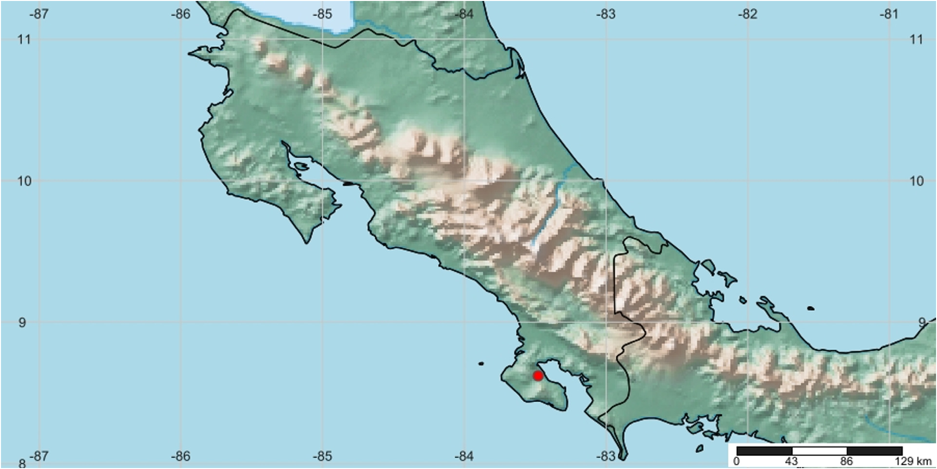
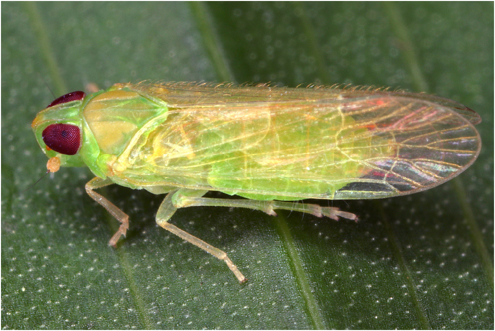
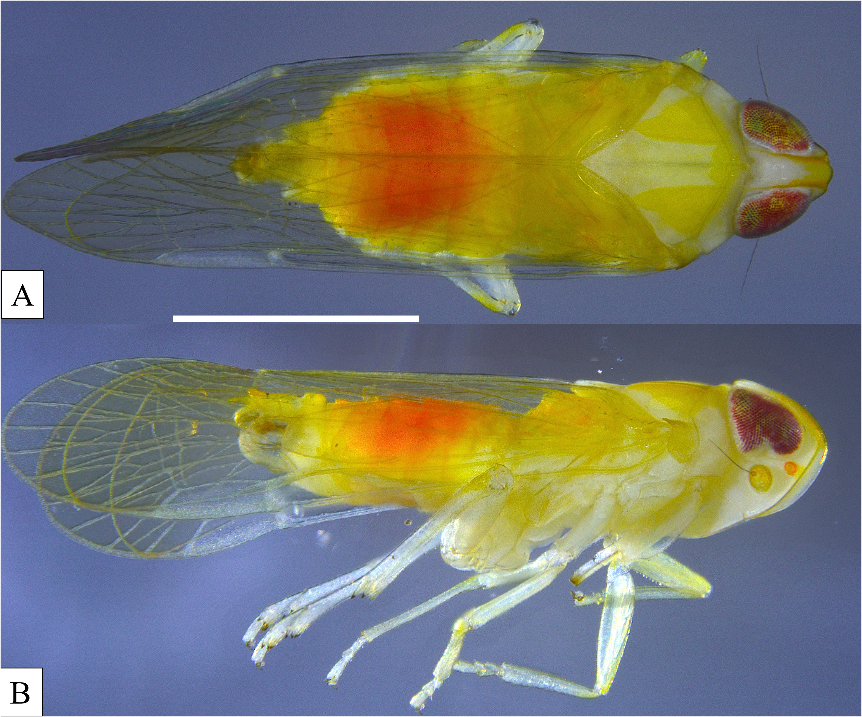
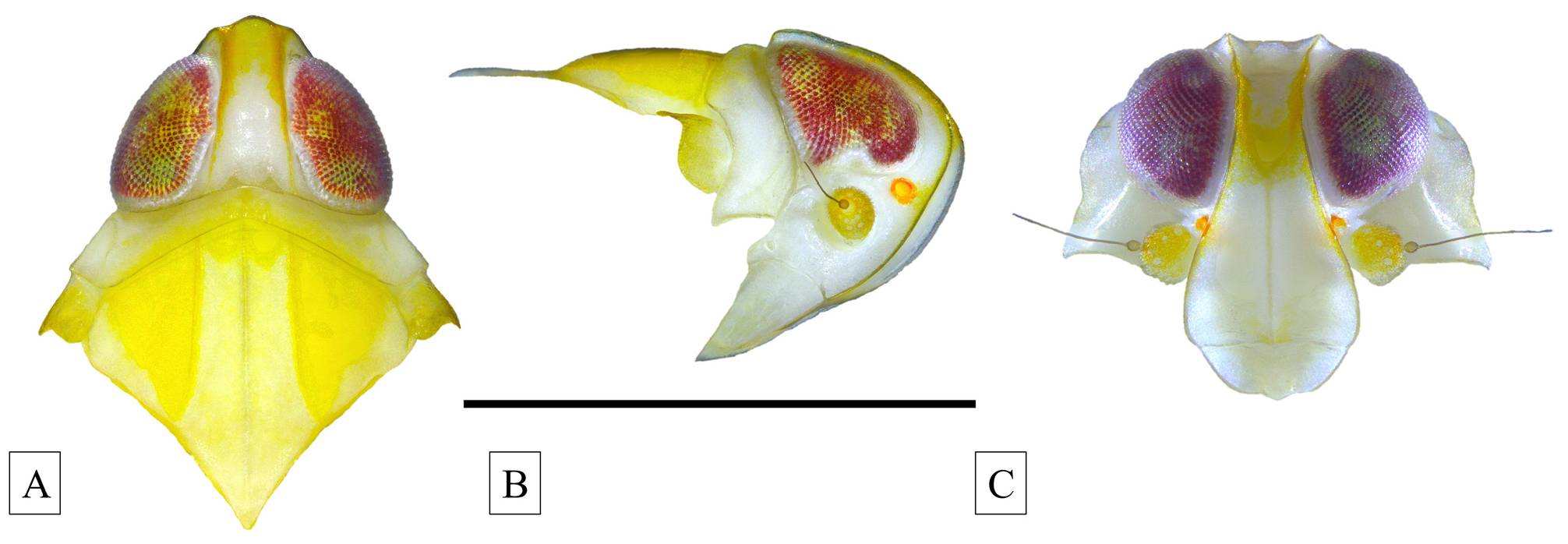
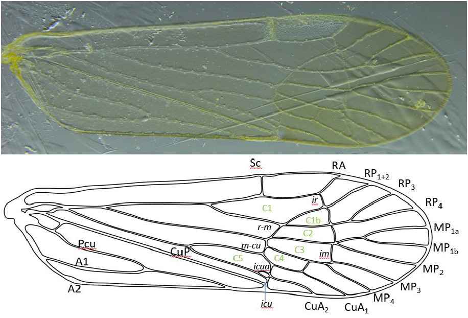

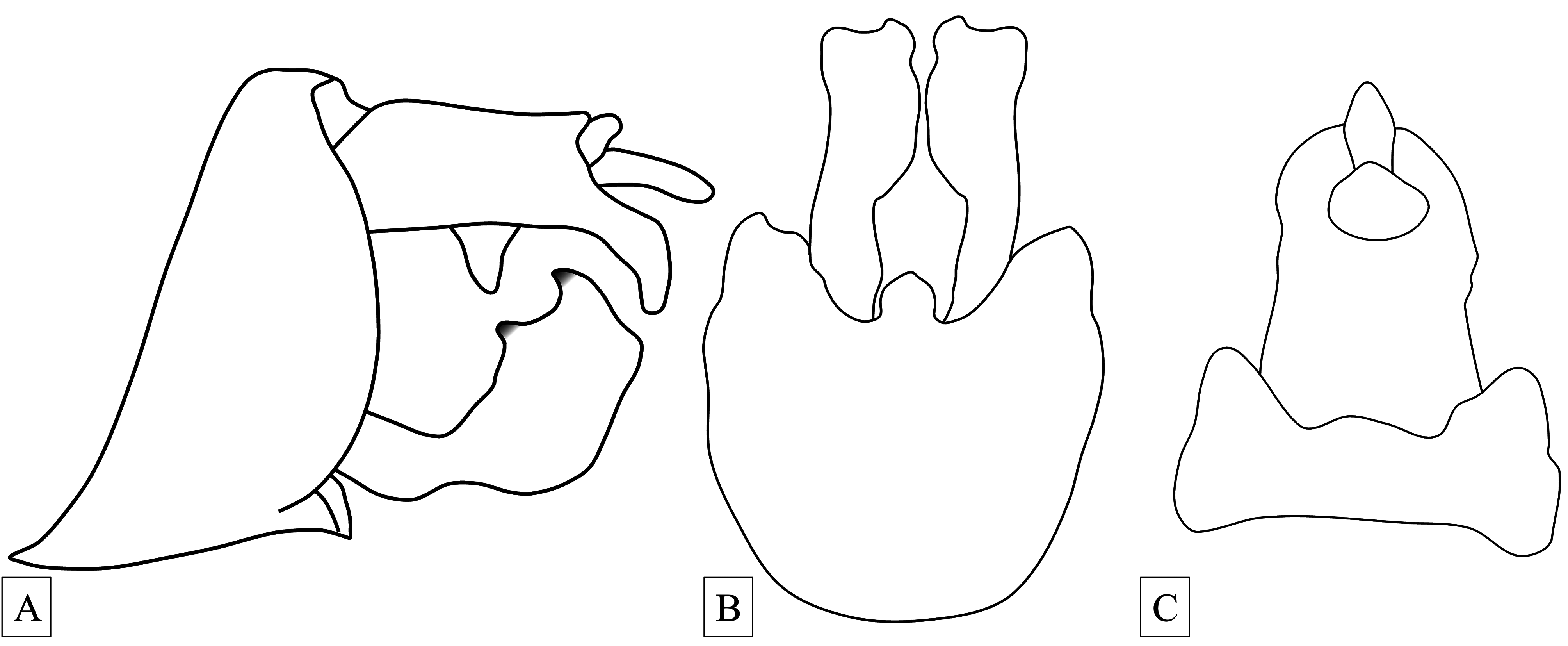
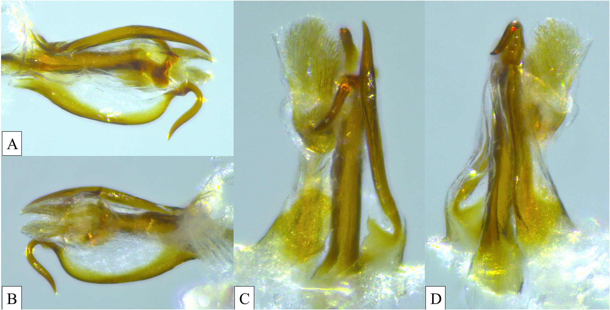
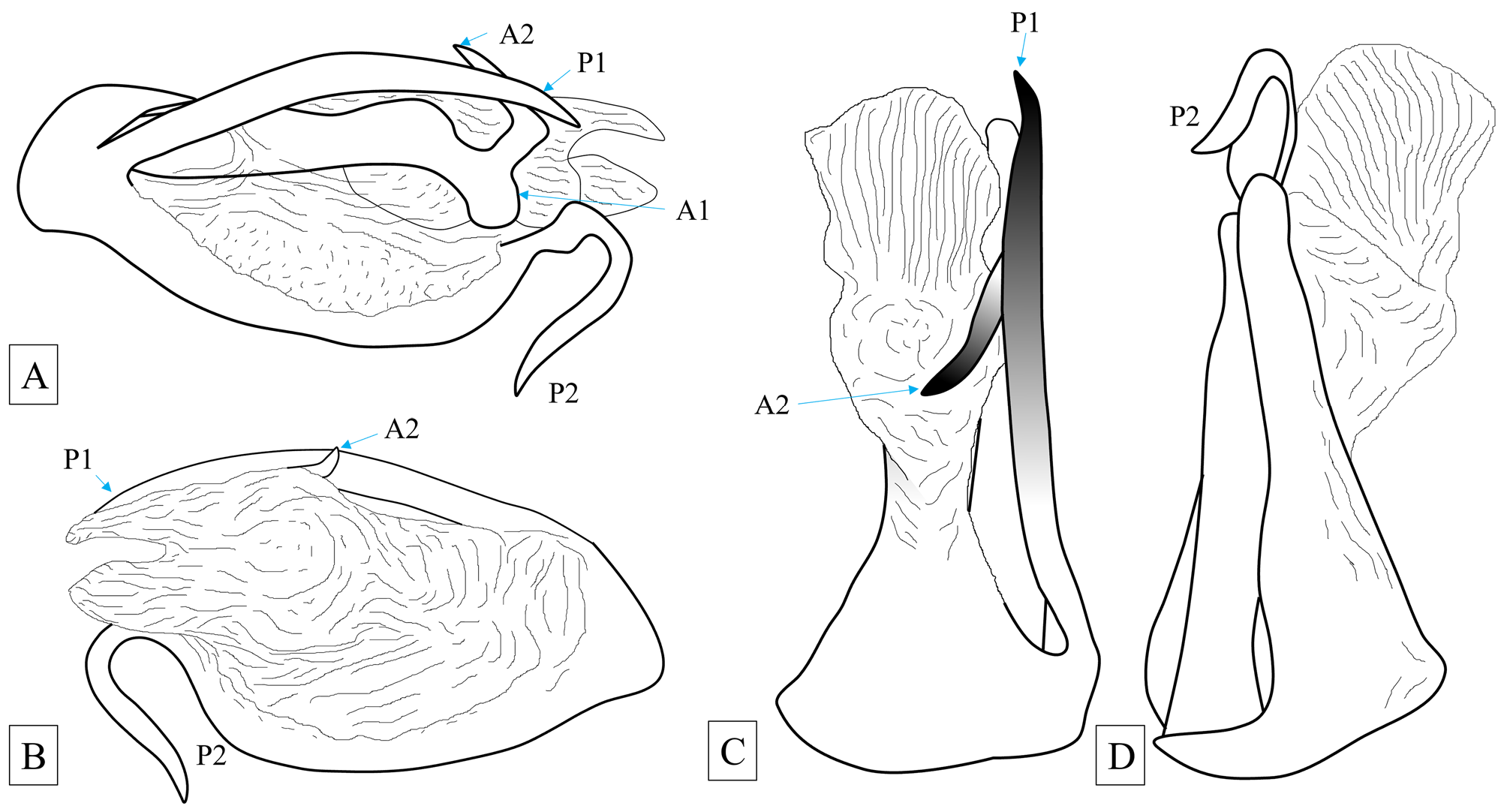
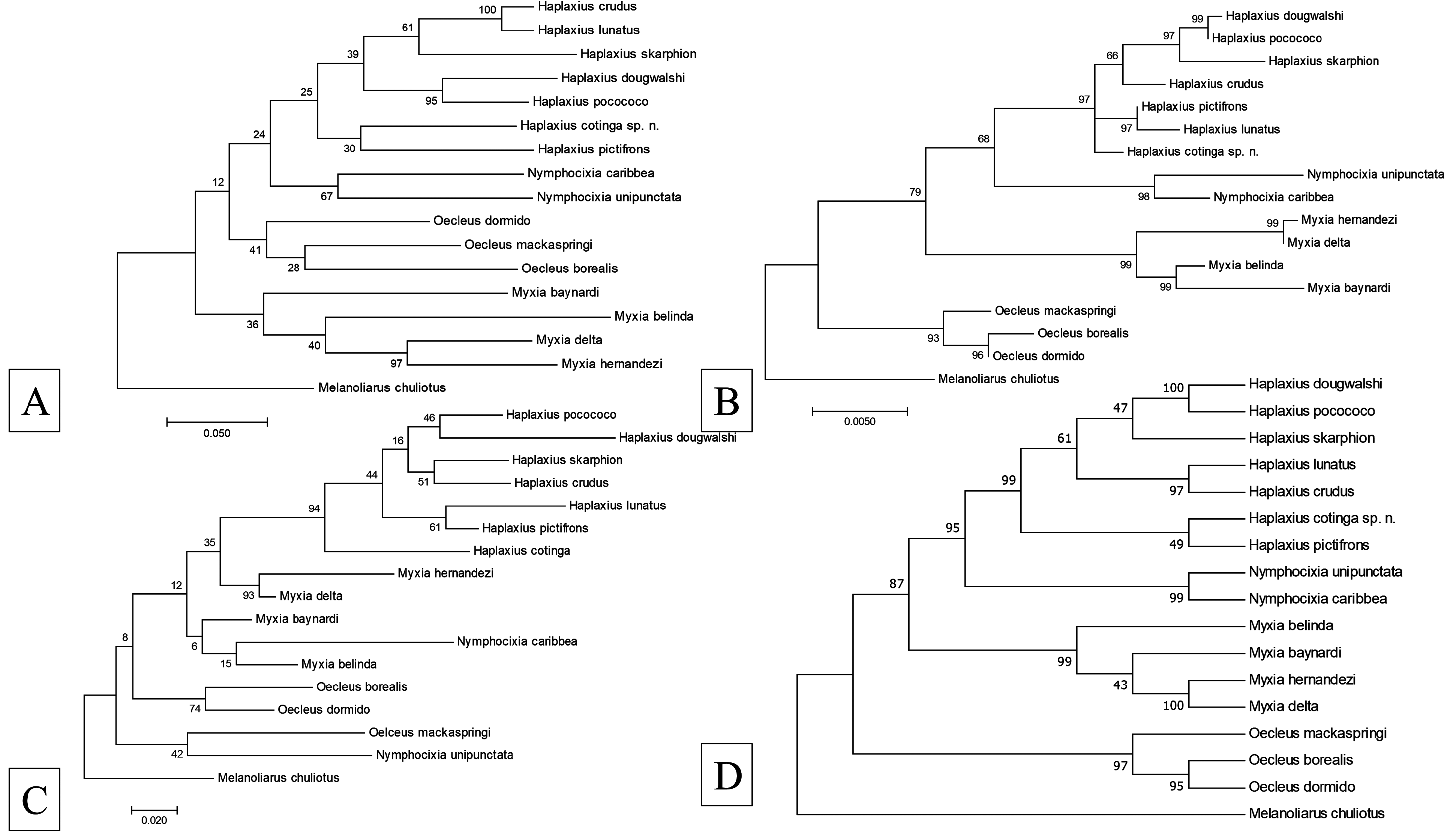
TABLE 3. Pairwise comparison showing estimates of evolutionary divergence between sequences based on the 18S rRNA gene for Haplaxius cotinga sp. n. demonstrating intrageneric (orange) and intergeneric (blue) variability; the number of base differences per site from between sequences are shown. Standard error estimate(s) are shown above the diagonal and were obtained by a bootstrap procedure (1000 replicates).
| 1 | 2 | 3 | 4 | 5 | 6 | 7 | 8 | 9 | 10 | 11 | ||
|---|---|---|---|---|---|---|---|---|---|---|---|---|
| 1 | Haplaxius cotinga sp. n. | 0.002 | 0.002 | 0.004 | 0.003 | 0.003 | 0.004 | 0.003 | 0.004 | 0.003 | 0.004 | |
| 2 | Haplaxius pocococo | 0.007 | 0.002 | 0.004 | 0.004 | 0.004 | 0.004 | 0.004 | 0.004 | 0.004 | 0.004 | |
| 3 | Haplaxius crudus | 0.004 | 0.005 | 0.004 | 0.004 | 0.004 | 0.004 | 0.003 | 0.004 | 0.003 | 0.003 | |
| 4 | Oecleus borealis | 0.019 | 0.025 | 0.021 | 0.002 | 0.001 | 0.004 | 0.004 | 0.004 | 0.004 | 0.004 | |
| 5 | Oecleus mackaspringi | 0.016 | 0.022 | 0.019 | 0.006 | 0.002 | 0.004 | 0.004 | 0.004 | 0.004 | 0.004 | |
| 6 | Oecleus dormido | 0.018 | 0.023 | 0.019 | 0.001 | 0.004 | 0.004 | 0.004 | 0.004 | 0.004 | 0.004 | |
| 7 | Nymphocixia unipunctata | 0.017 | 0.022 | 0.019 | 0.026 | 0.024 | 0.025 | 0.003 | 0.004 | 0.004 | 0.004 | |
| 8 | Nymphocixia caribbea | 0.013 | 0.016 | 0.013 | 0.024 | 0.021 | 0.022 | 0.010 | 0.004 | 0.004 | 0.004 | |
| 9 | Myxia hernandezi | 0.019 | 0.022 | 0.019 | 0.025 | 0.025 | 0.023 | 0.027 | 0.022 | 0.001 | 0.003 | |
| 10 | Myxia delta | 0.019 | 0.021 | 0.019 | 0.025 | 0.024 | 0.024 | 0.027 | 0.021 | 0.001 | 0.003 | |
| 11 | Myxia belinda | 0.019 | 0.023 | 0.019 | 0.023 | 0.022 | 0.022 | 0.026 | 0.022 | 0.009 | 0.009 |
TABLE 4. Pairwise comparison showing estimates of evolutionary divergence between sequences based on the COI gene for Haplaxius cotinga sp. n. demonstrating intrageneric (orange) and intergeneric (blue) variability; the number of base differences per site from between sequences are shown. Standard error estimate(s) are shown above the diagonal and were obtained by a bootstrap procedure (1000 replicates).
| 1 | 2 | 3 | 4 | 5 | 6 | 7 | 8 | 9 | 10 | 11 | ||
|---|---|---|---|---|---|---|---|---|---|---|---|---|
| 1 | Haplaxius cotinga sp. n. | 0.015 | 0.015 | 0.015 | 0.016 | 0.015 | 0.016 | 0.015 | 0.017 | 0.017 | 0.016 | |
| 2 | Haplaxius pocococo | 0.153 | 0.014 | 0.015 | 0.016 | 0.015 | 0.017 | 0.016 | 0.016 | 0.016 | 0.016 | |
| 3 | Haplaxius crudus | 0.139 | 0.139 | 0.016 | 0.016 | 0.016 | 0.016 | 0.016 | 0.016 | 0.017 | 0.017 | |
| 4 | Oecleus borealis | 0.164 | 0.166 | 0.186 | 0.015 | 0.015 | 0.017 | 0.016 | 0.017 | 0.017 | 0.017 | |
| 5 | Oecleus mackaspringi | 0.188 | 0.168 | 0.181 | 0.159 | 0.014 | 0.017 | 0.016 | 0.016 | 0.016 | 0.017 | |
| 6 | Oecleus dormido | 0.166 | 0.161 | 0.168 | 0.146 | 0.141 | 0.016 | 0.016 | 0.016 | 0.015 | 0.016 | |
| 7 | Nymphocixia unipunctata | 0.164 | 0.190 | 0.175 | 0.201 | 0.197 | 0.177 | 0.015 | 0.017 | 0.016 | 0.017 | |
| 8 | Nymphocixia caribbea | 0.161 | 0.168 | 0.175 | 0.181 | 0.190 | 0.177 | 0.164 | 0.017 | 0.016 | 0.018 | |
| 9 | Myxia hernandezi | 0.221 | 0.193 | 0.192 | 0.206 | 0.177 | 0.190 | 0.223 | 0.204 | 0.013 | 0.015 | |
| 10 | Myxia delta | 0.208 | 0.190 | 0.193 | 0.206 | 0.192 | 0.177 | 0.197 | 0.203 | 0.120 | 0.017 | |
| 11 | Myxia belinda | 0.177 | 0.192 | 0.206 | 0.204 | 0.199 | 0.206 | 0.232 | 0.212 | 0.186 | 0.192 |
| FSCA |
Florida State Collection of Arthropods, The Museum of Entomology |
No known copyright restrictions apply. See Agosti, D., Egloff, W., 2009. Taxonomic information exchange and copyright: the Plazi approach. BMC Research Notes 2009, 2:53 for further explanation.