
Macrobrachium dienbienphuense Dang & Nguyen, 1972
|
publication ID |
https://doi.org/10.5281/zenodo.204065 |
|
DOI |
https://doi.org/10.5281/zenodo.5673775 |
|
persistent identifier |
https://treatment.plazi.org/id/221C87DC-FF93-6846-33CC-C95CFD6059FF |
|
treatment provided by |
Plazi |
|
scientific name |
Macrobrachium dienbienphuense Dang & Nguyen, 1972 |
| status |
|
Macrobrachium dienbienphuense Dang & Nguyen, 1972 View in CoL
( Figs. 3 View FIGURE 3 , 4 View FIGURE 4 )
Macrobrachium dienbienphuense Dang & Nguyen, 1972: 4 View in CoL , fig. 3; Dang 1980: 384, fig. 220; Liu et al. 1990: 121, fig. 19; Cai & Dai 1999: 223, fig. 9; Cai et al. 2004: 634, figs. 14h, 1, 20; Li et al. 2007: 84 (part).
Macrobrachium longidigitum Dai, 1984: 248 View in CoL , figs. 18–22.
Material examined. Luang Prabang Province: Mekong River, Pakxeng Village, 2 males (CL 12.4 mm, 14.6 mm), 1 ovig. female (CL 13.0 mm), Feb 2001, coll. O. Lasasimma; Xuang River, Na Pho Village, St. 3, 1 ovig. female (CL 10.0 mm), 27 Mar 2007, hand net, coll. S. Ito et al.; Xuang River, Na Pho Village, St. 2, 1 male (CL 10.5 mm), 27 Apr 2008, hand net, coll. S. Ito et al.; Morning market, Luang Prabang city, 24 males (CL 8.6–17.7 mm), 5 females (CL 7.2–10.0 mm), 26 Feb 2009, coll. S. Ito et al.
Diagnosis. Rostrum ( Figs. 3 View FIGURE 3 , 4 View FIGURE 4 a) slightly longer than half length of carapace, barely reaching or slightly overreaching end of antennular peduncle, dorsal margin usually convex, armed with 11–13 teeth including 3–5 situated posterior to orbital margin, ventral margin with 2 or 3 teeth; antennal spine sharp, arising just behind lateral margin, apex extending well beyond antennal lobe; hepatic spine slightly smaller than antennal spine, situated posteriorly, below level of antennal spine.
Sixth abdominal somite 1.4–1.5 times as long as fifth, pre-anal carina sharp, triangular in shape. Telson 1.4–1.7 times as long as sixth abdominal somite, posterior margin triangular, with moderately developed median projection and 2 pairs of ordinary sub-terminal spines in addition to 2 pairs of dorsolateral spines, anterior pair of latter ones situated near mid-length.
Epistome ( Fig. 4 View FIGURE 4 b) with anteromedian projection (tri-lobed), but occasionally anteromedian lobe indistinct. Antennal scale 0.6–0.7 times as long as carapace, 2.7–3.0 times as long as wide. Third maxilliped reaching midlength of antennal scale, distal segment slightly shorter than penultimate one.
First pereopod extending beyond antennal scale by half length of carpus and onwards, fingers 0.7–0.8 times as long as palm. Second pereopods ( Figs. 3 View FIGURE 3 , 4 View FIGURE 4 a, c) distinctly unequal in length but similar in shape, palm and fingers (also occasionally carpus and ventral part of merus) covered with long velvety setae: major leg as long as to noticeably longer than total body length in fully grown male, extending beyond antennal scale by half length of merus and onwards; merus not inflated, 3.75–4.25 times as long as high, sub-equal in length to palm, covered with numerous spinules over its length; carpus elongated and sub-cylindrical, 2.4–2.9 times as long as high, with minute spinules on lateral surface; palm comparatively slender, sub-equal or shorter than fingers, 3.1–3.8 times as long as wide; fingers markedly gaped when closed, with 18–22 tuberculate teeth on cutting edges; minor leg overreaching antennal scale by chela, fingers sub-equal in length to palm. Third pereopod ( Fig. 4 View FIGURE 4 d, e) with propodus 2.3–3.0 times as long as dactylus, with 5 or 6 spines along posterior margin.
Appendix masculina ( Fig. 4 View FIGURE 4 f) about twice length of appendix interna, with numerous stiff setae on anterolateral margin as well as mesial surface. Exopod of uropod sub-equal or slightly longer than endopod, movable spine on diaeresis ( Fig. 4 View FIGURE 4 g) as long as or slightly shorter than lateral projection.
Egg. Eggs oblong, moderately large in size, non-eyed eggs measuring 0.90–1.0 x 1.35–1.50 mm
Remarks. Li et al. (2007) considered, as mentioned above, that M. dienbienphuense is a senior synonym of M. amplimanus , M. eriocheirum , and M. pilosum . In Laotian specimens examined so far, the merus of the second pereopod of M. dienbienphuense is nearly cylindrical ( Figs. 3 View FIGURE 3 , 4 View FIGURE 4 a) rather than inflated in those attributed to M. amplimanus or M. eriocheirum ( Figs. 2 View FIGURE 2 c, 6a, b). Moreover, the carpus of the same leg of M. dienbienphuense is elongated, forming a sub-cylindrical structure, about twice and a half or more as long as high rather than a cupshaped carpus shorter than two times of its maximum high ( Table 2 View TABLE 2 ). The fingers of the second pereopod in M. dienbienphuense also have more numerous teeth than those in its two congeners (18–22 vs. 12–17 teeth). Although females and immature males were often difficult to speciate, the adult male characters are diagnostic.
from the Luang Prabang Province, northern Laos.
Characters M. amplimanus M. dienbienphusense M. eriocheirum
Major second pereopod
To clarify morphological observations, a 472-bp region of the16S ribosomal RNA gene in mitochondrial DNA was sequenced, and the result of analyses yielded the three genetically different clades shown in Fig. 5 View FIGURE 5 . Although specimens within Clade B are genetically divergent (0.5–1.0%), the nucleotide divergences between Clade B and Clade C are much higher (3.3–3.5%) than this level. Hence, the DNA gene analysis, coupled with the morphological features, confirmed the recognition of three genetically distinct groups: i.e., Clade A attributed to M. amplimanus ; Clade B, M. eriocheirum ; and Clade C, M. dienbienphuense .
Cai & Dai (2004) noted that the second pereopod of M. pilosum are densely covered with velvety setae on the fingers through the merus. Specimens collected from the Luang Prabang Province, northern Laos, often have sparse velvety setae on an anterior part of the merus of the second pereopod (see Cai & Dai 2004; Figs. 10 View FIGURE 10 , 11 View FIGURE 11 ), but we could not find any individuals bearing dense setae on the entire length of the merus, as it has been shown in the typical M. pilosum . Moreover, the second major chela of our specimens had proportionately longer fingers (longer than palm) and also a relatively long rostrum reaching the anterior end of the antennular peduncle. The identity of M. pilosum is needed to be re-examined in future studies.
Distribution. Northern Vietnam, Yunnan Province in south-western China, Thailand, and Laos ( Dang 1980; Cai & Dai 1999; Cai et al. 2004; present study).
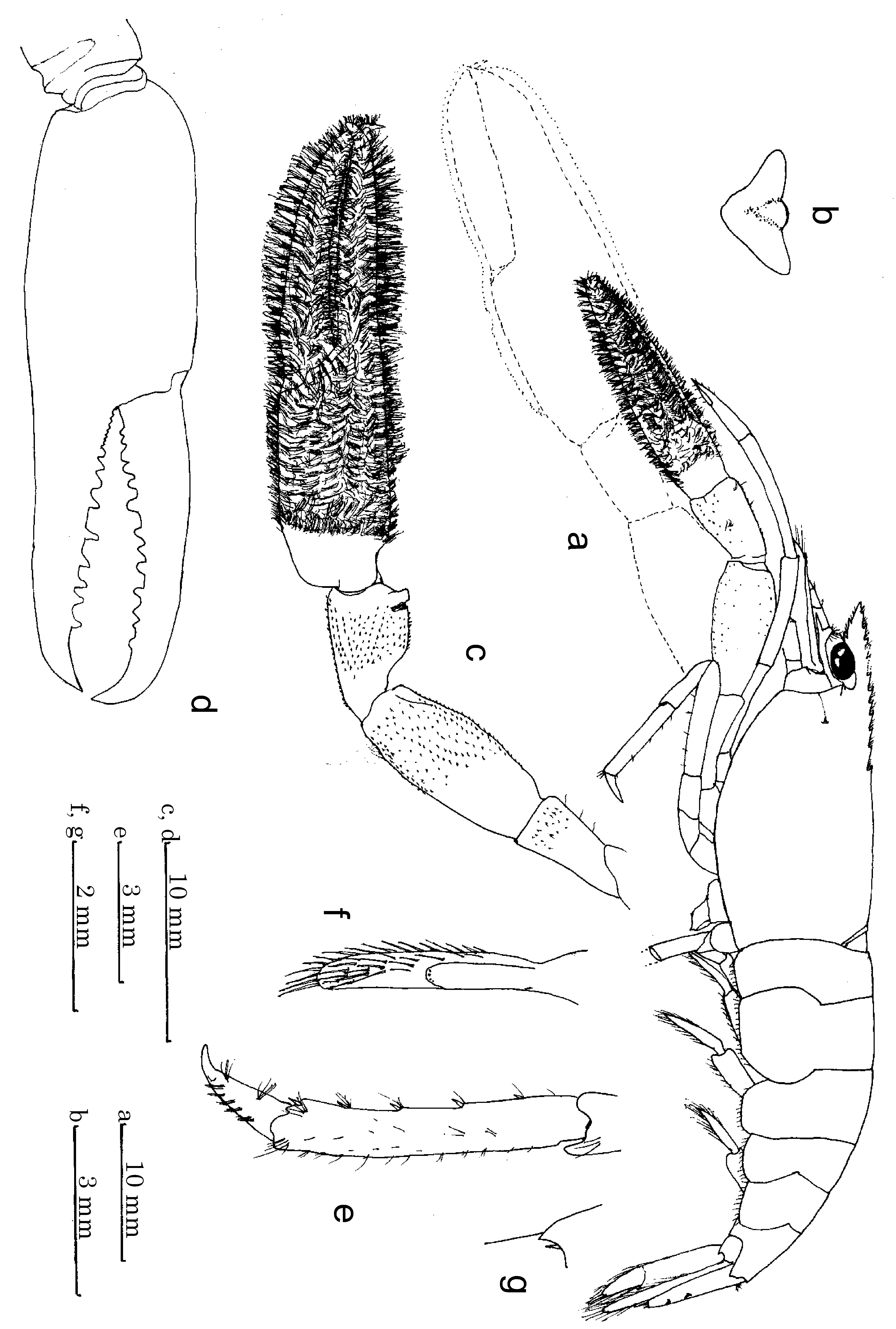
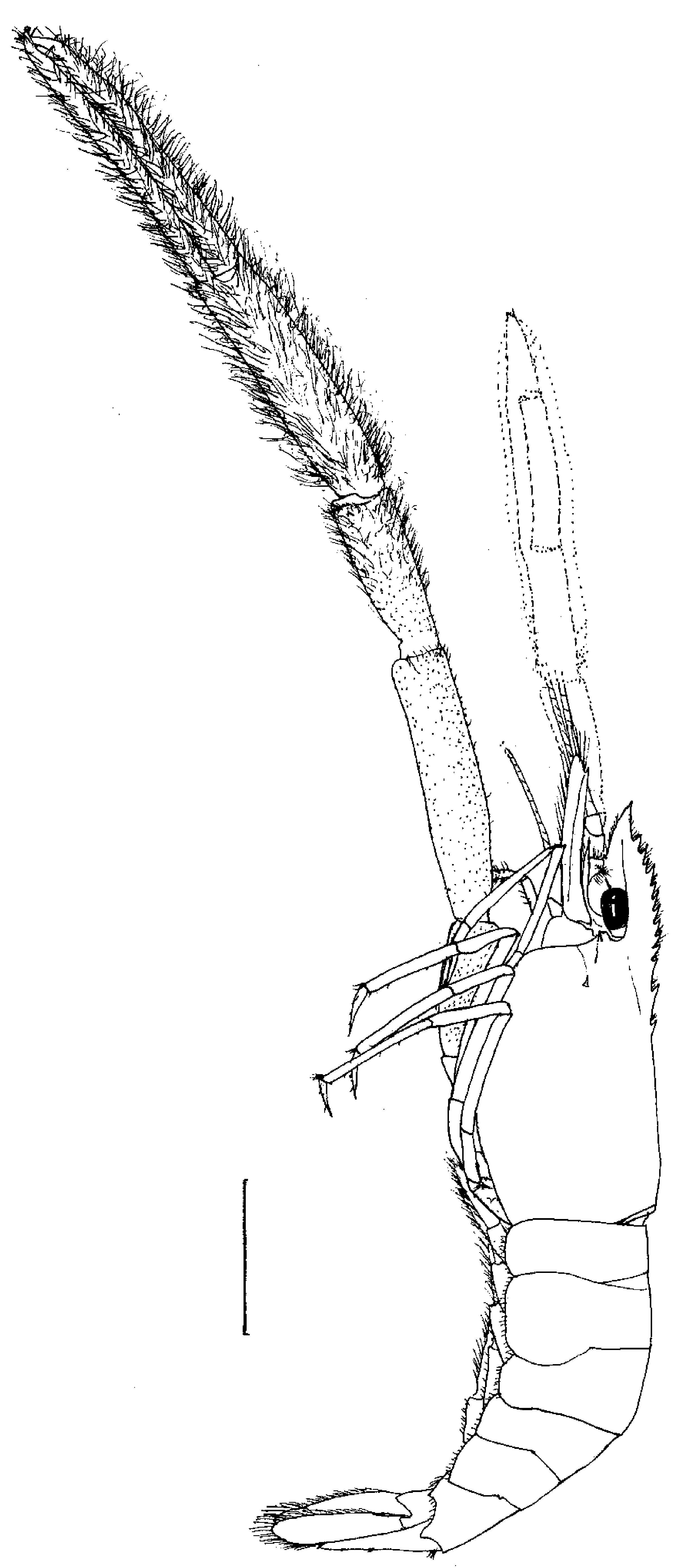

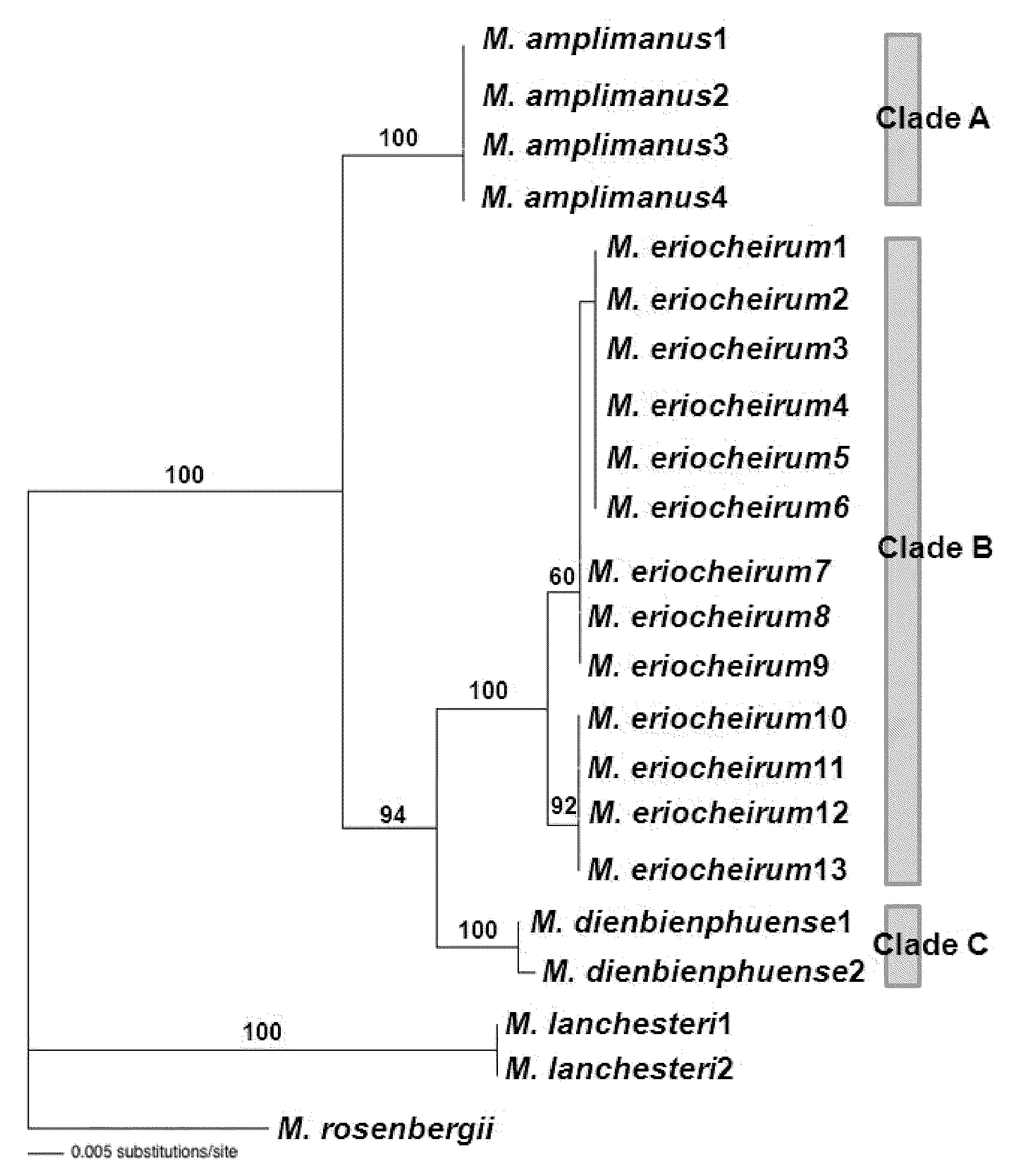
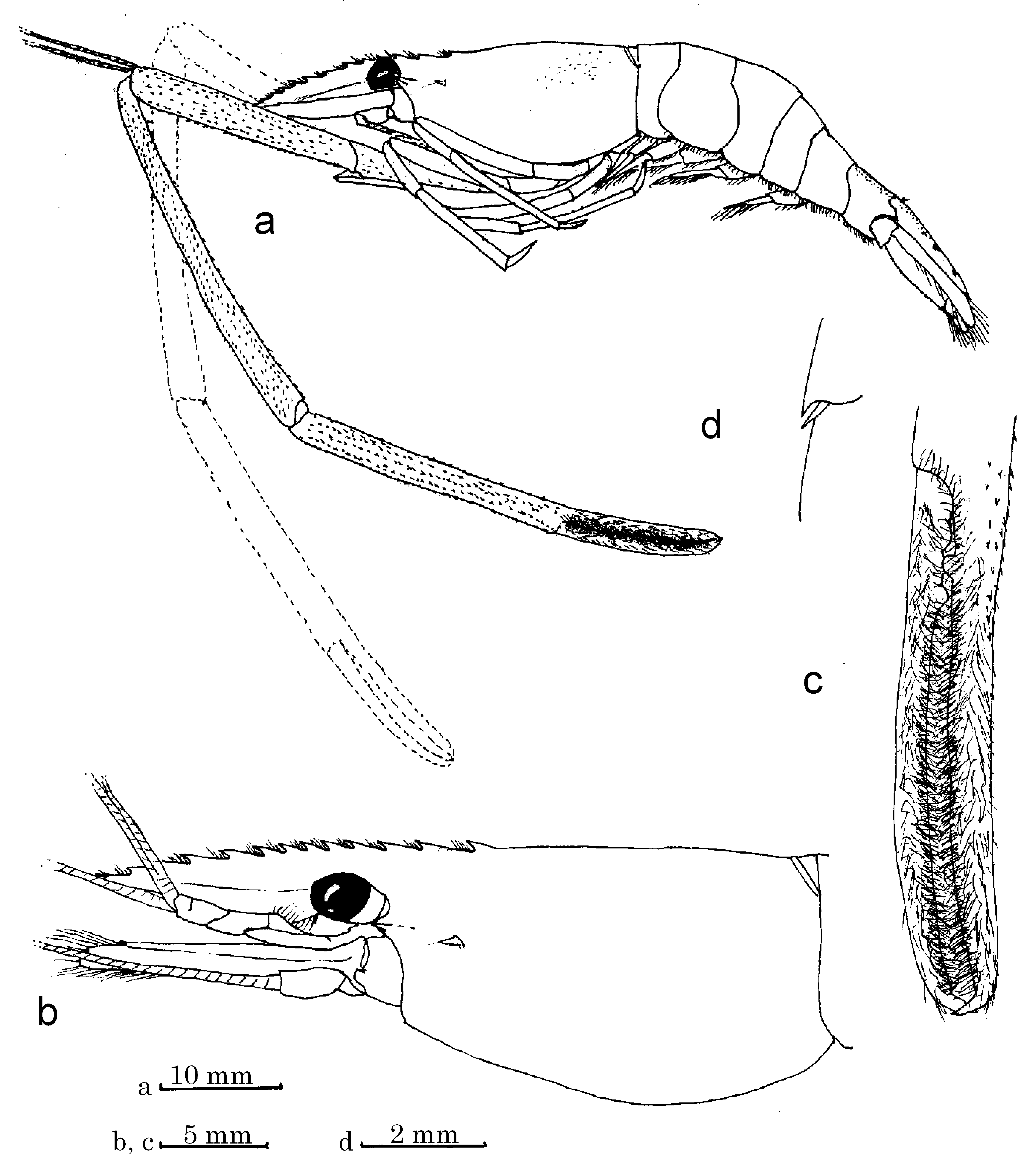
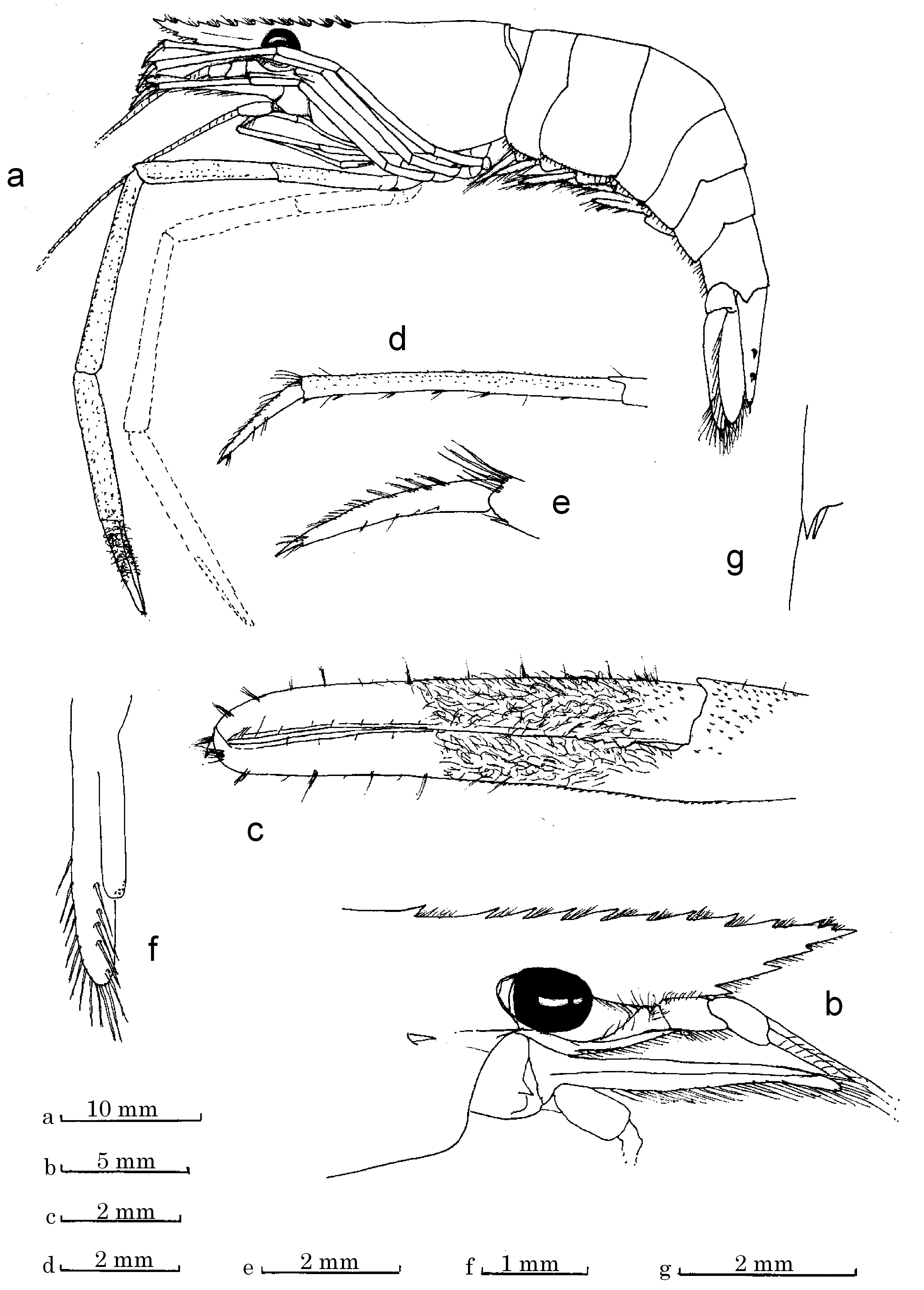
TABLE 2. Comparison of specimens of three species belonging to the “ Macrobrachium dienbienphuense ” group collected
| merus | inflated, length 2.3–3.0 x high | sub-cylindrical, length 3.75–4.25 x high | slightly inflated, length 2.9–3.0 x high |
|---|---|---|---|
| carpus | cup-shaped, length 1.0 x high | sub-cylindrical, length 2.4–2.9 x high | sub-cup-shaped, length 2.0 x high |
| palm fingers | length 1.6–2.2 x broad narrΟʷ ǥapɵ﹐ ₁ɜ–₁7 ɭɵɵɭh | length 3.1–3.8 x broad ʷiđɵ ǥapɵ﹐ ₁⁸–²² ɭɵɵɭh | length 2.2–2.8 x broad narrow gape, ₁₁–₁7 ɭɵɵɭh |
| Egg size (mm) | 0.9–1.1 x 1.3–1.5 | 0.9–1.0 x 1.35–1.5 | 1.0–1.25 x 1.4–1.7 |
No known copyright restrictions apply. See Agosti, D., Egloff, W., 2009. Taxonomic information exchange and copyright: the Plazi approach. BMC Research Notes 2009, 2:53 for further explanation.
|
Kingdom |
|
|
Phylum |
|
|
Class |
|
|
Order |
|
|
Family |
|
|
Genus |
Macrobrachium dienbienphuense Dang & Nguyen, 1972
| Hanamura, Yukio, Imai, Hideyuki, Lasasimma, Oulaytham, Souliyamath, Pany & Ito, Sayaka 2011 |
Macrobrachium longidigitum
| Dai 1984: 248 |
Macrobrachium dienbienphuense
| Li 2007: 84 |
| Liu 1990: 121 |
| Dang 1980: 384 |
| Dang 1972: 4 |