
Spirostreptus digitus, Enghoff, 2023
|
publication ID |
https://doi.org/10.11646/zootaxa.5389.2.9 |
|
publication LSID |
lsid:zoobank.org:pub:4D3459E8-2985-49C9-A035-D3FA95438A5C |
|
DOI |
https://doi.org/10.5281/zenodo.10406991 |
|
persistent identifier |
https://treatment.plazi.org/id/1C297063-FF9F-FFB2-FF1D-F891FCA2F9BE |
|
treatment provided by |
Plazi |
|
scientific name |
Spirostreptus digitus |
| status |
sp. nov. |
Spirostreptus digitus View in CoL sp. nov.
Figs 1–2 View FIGURE 1 View FIGURE 2
Diagnosis. Differs from other species of Spirostreptus in the peculiar morphology of the gonopod coxa, particularly the straight (latero)apicad process ( ff) originating from the posterior side of the coxa ( Fig. 2E, F View FIGURE 2 ).
Etymology. Refers to the distal part of the gonopod coxa which somewhat resembles a fist with an extended (index) finger (Latin: digitus) and a laterally extended thumb. Noun in apposition.
Material studied.
Holotype: ♂ TANZANIA • Iringa Region, on David Moyer’s land E of Iringa; 7°45’S, 35°40’E; 1200–1500 m a.s.l.; 10 Jan. 1996; M. Andersen, P. Gravlund, A. Jakobsen leg.; open bushland with scattered trees, sloping towards Little Ruaha River; NHMD 1184565 . GoogleMaps
Paratypes: TANZANIA • 1 ♀; same collection data as holotype; NHMD 1184566 GoogleMaps • 1 ♂; Iringa region, Mufindi District, Mufindi ; 8°36’S, 35°17’E; 1800 m a.s.l.; Mar.–Apr. 1996; L. L. Sørensen leg.; pine plantation; NHMD 1184568 GoogleMaps .
Referred specimens (not types): TANZANIA • 3 juv.; same collection data as holotype; NHMD 1184567 GoogleMaps .
Description.
Holotype [Differences in male paratype in square brackets]
SIZE. Length ca. 170 [150] mm, vertical diameter 11.3 [11.7] mm. 62 podous rings, no apodous rings in front of telson.
COLOUR. After 27 years in alcohol, head above eyes dark brown, below eyes yellowish brown; antennae yellowish brown; collum dark brown; body rings with light-yellowish brown prozonites and blackish brown metazonites, posterior margin of metazonites amber; telson medium [yellowish] brown; legs medium [yellowish] brown.
HEAD ( Fig. 1 View FIGURE 1 ). Head capsule without peculiarities. Eyes medially reaching just beyond antennal socket, with ca. 55 [50] ommatidia in ca. 15 vertical and ca. 6 horizontal rows. Antennae reaching back to body ring 3 when stretched. Mandibles with small stipital lobes ( stl) ( Fig. 1B, C View FIGURE 1 ). Gnathochilarium ( Fig. 1B, D View FIGURE 1 ): gula ( gu) three-lobed; prementum (= prebasilar plate) ( pm) in one piece, three-lobed; mentum ( me) heptagonal, with a subcircular field of stout setae in distal half; stipites ( st) with a field of setae paralleling stipito-mental suture and another field of setae along distal half of lateral margin; lamellae linguales ( ll) setose all over.
COLLUM ( Fig. 1A View FIGURE 1 ). Smooth, with three strong lateral furrows. Lateral lobes rounded-rectangular, antero-lateral corner only very slightly expanded compared to female.
BODY RINGS. With horizontal suture at ozopore level. Prozonites with usual fine ring furrows in anterior part. Metazonites not vaulted, clearly striate up to a little above ozopore, dorsally smooth. Ozopore situated ¼–⅓ metazonite length behind suture. A single row of small sigilla, along part of the circumference paralleled by a row of even smaller ones. Stigmatal grooves not extended.
TELSON. Finely punctate. Preanal ring without a process. Anal valves with raised lips, i.e., “labiate” in the terminology of Hoffman (2011). Subanal scale broad triangular.
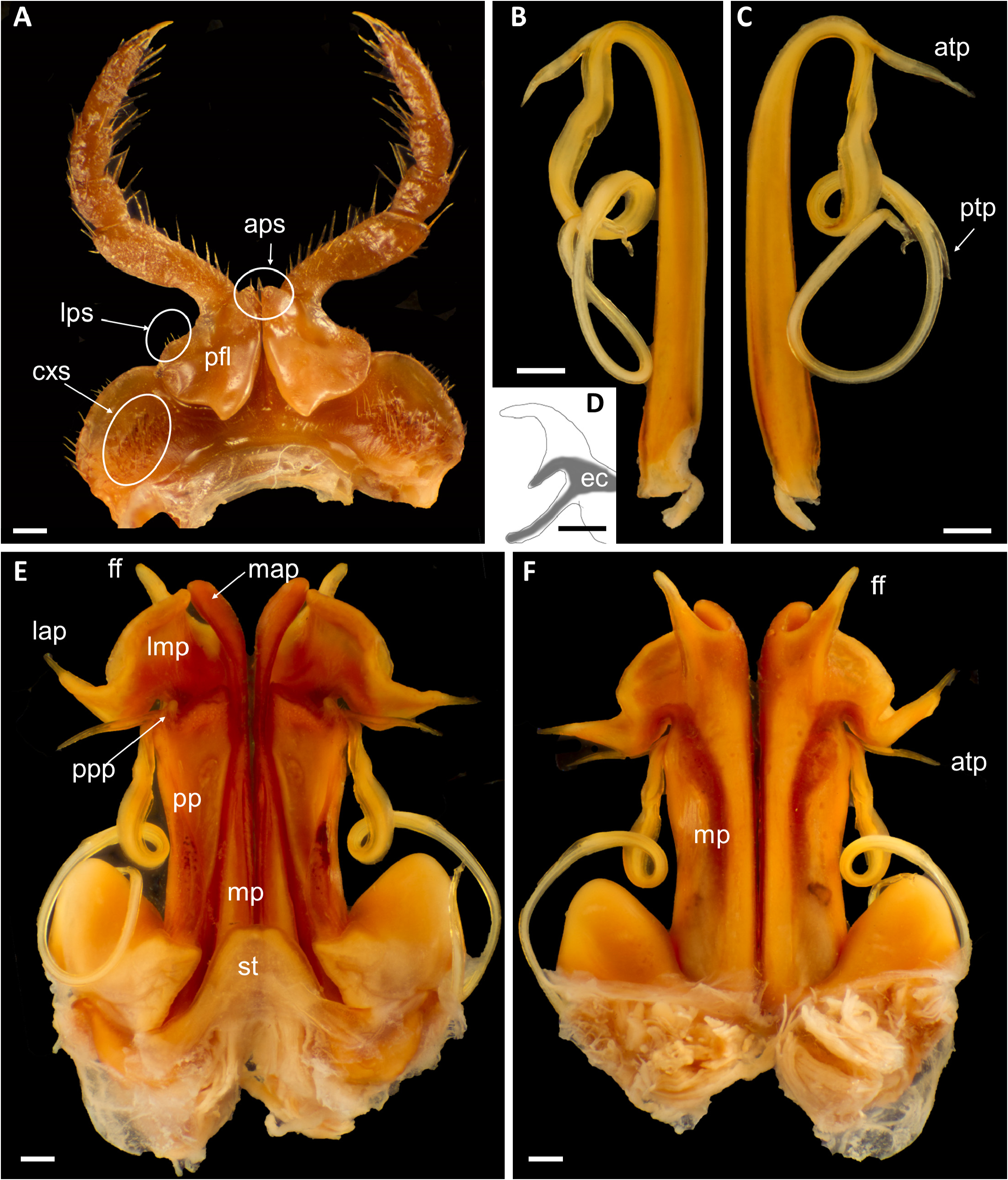
LEGS. Length 0.8 × body diameter. With postfemoral and tibial pads from leg-pair 3 until the seventh-last body ring, pads gradually smaller on last padded leg-pairs. First pair ( Fig. 2A View FIGURE 2 ): coxosternum with setae on lateral margin and roundish fields of setae ( cxs) on the anterior surface. Prefemur with a few apical setae ( aps) and a field of setae ( lps) at the lateral margin. Prefemoral lobes ( pfl) laterally produced into large semicircular process, distally (=dorsally) produced into smaller triangular process.
GONOPODS ( Fig. 2B–F View FIGURE 2 ). Sternum ( st) trapezoidal [broadly rounded triangular]. Proplica ( pp) ca. ¾ as long as metaplica; mesal margin straight; lateral margin raised to form longitudinal keel, parallel to mesal margin for ca. 40% of its length, then moderately diverging, apical part of proplica hence triangular; apical margin oblique, excavated to form a shallow transverse groove, laterally with a short, rounded process ( ppp) covering exit of telopodite from coxa; apical margin and lateral side of rounded process setose. Metaplica ( mp) complex; mesal margin straight until tip of proplica, then curving gently laterad; with high keel along it entire length, ending in rounded process ( map); a large lateroapical structure ( lmp) separated from rounded process by obvious incision, lmp roughly triangular, its oblique distal margin swollen, its lateral corner drawn out into straight, pointed, lateroapical process ( lap); a long, straight (latero)apicad process ( ff) originating from posterior side of lmp and overreaching rest of gonopod. Telopodite simple; antetorsal process ( atp) thin, straight, smooth, directed laterad; torsotope extended, situated just after antetorsal process; posttorsal telopodite whiplike, making a 270° turn, thereafter with a short, pointed posttorsal process ( ptp); tip of telopodite ( Fig. 2D View FIGURE 2 ) trifid, the efferent canal ( ec) branching into two of the prongs.
Female paratype
Length ca. 170 mm, vertical diameter 13.7 mm. 64 podous rings, no apodous rings in front of telson. Similar to male in non-sexual characters. Mandibular stipites without lobes. Anterolateral corner of lateral lobes of collum slightly less expanded.
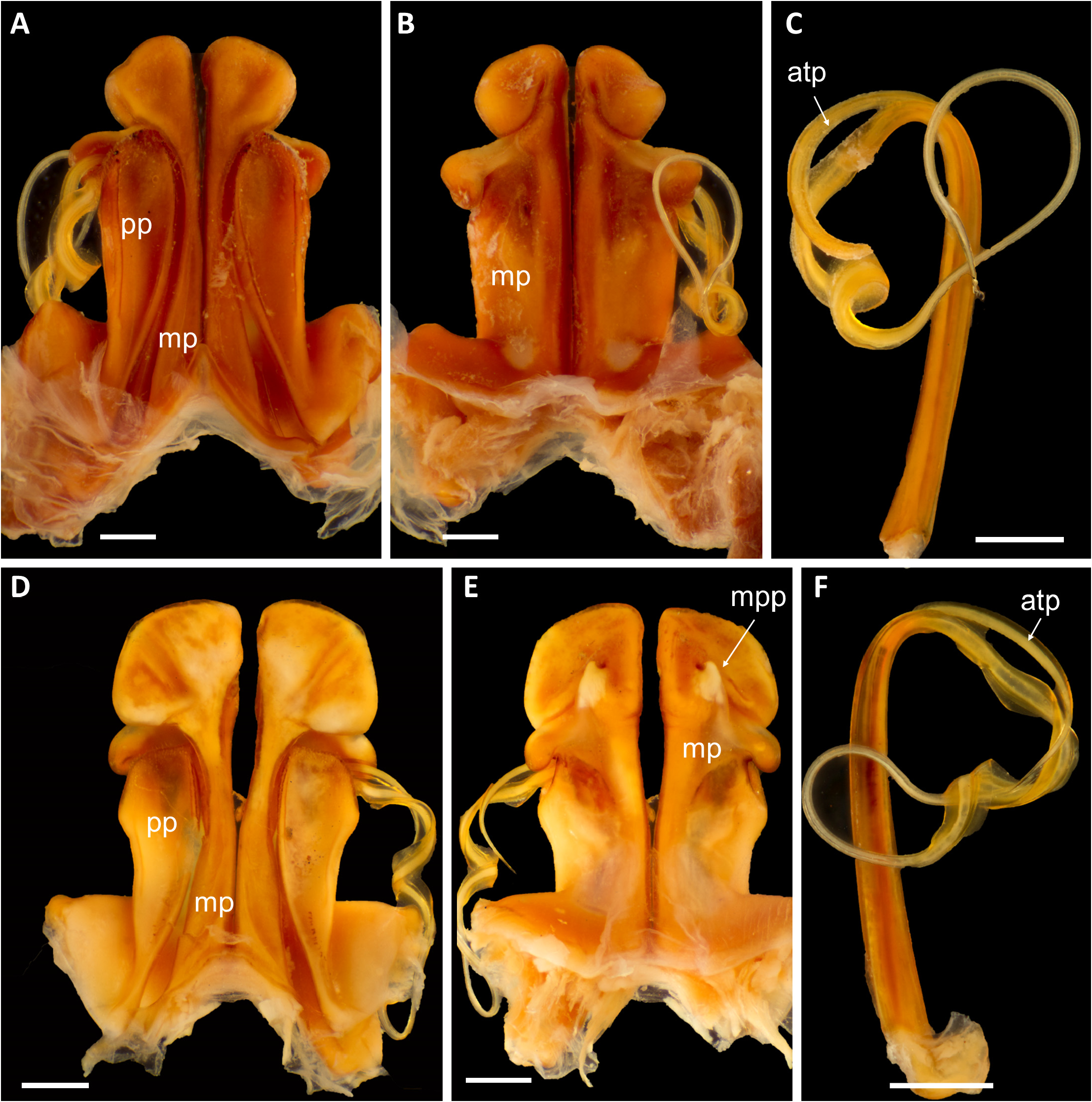
Remarks. The new species fulfils the above differential diagnosis of Spirostreptus which is built on the key to spirostreptinine genera in Hoffman (2008) and the broader diagnosis in Mwabvu et al. (2009a). The other species of Spirostreptus form quite a homogeneous group in terms of gonopod shape, see Mwabvu et al. (2009a) and Fig. 3 View FIGURE 3 . Spirostreptus digitus sp. nov., in contrast, stands out, notably by the (latero)apicad process ( ff) originating distally on the posterior side of the coxa; such a structure is unknown from other Spirostreptus species, except perhaps in S. tripartitus where the “metaplica prong” illustrated by Mwabvu et al. (2009a: fig. 4b), see also Fig. 3E View FIGURE 3 : mpp, may be homologous with ff in S. digitus sp. nov. Also the lateroapical process ( lap) of the gonopod coxa of S. digitus sp. nov. lacks a counterpart in other congeners; in this respect the new species more resembles species of the genus Archispirostreptus ( Mwabvu et al. 2010) . See discussion.

No known copyright restrictions apply. See Agosti, D., Egloff, W., 2009. Taxonomic information exchange and copyright: the Plazi approach. BMC Research Notes 2009, 2:53 for further explanation.
|
Kingdom |
|
|
Phylum |
|
|
Class |
|
|
Order |
|
|
Family |
|
|
Tribe |
Spirostreptini |
|
Genus |