
Simulium ( Hebridosimulium ) tuberculum Craig., 2006
|
publication ID |
https://doi.org/10.11646/zootaxa.1380.1.1 |
|
publication LSID |
lsid:zoobank.org:pub:ADA6B48B-CF5D-43A2-8E66-CA946A79A8F8 |
|
persistent identifier |
https://treatment.plazi.org/id/1C1B2B5D-FF97-FFC5-8748-FC9D48DBFA85 |
|
treatment provided by |
Felipe |
|
scientific name |
Simulium ( Hebridosimulium ) tuberculum Craig. |
| status |
sp. nov. |
Simulium ( Hebridosimulium) tuberculum Craig. View in CoL n. sp.
( Figs. 5d View FIGURE 5 , 8d View FIGURE 8 , 9e View FIGURE 9 , 11e View FIGURE 11 , 13e View FIGURE 13 , 16e View FIGURE 16 , 18e View FIGURE 18 , 20e View FIGURE 20 )
Hebridosimulium jolyi: Grenier and Rageau (1961: 96) View in CoL not Roubaud 1906.
Simulium ( Hebridosimulium) jolyi: Crosskey (1967:27) View in CoL in part not Roubaud 1906.
Types
Holotype. Adult : pinned reared female, dried from alcohol. Label data— “ Simulium ( Hebridosimulium) tuberculum , VANUATU, Efate, Ewor R., La Cressionnière, 16.vi.1981, Coll. D. A. Craig ”, “ Holotype ”. Pupa and cocoon as subsidiary material ( BM).
Paratypes. Adults: Six pinned reared females (4 BM, 2 BPBM), one reared male ( BM). Label data— as for Holotype, but with “ Paratype ”. Six reared females, two males, label data— “ S. ( H.) tuberculum, Ewor River, La Cressionnière. S17.71728° E168.56946°, alt. 24m, 5.x.2004. Coll. D. A. & R. Craig. Paratype ” ( BM, BPBM). Alcohol: label data— as for GoogleMaps Holotype, but with “ 21.vi.1981 ” and ‘PARATYPE’ (larvae; BM, BPBM. Pupae, reared adults; DAC) .
Diagnosis
Pupa: thoracic cuticle with sparse, unevenly distributed granules. Cocoon: relatively small and not completely covering pupa, with tendency to be flared basally. Larva: lateral head spot beneath stemmata absent, dorsolateral tubercles present.
Description
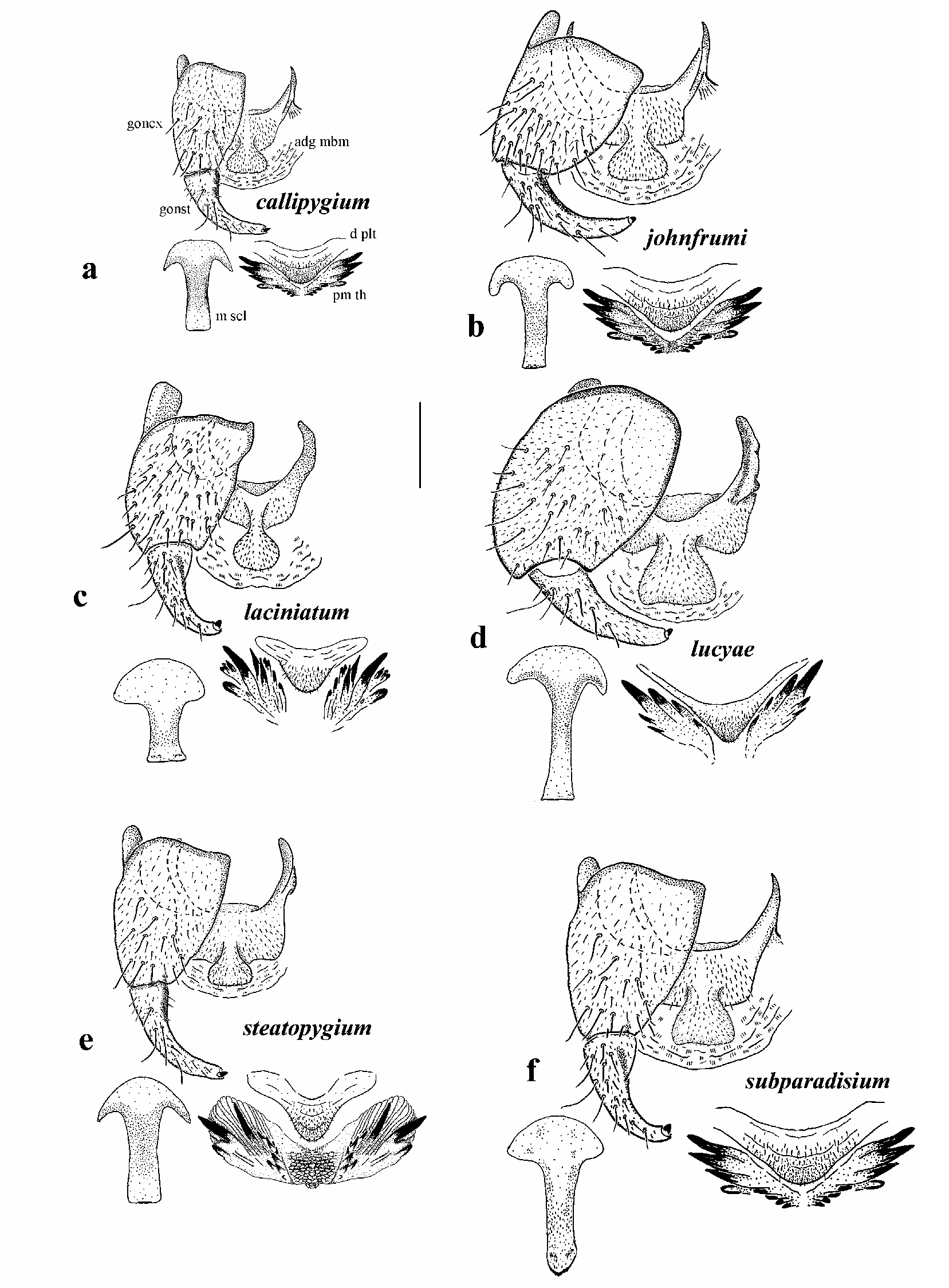
Adult female (based on numerous specimens). Body: head and abdomen very dark brownish black, thorax evenly light brown; total length 2.1–2.6 mm. Head: width 0.72–0.83 mm; depth 0.57 mm; postocciput black, vestiture of dense short black hairs; frons paler ventromedially; frons-head ratio (narrowest width of frons: greatest width of head) 1.0:4.0. Eye: interocular distance 0.18–0.21 mm; ommatidia 0.026 mm in diameter; 35 rows up and across at mid eye. Clypeus: 0.22 mm wide; concolourous with ventral region of frons; vestiture of black and pale hairs. Antenna: length 0.48 mm; flagellomeres light brown, scape and pedicel pale yellow. Mouthparts: 0.3 length of head depth; mandible shorter than lacinia, poorly sclerotized with 20 inner teeth, larger distally, poorly developed basally; lacinia with 11 inner teeth and 11 outer teeth; maxillary palpus, all articles evenly brown, proportional length of 3rd, 4th, and 5th articles 1.0:0.8:1.3; sensory vesicle ovoid, less than 0.3 times width of 3rd article, opening 0.5 times width of vesicle. Thorax: length 1.1–1.2 mm; width 0.94 mm; postpronotal lobes paler than scutum; scutum evenly medium brown, vestiture of very sparse, fine pale hairs, with few longer darker hairs posteriorly; scutellum pale, vestiture of few black hairs and pale hairs laterally, apical angle 120°, rounded and bare; postnotum concolourous with scutum; pleural membrane, pale brown and with markedly fine pale hairs (not observable at 50x in alcohol). Wing: length 1.6–2.1 mm; width 0.9–1.1 mm. Legs: as for S. johnfrumi . Abdomen: as for S. johnfrumi . Genitalia ( Fig. 5d View FIGURE 5 ): similar to S. johnfrumi ; sternite VIII pale, not deeply indented; hypogynial valves lightly pigmented with sparse vestiture; median edges markedly concave and divergent, membranous apex ridged, markedly directed medially; genital fork with stem thickened, posterolateral arms lightly pigmented, anteriorly directed apodeme of lateral arm rounded and smaller than posterior portion; anal lobes with anterior extension not markedly developed, median depression not strongly developed; cercus elongated.
Adult male (based on 9 reared specimens). Generally as for S. johnfrumi , but smaller. Body: total length 1.9–2.0 mm. Head: width 0.77–0.82 mm; depth 0.47–0.53 mm. Eyes: upper ommatidia orange, 0.039 mm in diameter, ca. 13 across and 16 down; lower ommatidia dark brown, 0.018 mm in diameter, ca. 24 across and down. Clypeus: brown, paler medially; 0.2 times as wide as head; vestiture of long fine pale hairs. Antenna: total length 0.44 mm; flagellomeres pale brown, scape and pedicel yellow. Mouthparts: length 0.34 times head depth; mandibles insubstantial, finely tapered with apical hairs; lacinia broad basally, finely tapered apically with terminal hairs; maxillary palpus dark brown, 0.38 mm long, proportional lengths of 3rd, 4th, and 5th articles 1.0:0.8:1.1, 3rd article darker and markedly hairy, sensory vesicle spherical, occupying 0.33 times width of article, opening 0.25 times width of vesicle. Thorax: as for S. johnfrumi , but smaller, length 0.86–0.93 mm; width 0.8 mm. Wing: 1.6–1.9 mm in length, 0.77–0.83 mm at maximum width. Legs: essentially as for S. johnfrumi , but hind leg with femur and tibia not as swollen and flattened, width to length ca. 0.22, and pretarsal claw with ca. 23 grappling hooks dorsally. Abdomen: dark brown; basal scale well developed and dark, hairs markedly long, fine and pale, extended beyond 2nd segment; 1st and 2nd segment paler brown, remainder dark brown; vestiture of sparse pale hairs. Genitalia: as for S. johnfrumi (e.g., Figs. 6b View FIGURE 6 ).
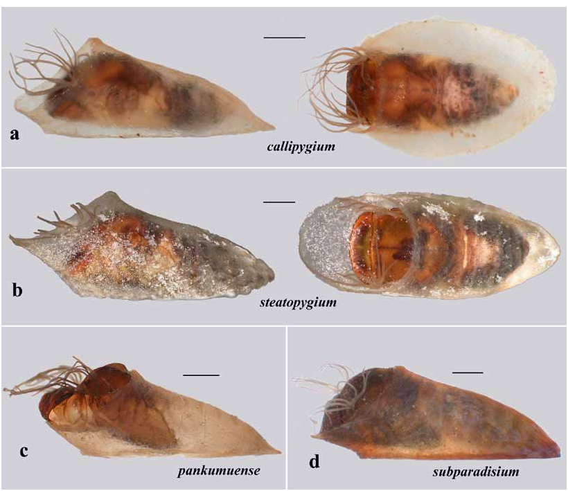
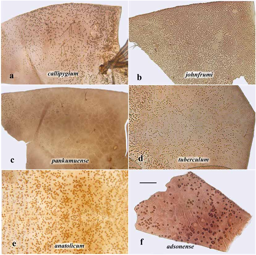
Pupa (based on numerous specimens). Generally as for S. johnfrumi , but with coarser thoracic granules ( Fig. 8d View FIGURE 8 ). Body length: male 2.2–2.4 mm, female 2.6–2.9 mm. Gill ( Fig. 9e View FIGURE 9 ): petioles moderately elongate except for 1 ventral pair, but variable; 6 dorsal filaments subequal in length, 0.6–0.8 length of ventral filaments; branching pattern of filaments (2+2)+2+2+2; annulations fine. Cocoon: shoe shaped, as for S. johnfrumi ; in males flared basally more often than not, less so in females.
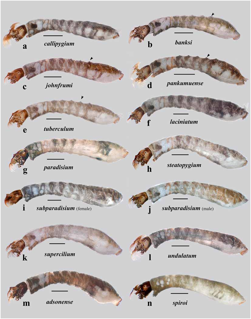
Larva (based on numerous mature last-instar larvae). Body ( Fig. 11e View FIGURE 11 ): generally as for S. johnfrumi ; male yellowish, females grayish; total length, male 4.7–5.9 mm, female 5.6–6.4 mm. Head ( Fig. 13e View FIGURE 13 ): overall as for S. johnfrumi but slightly darker; male length 0.74–0.83 mm, width 0.58–0.66, distance between antennal bases 0.32–0.34; female length 0.73–0.86 mm, width 0.55–0.65, distance between antennal bases 0.35–0.37; lateral margins of head more convex than for S. johnfrumi ; head-spot pattern less distinct than for S. johnfrumi , lacking distinct lateral head spot beneath stemmata (similar to S. johnfrumi from Erromango); ecdysial lines markedly broadly rounded at maximum width; posterior margin of apotome very slightly emarginate; postocciput finely extended between cervical sclerite and apotome. Antenna as for S. johnfrumi . Labral fan: anterior palatal bar on stalk markedly developed; 38–41 rays, 1.0 mm in length, 5 or 6 rays less substantial, distinct pattern of microtrichia, with 6 smaller, decreasing in size between those longer. Postgenal cleft ( Fig. 16e View FIGURE 16 ): arrowhead shaped, 1.5 times deeper than wide, posteroventral elongated muscle spots neutral. Postgenal bridge: 0.5 times as long as cleft depth; genae and postgenae evenly light brown. Hypostoma ( Fig. 18e View FIGURE 18 ): overall cone-shaped; ratio 4.7; median and lateral teeth distinct, subequal in length, latter slightly scalloped medially, sublateral teeth usually small, but individual teeth variably occasionally larger; paralateral teeth not apparent, lateral serrations barely visible; 8 or 9 hypostomal setae per side, medial setae markedly substantial. Mandible ( Fig. 20e View FIGURE 20 ): apical and subapical teeth substantial as are the few spinous teeth, gap marked; sensillum fused to serration; blade region smooth and convex. Abdomen: as for S. johnfrumi , but lighter in colour. Posterior circlet: directed slightly ventrally; 160 rows, 23–26 hooks (total ca. 3,800).
Additional material examined
Efate. Fourteen slide mounts, including 11 “plesiotypes” with round green labels, variously labeled, Klem (sic) River, 26.v.1958, Coll. J. Rageau (larvae, pupae, adults; PI) . Klem (sic) River, 26. v. 1958, 5.vi.1958, Coll. J. Rageau (larvae — numerous, pupae, adults; BM, PI) . Klehm Cascade , 17.vi.1981. Coll. D. A. Craig (larvae; DAC) . Ewor River , La Cressionnière, alt. 10m, 16.v.1981. Coll. D. A. Craig (reared adults; DAC) ; 21.vi.1981 (larvae, pupae; ANIC, BM, BPM, DAC, HT, PI) ; 23.ii.1985, Coll. B. S. Batson (larvae; DAC) . Ewor River , La Cressionnière. S17.71728° E168.56946°, alt. 24m. 5-x- 2004. Coll. D. A. & R. Craig (larvae (some in Carnoy’s), pupae, reared adults; BPBM, DAC, LCNZ, ROM) GoogleMaps . Mele Cascade. S17.67778° E168.25473°. alt. 65m. 6.x.2004. Coll. D. & R. Craig. (larvae, pupae, reared adults; DAC) GoogleMaps .
Etymology Named for the dorsolateral abdominal tubercles on the larva.
Distribution VANUATU: Efate.
Comments
Problems associated with the work of Grenier & Rageau (1961) on Efate material were discussed earlier. Simulium tuberculum is, in great part, that described by these authors as S. jolyi, Efate. Although S. johnfrumi is very similar to S. tuberculum , it differs markedly by lacking lateral head spots in the larva and possessing coarser dorsal thoracic granules in the pupa. The highly variable pupal gill-filament branching patterns might indicate that S. tuberculum is a complex of species.
Ontogenetically, the four pairs of dorsolateral tubercles on abdominal segments II–V are derived. Earlier instars lack them and they develop from approximately the 5th instar onwards. This applies to all the tuberculum species subgroup larvae.
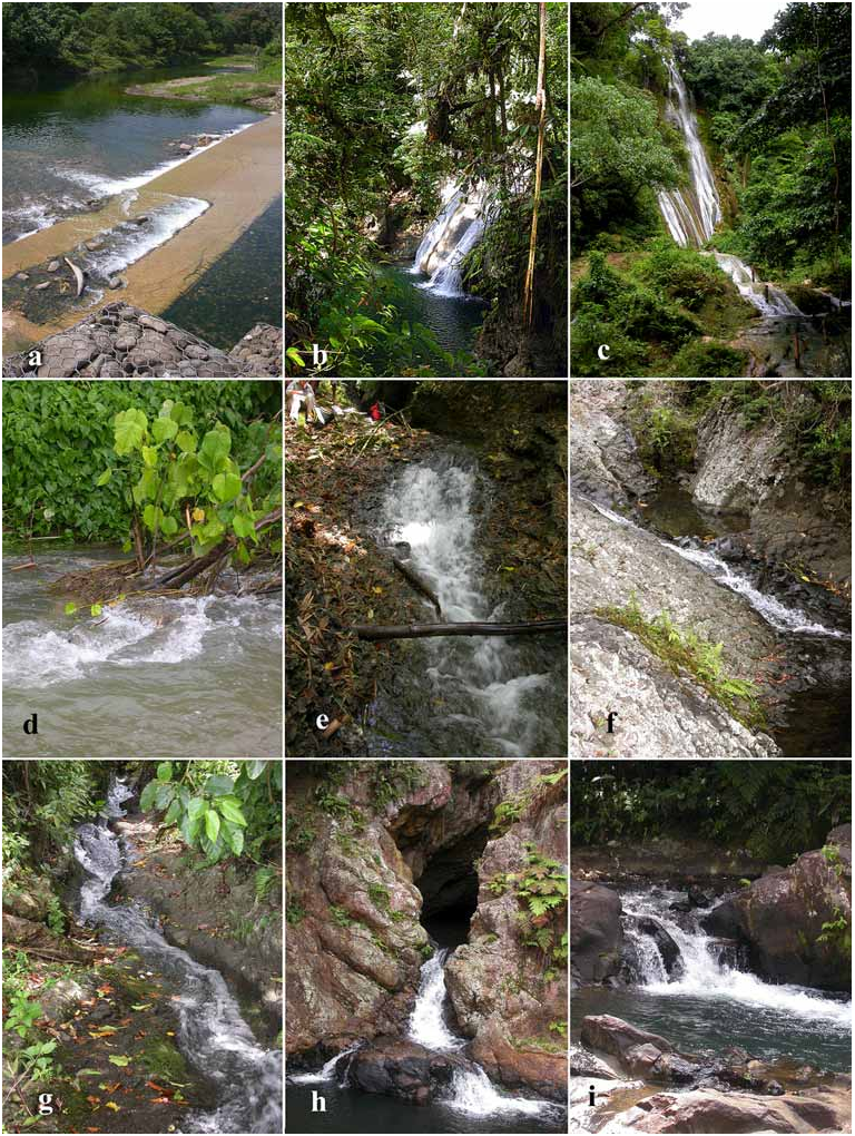
Many streams in Efate flow over raised fossilized coral and the water is hard (Table 1), with calcium deposits or travertine (tufa) ( Hynes 1970) common. Both the Mele (or Klehm) Cascade and the Ewor River at La Cressionnière have such ( Figs. 23g View FIGURE 23 , 24c View FIGURE 24 ). Larvae could be taken in astronomical numbers from travertine surfaces and vegetation in fast flow. Pupae were mainly taken from trailing vegetation, with the cocoon normally encrusted with travertine (e.g., Fig. 7b View FIGURE 7 ). At La Cressionnière, large collections by DAC, June, 1981, were entirely of S. tuberculum ; however, in January, 1985, B. S. Batson collected a few S. steatopygium larvae in an otherwise monospecific population of S. tuberculum . Rageau’s material from the Klehm (sic) River (or Mele Cascade) in May/ June 1958 was almost entirely S. tuberculum . Again, in 2004, larvae from La Cressionnière were solely S. tuberculum , suggesting seasonal differences in species as well as probable habitat preferences.


No known copyright restrictions apply. See Agosti, D., Egloff, W., 2009. Taxonomic information exchange and copyright: the Plazi approach. BMC Research Notes 2009, 2:53 for further explanation.
|
Kingdom |
|
|
Phylum |
|
|
Class |
|
|
Order |
|
|
Family |
|
|
Genus |
Simulium ( Hebridosimulium ) tuberculum Craig.
| Craig, Douglas A., Currie, Douglas C., Hunter, Fiona F. & Spironello, Mike 2006 |
Simulium ( Hebridosimulium ) jolyi : Crosskey (1967:27)
| Crosskey, R. W. 1967: ) |
Hebridosimulium jolyi :
| Grenier, P. & Rageau, J. 1961: ) |