
Xenochironomus canterburyensis (Freeman, 1959)
|
publication ID |
https://doi.org/ 10.11646/zootaxa.3646.2.1 |
|
publication LSID |
lsid:zoobank.org:pub:A574552A-1208-4AB6-B3FA-9D6AAEFEB1E2 |
|
DOI |
https://doi.org/10.5281/zenodo.6162989 |
|
persistent identifier |
https://treatment.plazi.org/id/142A4E4C-B654-AB4E-FF34-C59133E9FE64 |
|
treatment provided by |
Plazi |
|
scientific name |
Xenochironomus canterburyensis (Freeman, 1959) |
| status |
|
Xenochironomus canterburyensis (Freeman, 1959) View in CoL
Chironomus (Dicrotendipes) canterburyensis Freeman 1959: 425 ; Forsyth 1971: 128.
Xenochironomus canterburyensis (Freeman, 1959) ; Forsyth and McCallum 1978a: 331, 1978b: 795, Forsyth 1979: 467.
Material examined. NEW ZEALAND, Ngapuna, Lake Rotorua, North Island, NZ-17.1, 5–14. xii.1973, 1 male, leg.: Jon Martin, D. J. Forsyth (UMGD). Lake Wakatipu, 4 km Queenstown, South Island, NZ-61.1, 21.i.1978, in spider web, 1 male, leg.: Jon Martin (UMGD). Ngapuna, Lake Rotorua, North Island, NZ-17.1, 5–14. xii.1973, 1 female, leg.: Jon Martin, D. J. Forsyth (UMGD). Te Anau, Lake Te Anau, South Island, NZ-46.1, 6. i.1974, 1 female, leg: Jon Martin (UMGD). Ngapuna, Lake Rotorua, North Island, NZ-17.1, 5–14. xii.1973, 1 pupa, leg.: Jon Martin, D. J. Forsyth (UMGD). Ngapuna, Lake Rotorua, North Island, NZ-17.1, 5–14. xii.1973, 1 larva, leg.: Jon Martin, D. J. Forsyth (UMGD).
Diagnosis. Xenochironomus canterburyensis can be separated from other species in the genus by the following characteristics: male, apex of anal point not surpassing of apex of inferior volsella; median setae of anal tergite divided into two areas; several setae of the inferior volsella and superior volsella pediform. Pupa, Tergite II without bands or groups of black shagreen, sternites I and II with posterior bands of clear spines and pedes spurii A absent in segment IV. Larva, with labral sclerite 1 undivided; mentum with median tooth sutured; number of teeth on the mentum may be either odd or even and teeth may have different shapes depending on the instar.
Description. Male (n = 1–2)
Color. Light brown. Thorax with scutum, scutellum and pronotum brown. Legs light brown, with apex of tarsomere 1 and tarsomere 2–5 dark brown.
Total length 6.23–7.56 mm. Wing length 3.19–3.88 mm, width 0.85–0.92 mm. Total length/wing length 1.95. Wing length/profemur length ratio 2.41–2.74.
Head. AR 3.40–3.43. Apical flagellomere 1109–1311 Μm long Temporals 9. Clypeus with 13 setae. Palpomere lengths 1–5 (in µm): 58–60; 56–72; 281; 229–242; 311–363.
Thorax. Ac approximately 19; Dc 11–14; Pa 5–7; Scts 10.
Wing. VR 1.04–1.08. Brachiolum with 2 setae. R with 31 setae, R1 with 23 setae and R4+5 with 29 setae, remaining veins bare. Squama with 10 setae.
Legs. Length (µm) and proportions of leg segments as in Table 1 View TABLE 1 .
Hypopygium. ( Fig. 1 View FIGURE 1 A–C) Laterosternite IX with 5 setae. Anal tergite with 40–45 setae divided into two areas. Anal point 44–69 Μm long; 54–74 Μm wide at the base. Phallapodeme 138–162 Μm long; transverse sternapodeme 121–160 Μm long. Superior volsella 94–125 Μm long, 21–32 Μm wide at the base. Inferior volsella 151–202 Μm long, 27–33 Μm wide at the base and 33–46 Μm wide at the apex. Gonocoxite 242–277 Μm long. Gonostylus 249–334 Μm long. HR 0.83–0.97. HV 2.26–2.50.
Female (n = 1–2)
Similar to male, except as follows:
Total length 6.17–7.35 mm. Wing length 3.88–4.64 mm, width 1.22–1.50 mm. Total length/wing length 1.59. Wing length/profemur length ratio 2.72–2.93.
Head: AR 0.38–0.45. Flagellomere lengths 1–5 (µm): 149–157; 114–129; 119–129; 121–125; 193–243. Clypeus with 22 setae. Palpomere lengths 1–5 (µm): 80–84; 70–80; 260–286; 249–261; 369–407.
Thorax. Ac 15; Dc 18; Pa 9–10; Scts 22–24.
Wing. VR 1.14–1.15. Brachiolum with 3 setae. R with 29–33 setae, R1 with 33–38 setae, R4+5 with 45–46 setae; remaining veins bare. Squama with 24 setae.
Legs. Length (µm) and proportions as in Table 2 View TABLE 2 .
Genitalia. Seminal capsule ovoid, 106–124 µm long and 84–97 µm wide. Spermathecal duct straight. Notum 221 µm. Gonocoxapodeme VIII 111–249 µm. Gonapophysis VIII divided into two lobes; ventrolateral lobe strongly slender and long, extends to the edge of the dorsomesal lobe. Gonocoxite IX without setae. Coxoesternapodeme relatively straight. Postgenital plate relatively well developed, triangular, 23 µm long. Cerci large, 149–185 µm long and 176 µm wide.
Pupa (n = 1)
Total length 7 mm. Exuviae light brown.
Cephalothorax. In poor state of conservation.
Abdomen. ( Fig. 2 View FIGURE 2 A) Tergite I bare, TII–VII with shagreen in median region. Tergite II with anterior and posterior, transverse bands of strong dark shagreen. Tergite III with anterior band and transverse shagreen. Anterior tergites IV–VI with two bands of dark spines medially interrupted. Sternites I and II with spines. Tergite II with continuous row of hooks 417–488 µm long. Pedes spurii B absent on segment II. Pedes spurii A absent on segment IV. Paratergites V and VI with small thin spines on the posterior region. Segment I with one LS, II–V with two LS, VI–VII with four lateral taeniae, VIII with five lateral taeniae. Anal lobe with small spines on the middle region, with complete fringe and more than 50 setae on each side.
Fourth instar larva (n = 1)
Head. Width 0.57 mm, length 0.70 mm. IC 0.81. Labral sclerite 1 undivided; labral sclerite 2 with rostrum anterior. Pecten epipharyngis and premandible unobserved. Antenna 126 µm long, with 5 segments ( Fig. 2 View FIGURE 2 B); basal segment 74 µm long; AR 1.42; ring organ near the base of antenna, blade shorter than flagellum, 43 µm long. Accessory blade short. Mandible 185 µm long, with apex dark and three inner teeth, seta subdentalis long ( Fig. 2 View FIGURE 2 C). Mentum 150 µm wide, with broad sutured median tooth and 10 dark lateral teeth ( Fig. 2 View FIGURE 2 D); ventromental plates 137 µm wide, middle separated by half the width of the median tooth. Setae submenti long (53 µm).
Abdomen. In poor state of conservation.
Comments. The larvae of X. canterburyensis are associated with Mollusca Hyridella menziesi (Gray, 1843) .
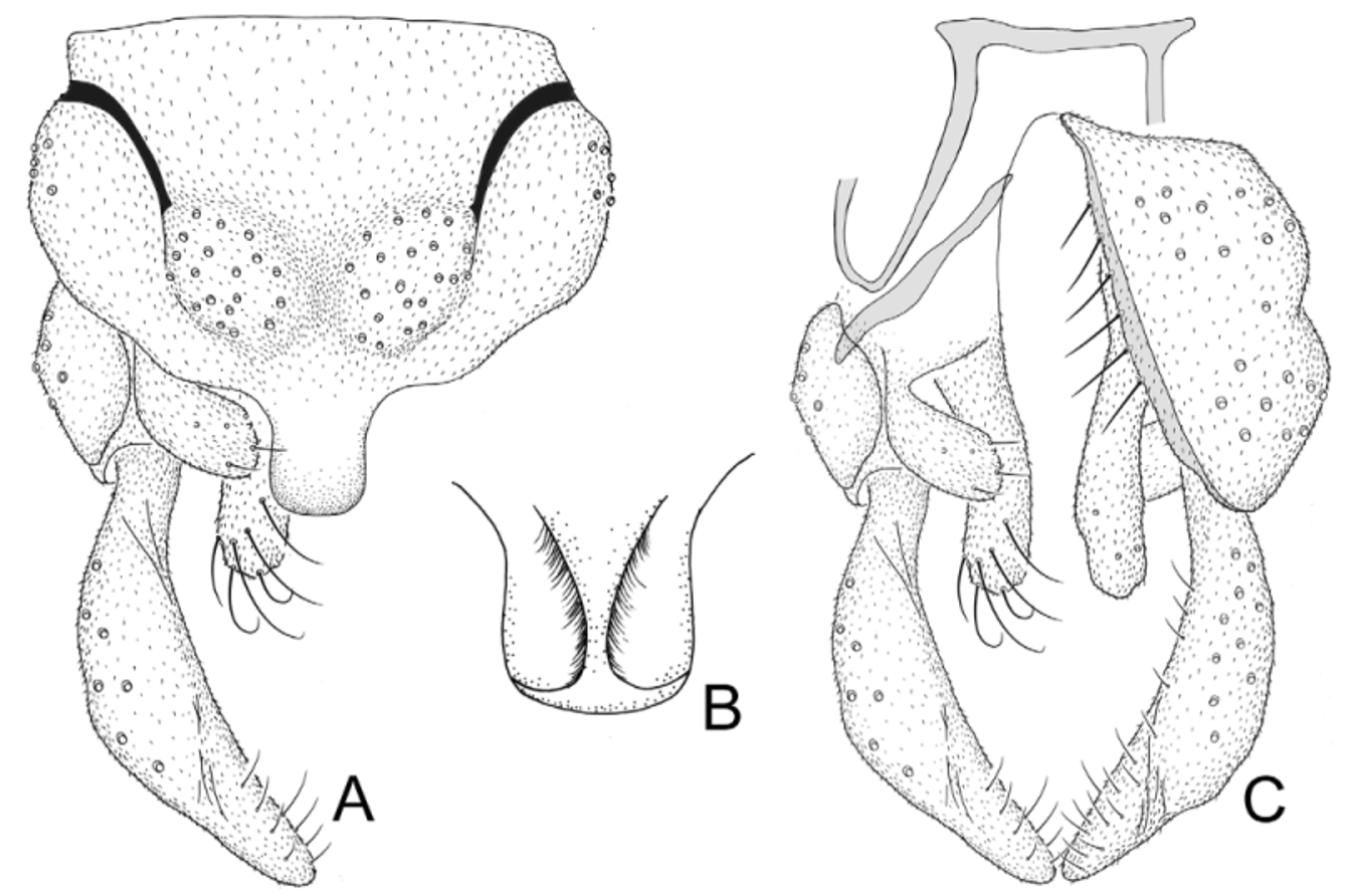
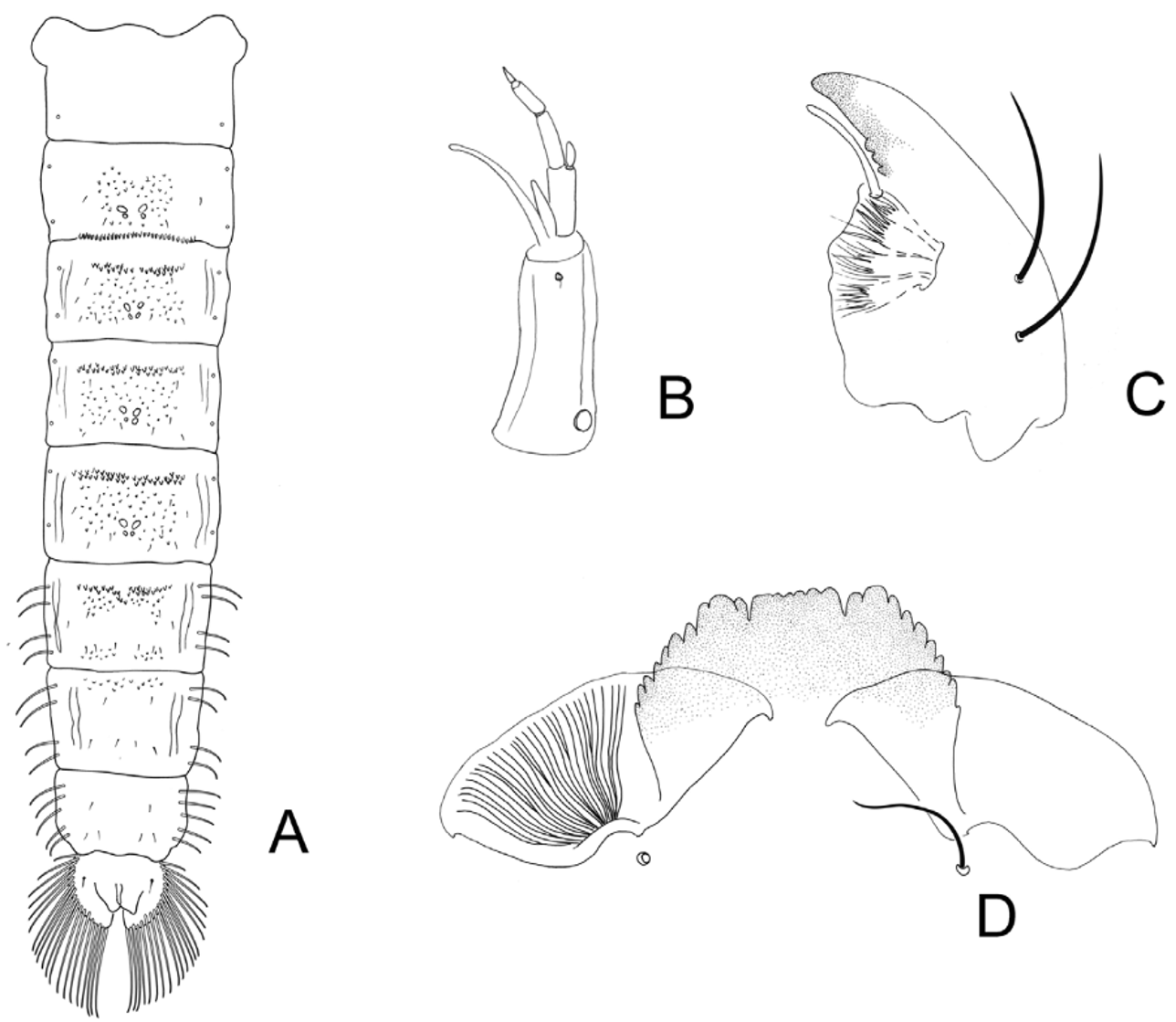
TABLE 1. Length (in μm) and proportions of legs of Xenochironomus canterburyensis, male (n = 1 – 2).
| Fe | ti | ta1 | ta2 | ta3 | |
|---|---|---|---|---|---|
| P1 | 1325–1420 | 1125–1310 | 1675 | 805 | 725 |
| P2 | 1215–1350 | 1155–1300 | 670–750 | 390–435 | 275–330 |
| P3 | 1490–1640 | 1550–1785 | 1055–1130 | 590–655 | 430–480 |
TABLE 2. Length (in μm) and proportions of legs of Xenochironomus canterburyensis, female (n = 1 – 2).
| fe | ti | ta1 | ta2 | ta3 |
|---|---|---|---|---|
| P1 1430–1585 | 1185–1405 | 1825–1910 | 865–910 | 730–840 |
| P2 1260–1460 | 1220–1415 | 705–800 | 370–450 | 285–350 |
| P3 1545–1780 | 1645–1910 | 1125–1235 | 615–700 | 465–530 |
No known copyright restrictions apply. See Agosti, D., Egloff, W., 2009. Taxonomic information exchange and copyright: the Plazi approach. BMC Research Notes 2009, 2:53 for further explanation.
|
Kingdom |
|
|
Phylum |
|
|
Class |
|
|
Order |
|
|
Family |
|
|
Genus |
|
Kingdom |
|
|
Phylum |
|
|
Class |
|
|
Order |
|
|
Family |
|
|
Genus |