
Yangambia, SCHOUTEDEN, 1942
|
publication ID |
https://doi.org/10.1111/zoj.12311 |
|
persistent identifier |
https://treatment.plazi.org/id/142A4050-DE11-FFA7-91C6-E41CFA75FA03 |
|
treatment provided by |
Marcus (2021-08-29 06:21:27, last updated by Plazi 2023-11-05 13:21:54) |
|
scientific name |
Yangambia |
| status |
|
YANGAMBIA SCHOUTEDEN View in CoL
Figures 8 View Figure 8 , 10I View Figure 10 , 11I View Figure 11 , 17 View Figure 17 AH–AK, 21C, D, 24
Yangambia Schouteden, 1942b: 5 View in CoL (gen. nov.; type species Yangambia vesiculata View in CoL by monotypy); Schouteden, 1945: 116 (syn.); Carvalho, 1952: 60 (cat.); Carvalho, 1955: 41 (key to gen.); Carvalho, 1957:150 (cat.); Odhiambo, 1962: 307 (disc.); Villiers, 1952: 189 (descr.); Schuh, 1995: 533 (cat.); Schuh, 2002 –2013 (cat.); Namyatova et al., in press (phylogeny).
Idioaspis China, 1944: 186 (gen. nov.; type species Idioaspis macarangae by monotypy, syn. by Schouteden, 1945: 116); China, 1944: 174 (key to gen.); Carvalho, 1952: 60 (cat.); ( Carvalho, 1957: 150 (cat.); Schuh, 1995: 533 (cat.); Schuh, 2002 –2013 (cat.).
Diagnosis: Yangambia belongs to the Odoniella - complex (see discussion after tribe) and it can be distinguished from other genera of this group by: scutellum divided into six parts dorsally ( Fig. 11I View Figure 11 ), scutellum impunctate ( Fig. 11I View Figure 11 ), humeral angles of pronotum distinctly flattened and serrate ( Fig. 10I View Figure 10 ); flattened dark setae on hemelytra in parches, and spermathecal gland on DLP placed medially near posterior margin ( Fig. 21C View Figure 21 ).
Redescription: Male: Body length 4–5 mm. COLORA- TION ( Fig. 8 View Figure 8 ). Mostly whitish yellow to yellow, sometimes with pale brown to brown markings. TEXTURE. Head with two pairs of tubercles between eyes; flattened areas on vertex indistinct; antenna and tibiae with tubercles at base of setae; pronotum with punctures mixed with wrinkles, with four large tubercles on collar and 10 large tubercles on posterior part of pronotum ( Fig. 10I View Figure 10 ); scutellum without tubercles and punctures, with shallow wrinkles ( Fig. 11I View Figure 11 ); pair of punctures between mesoscutum and scutellum, pair of punctures between mesoscutum and scutellum, punctures on clavus and on R + M absent; striations on lateral margins of scutellum present only anteriorly; semicircular depression between scutellum and mesoscutum absent. VESTITURE. Body clothed with setae, shorter than with of hind tibia; head with rarely distributed simple or flattened pale setae, dorsal side of head without setae; antenna with mixture of pale adpressed and spine-like suberect setae; setae on pronotum and scutellum absent; thoracic pleura with rarely distributed, short, pale, adpressed setae; setae on hemelytron dark and flattened, forming patches, simple setae on posterior part of corium and on cuneus present; legs mostly with pale spine like suberect setae, not very dense, tarsi with adpressed pale setae; black spinules on femora absent, tibia with spinules placed irregularly (fig. 19F in Namyatova et al., in press); abdomen mostly clothed with short adpressed pale setae and flattened setae on apical segments. STRUCTURE. Head. Distance between eye and pronotum shorter than eye diameter; occipital region not delimited with depression; longitudinal depression on vertex absent or very short; eyes stylate, directed outwards and forwards, subequal to 1/6 th of head width; distance between antennal fossa twice as long as antennal fossa diameter; frons only slightly swollen ( Fig. 10I View Figure 10 ), without paired outgrowths or only with pair of very shallow tubercles, without ridges or longitudinal depression; anterior view of head c. 1.9× as wide as high; eye height subequal to distance from eye to apex of clypeus; antennal fossa oval, diameter subequal to 2/3 rd of eye height, not raised (as in fig. 3B in Namyatova et al., in press), inferior margin placed near inferior margin of eye; base of clypeus placed near inferior margin of eye, delimited with depression; head flat in lateral view, gula shorter than buccula length, straight. Labium. Reaching middle of mesosternum or slightly surpassing it; LSI twice as long as wide; LSII c. 2.5× as long as wide, subequal to LSI; LSIII 2.5× as long as wide, subequal to LSII; LSIV c. 4× as long as wide, c. 1.5× as long as LSIII. Antenna. Reaching base of cuneus or slightly surpassing it; ASI c. 1.5× as long as wide (as in fig. 8E in Namyatova et al., in press), subequal to 1/4 th of head width; ASII c. 6× as long as segment I, slightly shorter than head and pronotum combined, slightly widened towards apex (as in fig. 8E in Namyatova et al., in press), without swelling basally and medially; ASIII c. 0.7× as long as ASII, widened towards apex; ASIV c. 0.7× as long as ASIII, clavate. Thorax. Collar distinct, fused with callosite region medially, flat ( Fig. 10I View Figure 10 ); calli separated, flat; depression delimiting calli posteriorly absent ( Fig. 10I View Figure 10 ); humeral angles of pronotum strongly dilated, serrate, with six lobes ( Fig. 10I View Figure 10 ); posterior margin of pronotum distinctly concave, forming right angles ( Fig. 10I View Figure 10 ); scutellum distinctly swollen, covering base of pronotum, of irregular shape, divided into six parts, obtuse apically ( Fig. 11I View Figure 11 ), covering base of pronotum, with longitudinal depression medially, without outgrowth medially; metepimeron enlarged, twice as high as wide, angulate and subtriangular (as in Fig. 13E View Figure 13 ); metasternum with medial projection reaching abdominal segment II (as in fig. 17A in Namyatova et al., in press). Hemelytron. Costal margin of hemelytron slightly rounded; claval commissure c. 0.2–0.3× as long as scutellum, straight; R + M distinct only anteriorly, not reaching posterior margin of corium; medial fracture strongly inclined towards midline; cuneus approximately twice long as wide, c. 0.7× as long as pronotum, medial margin slightly concave (as in fig. 13B in Namyatova et al., in press); membrane cell not surpassing apex of cuneus, forming right angle (as in fig. 13B in Namyatova et al., in press), c. 0.6–0.7× as long as pronotum; auxiliary vein absent or short; distance from cell to apex of membrane as long as or slightly shorter than cell length. Legs. Forecoxae contiguous (as in fig. 17A in Namyatova et al., in press); femora not swollen apically, straight; foretibia shorter than head and pronotum combined; tibia without swellings; segment I of hind tibia of as long as segment II and shorter than segment III; apical half of claw curved; basal tooth on claw elongate, slightly concave (as in Fig. 13J View Figure 13 ). Genitalia ( Fig. 17 View Figure 17 AH–AK). Genital capsule wider than long, with small tubercle at each side, ventral wall not shortened anteriorly; left paramere r-shaped, c. 1.5–2× times as long as right paramere; phallobase sclerite of primary gonopore heartshaped, without outgrowths; ductus seminis not sclerotized basally and with narrow circle sclerite around secondary gonopore; ductus seminis as long as phallotheca, with coils forming wide tube, attached to phallobase medially; sclerotized part of phallotheca narrow, occupying half of dorsal part, widened and rounded apically; without ridge or outgrowth(s); endosoma with a number of serrate spicules of irregular shape).
Female: Body length 4–5 mm. COLORATION, TEXTURE, VESTITURE AND STRUCTURE ( Fig. 8 View Figure 8 ). As in male. Genitalia ( Fig. 21C, D View Figure 21 ). DLP with single sclerotized ring, divided medially, with pair of striated areas; lateral oviducts attached at midpoint of striated areas, widely separated, placed near lateral margins of DLP; spermathecal gland placed posteriomedially, equidistant from lateral oviducts; posterior wall covered with small tubercles, without outgrowths or sclerotizations; base of second valvula slightly concave; ventral wall membranous.
Distribution: Democratic Republic of Congo, Cameroon, Ghana ( Fig. 24 View Figure 24 ).
Host plants: Yangambia macarangae is known from Macaranga horaefolia (Euphorbiaceae) ( China, 1944).
Carvalho JCM. 1952. On the major classification of the Miridae (Hemiptera). (With keys to subfamilies and tribes and a catalogue of the world genera). Anais da Academia Brasileira de Ciencias 24: 31 - 110.
Carvalho JCM. 1955. Keys to the genera of Miridae of the world (Hemiptera). Boletim do Museu Paraense Emilio Goeldi, Zool 11: 1 - 151.
Carvalho JCM. 1957. A catalogue of the Miridae of the world. Part I. Arquivos do Museu Nacional 44: 1 - 158.
China WE. 1944. New and little known West African Miridae (Capsidae) (Hemiptera-Heteroptera). Bulletin of Entomological Research 35: 171 - 191.
Odhiambo TR. 1962. Review of some genera of the subfamily Bryocorinae (Hemiptera: Miridae). Bulletin of the British Museum (Natural History). Entomology 2: 245 - 331.
Schouteden H. 1942 b. Yangambia nov. gen. Miridarum (Hem. Miridae). Revue de Zoologie et de Botanique Africaines 36: 5 - 7.
Schouteden H. 1945. Notes sur quelques Bryocorine d'Afrique (Hem. Revue de Zoologie et de Botanique Africaines 39: 115 - 117.
Schuh RT 2002 - 2013. On-line systematic catalog of plant bugs (Insecta: Heteroptera: Miridae). Available at: http: // research. amnh. org / pbi / catalog.
Villiers A. 1952. Hemipteres de l'Afrique Noire (Punaises et cigales). Initiations Africaines 9: 1 - 256.
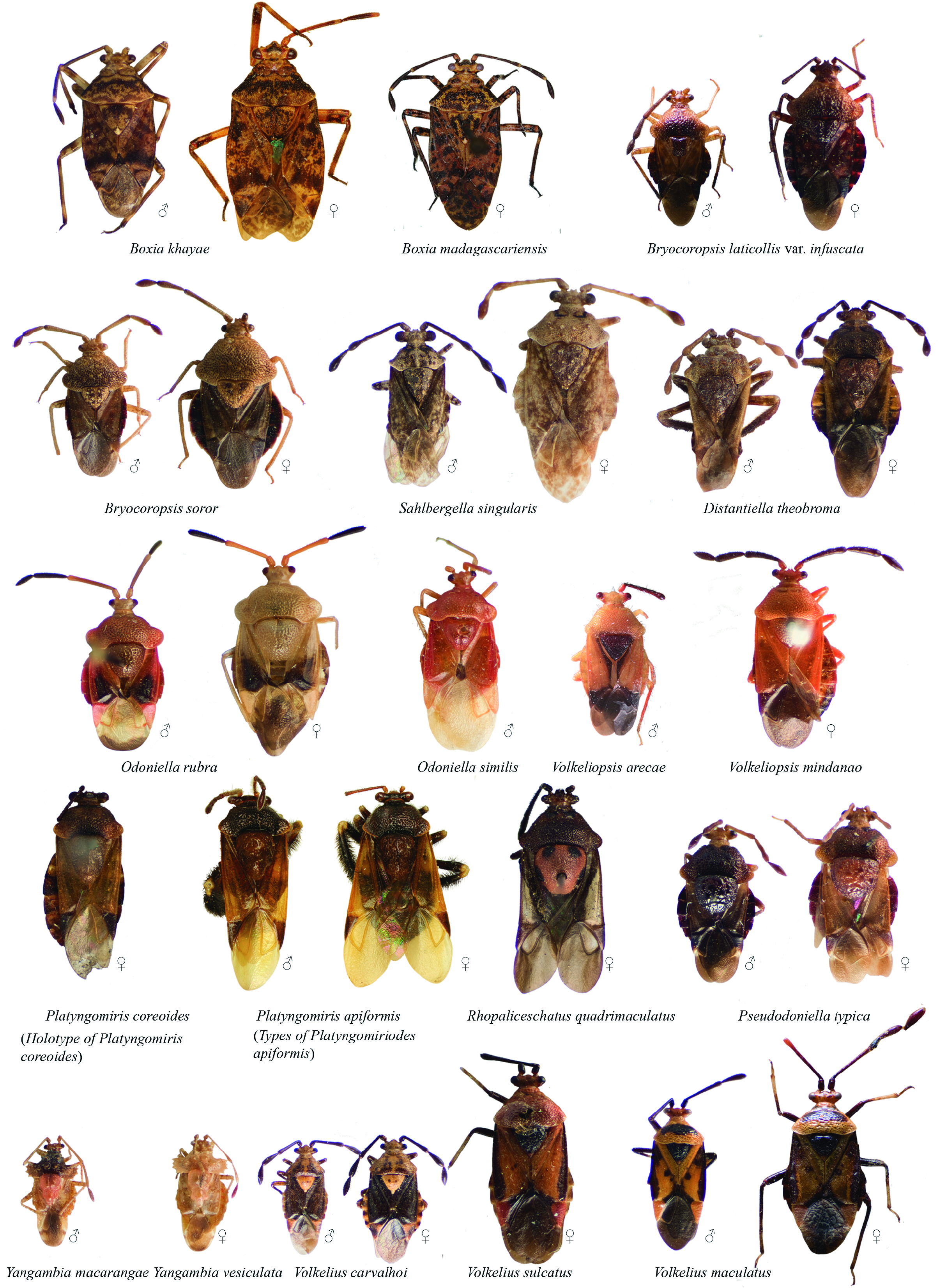
Figure 8. Habitus photographs. Boxia khayae ♂ AMNH_PBI 5065, ♀ type 19448; Boxia madagascariensis ♀ AMNH_PBI 19532; Bryocoropsis laticollis var. infuscata ♂ AMNH_PBI 18947, ♀ AMNH_PBI 18946; Bryocoropsis soror ♂ AMNH_PBI 5115, ♀ AMNH_PBI 5117; Sahlbergella singularis ♂ AMNH_PBI 19118, ♀ AMNH_PBI 19057; Distantiella theobromae ♂ AMNH_PBI 5016, ♀ AMNH_PBI 5019; Odoniella rubra ♂ AMNH_PBI 18951, ♀ AMNH_PBI 18958; Odoniella similis ♂ AMNH_PBI 5036; Volkeliopsis arecae ♂ HT AMNH_PBI 19516; Volkeliopsis mindanao ♀ AMNH_PBI 5237; Platyngomiris coreoides ♀ HT of P. coreoides AMNH_PBI 19643; ♂ HT of Platyngomiriodes apiformis AMNH_PBI 19471, ♀ PT of P. apiformis no USI label (BMNH); Platyngomiris quadrimaculatus ♀ AMNH_PBI 20253; Platyngomiris typicus ♂ AMNH_PBI ♀; Yangambia macarangae ♂ AMNH_PBI 34057, ♀ AMNH_PBI 20192; Yangambia vesiculata ♂ AMNH_PBI 19079, ♀ AMNH_PBI 19086; Volkelius carvalhoi ♂ AMNH_PBI 19309, ♀ AMNH_PBI 20197; Volkelius sulcatus ♀ PLT AMNH_PBI 5047; Volkelius maculatus ♂ AMNH_PBI 19043, ♀ AMNH_PBI 19386.
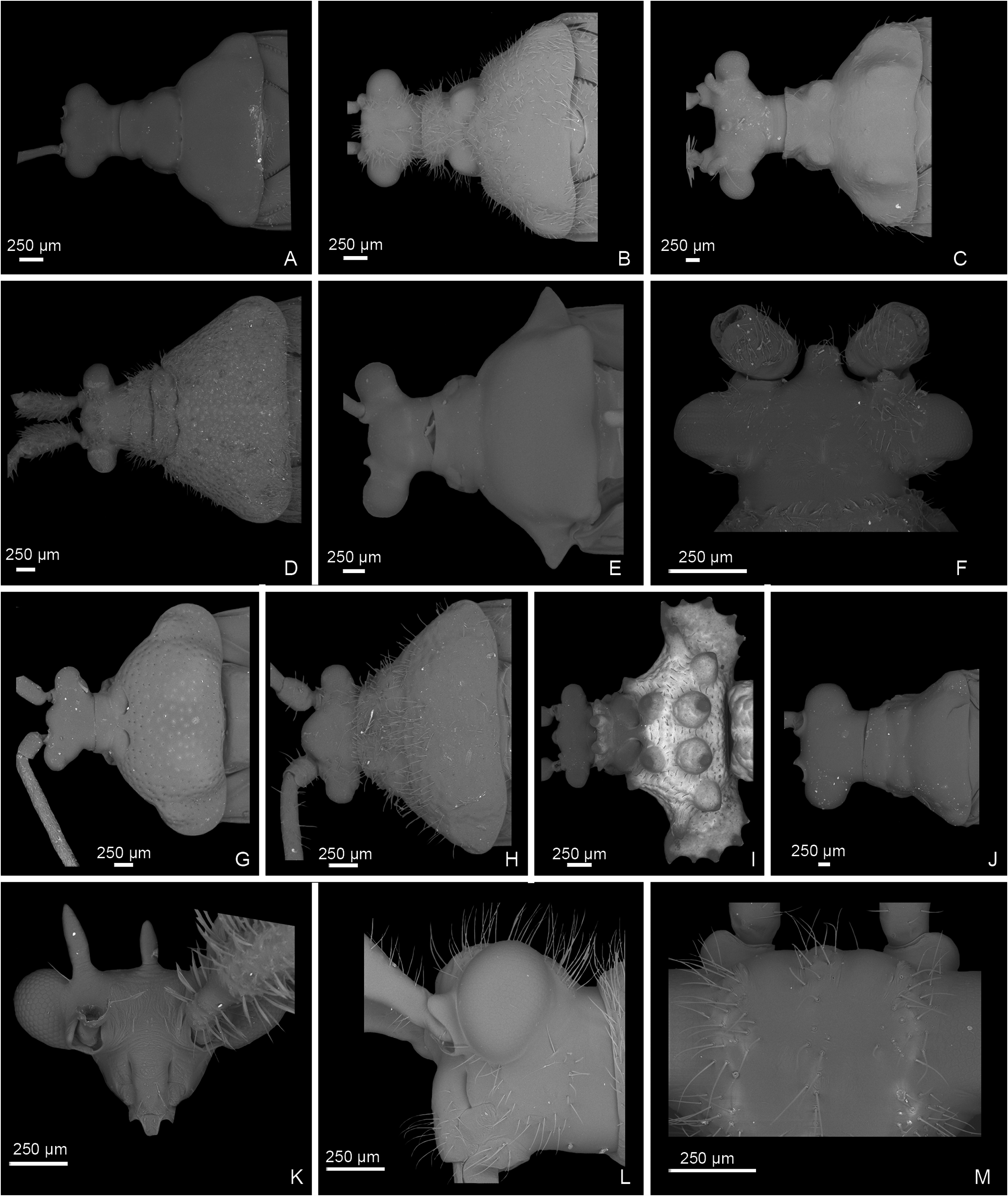
Figure 10. Scanning electron micrographs. Head and pronotum, dorsal view. A, Mansoniella nitida ♀ AMNH_PBI 46067; B, Poppiusia leroyi ♀ AMNH_PBI 5838; C, Chamus bellus ♀ AMNH_PBI 5223; D, Lycidocoris mimeticus ♀ AMNH_PBI 5043; E, Physophoropterella bondroiti ♂ AMNH_PBI 19110; G, Odoniellia reuteri ♂ AMNH_PBI 19090; H, Volkelius carvalhoi ♀ PT AMNH_PBI 19630; I, Yangambia vesiculata ♀ AMNH_PBI 19084; J, Ragwelellus suspectus ♂ AMNH_PBI 19629. Head, dorsal view. F, Sahlbergella tai ♀ AMNH_PBI 5106. Head, anterior view. K, Parachamus bellus ♀ AMNH_ PBI 5837. Head, lateral view. L, Poppiusia leroyi ♀ AMNH_PBI 5838. Frons, dorsal view. M, Poppiusia leroyi ♀ AMNH_PBI 5838.

Figure 11. SEM images. Scutellum, dorsal view. A, Chamus tuberculatus ♂ AMNH_PBI 5025; B, Volkeliopsis mindanao ♀ PT AMNH_PBI 45979; C, Boxia khayae ♂ AMNH_PBI 5065; D, Bryocoropsis soror ♂ AMNH_PBI 5115; E, Odoniella reuteri ♂ AMNH_PBI 19090; F, Pseudodoniella typical ♂ AMNH_PBI 45978; G, Sahlbergella tai ♀ AMNH_PBI 5106; H, Pseudodoniella pacifica ♂ AMNH_PBI 46080; I, Yangambia vesiculata ♀ AMNH_PBI 19084; J, Sahlbergella singularus ♂ AMNH_PBI 19053; K, Physophoropterella bondroiti ♂ AMNH_PBI 19110. Small tubercules on pronotum. L, Chamus tuberculatus ♂ AMNH_PBI 5025.
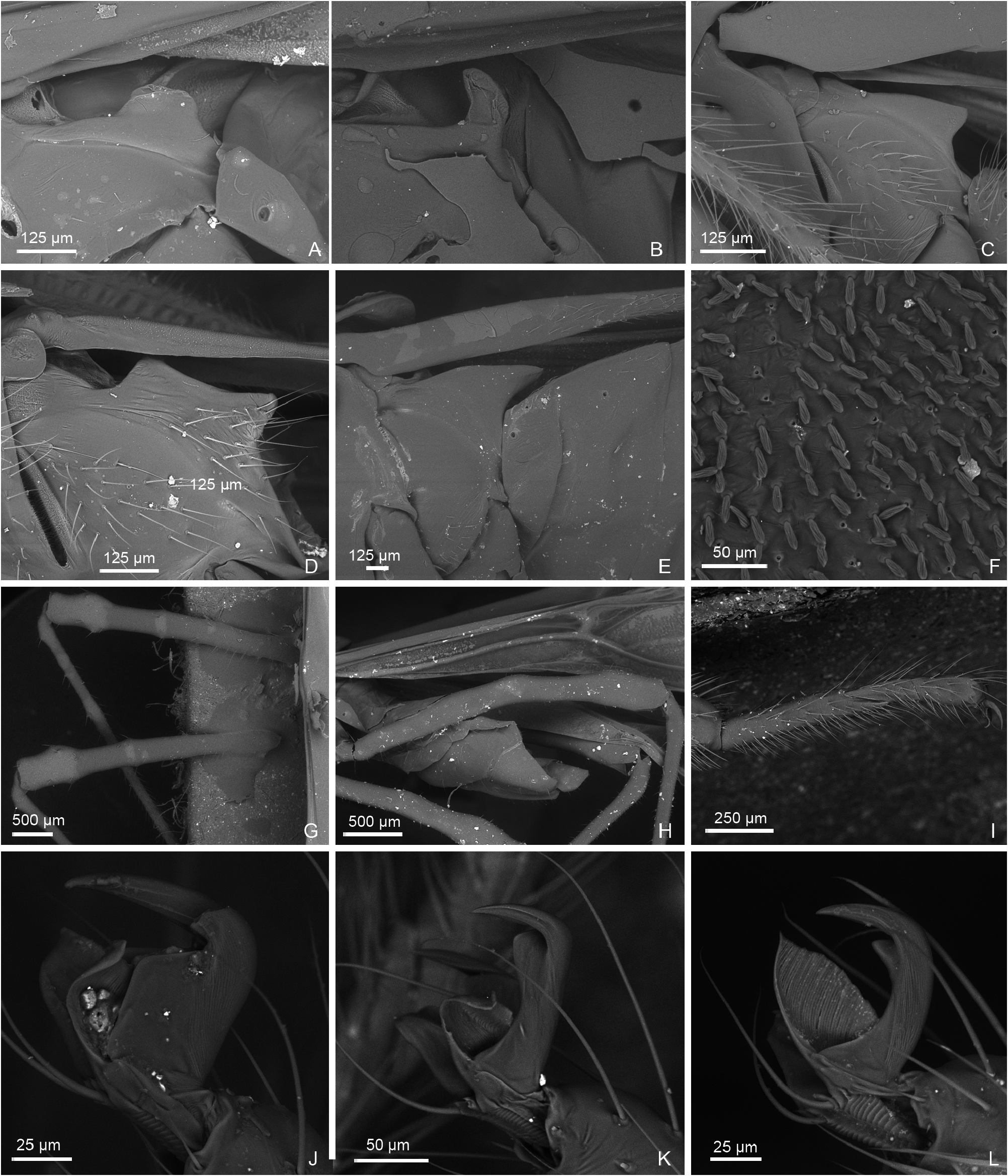
Figure 13. Scanning electron micrographs. Metepimeron. A, Helopletis pellucida sex unknown AMNH_PBI 19628; B, Afropeltis hyalospilosus ♂ AMNH_PBI 5137; C, Poppiusia leroyi ♀ AMNH_PBI 5838; D, Pachypeltis reuteri ♂ AMNH_PBI 45980; E, Odoniella reuteri ♂ AMNH_PBI 19194. Setae on hemelytron. F, Boxia khayae ♂ AMNH_PBI 5065. Fore- and middle femora. G, Physophoropterella bondroiti ♂ AMNH_PBI 19110. Hind femur. H, Helopeltis pellucida sex unknown AMNH_PBI 19628. Hind tarsus. I, Physophoropterella bondroiti ♂ AMNH_PBI 19110. Claw, lateral view. J, Chamus sp. sex unknown, not databased; K, Pachypeltis brevirostris sp. nov. sex unknown, not databased; L, Helopeltis bradyi sex unknown, not databased.

Figure 17. Male genitalia, dorsal view. Physophoroptera mirabilis AMNH_PBI 5070. A, aedeagus; B, right paramere; C, left paramere; D, genital capsule. Physophoropterella bondroiti no USI. E, aedeagus; AMNH_PBI 5072. F, right paramere; G, left paramere; H, genital capsule. Poppiusia leroyi AMNH_PBI 19314. I, aedeagus; J, right paramere; K, left paramere; L, genital capsule. Pseudodoniella typica no USI. M, aedeagus; N, right paramere; O, left paramere; P, genital capsule. Ragwelellus suspectus no USI. Y, aedeagus; Z, right paramere; AA, left paramere; AB, genital capsule. Ragwelellus vittatus no USI. U, aedeagus; V, right paramere; W, left paramere; X, genital capsule. Volkeliopsis arecae AMNH_PBI 19427. I, phallobase; J, endosoma and theca; K, right paramere; L, left paramere; M, genital capsule. Villiersicoris sessensis AMNH_PBI 19458. Q, aedeagus; R, right paramere; S, left paramere; T, genital capsule. The smaller scale is for genital capsule, the larger scale is for aedeagi and parameres. Yangambia macarangae AMNH_PBI 5006. AG, aedeagus. no USI. AH, right paramere; AI, left paramere; AJ, genital capsule. The smaller scale is for genital capsule, the larger scale is for aedeagi and parameres.

Figure 21. Female genitalia, bursa copulatrix. Pseudodoniella pacifica AMNH_PBI 45974. A, dorsal labiate plate. AMNH_PBI 20192. B, posterior wall. Yangambia vesiculata AMNH_PBI 19084. C, dorsal labiate plate; D, posterior wall. Platyngomiris coreoides AMNH_PBI 19643. E, dorsal labiate plate; F, posterior wall. Ragwelellus indonesicus AMNH_PBI 46074. G, dorsal labiate plate; H, posterior wall. Poppiusia leroyi AMNH_PBI 5049. I, dorsal labiate plate; J, posterior wall. Ragwelellus suspectus no USI. K, dorsal labiate plate; L, posterior wall. Ragwelellus magnificus AMNH_PBI 19384. M, posterior wall. Ragwelellus vittatus no USI. N, dorsal labiate plate; O, posterior wall. Sahlbergella singularis AMNH_PBI 20212. P, dorsal labiate plate; Q, posterior wall. Sahlbergella tai PT AMNH_PBI 5106. R, dorsal labiate plate. Volkeliopsis mindanao AMNH_PBI 5237. S, dorsal labiate; T, posterior wall.

No known copyright restrictions apply. See Agosti, D., Egloff, W., 2009. Taxonomic information exchange and copyright: the Plazi approach. BMC Research Notes 2009, 2:53 for further explanation.
|
Kingdom |
|
|
Phylum |
|
|
Class |
|
|
Order |
|
|
Family |
Yangambia
| Namyatova, Anna A. & Cassis, Gerasimos 2016 |
Idioaspis
| Carvalho JCM 1957: 150 |
| Carvalho JCM 1952: 60 |
| Schouteden H 1945: 116 |
| China WE 1944: 186 |
| China WE 1944: 174 |
Yangambia
| Odhiambo TR 1962: 307 |
| Carvalho JCM 1957: 150 |
| Carvalho JCM 1955: 41 |
| Carvalho JCM 1952: 60 |
| Villiers A 1952: 189 |
| Schouteden H 1945: 116 |
| Schouteden H 1942: 5 |