
Pachypeltis, SIGNORET, 1858
|
publication ID |
https://doi.org/10.1111/zoj.12311 |
|
persistent identifier |
https://treatment.plazi.org/id/142A4050-DE09-FFB0-91FC-E006FEBCFA66 |
|
treatment provided by |
Marcus |
|
scientific name |
Pachypeltis |
| status |
|
PACHYPELTIS SIGNORET View in CoL
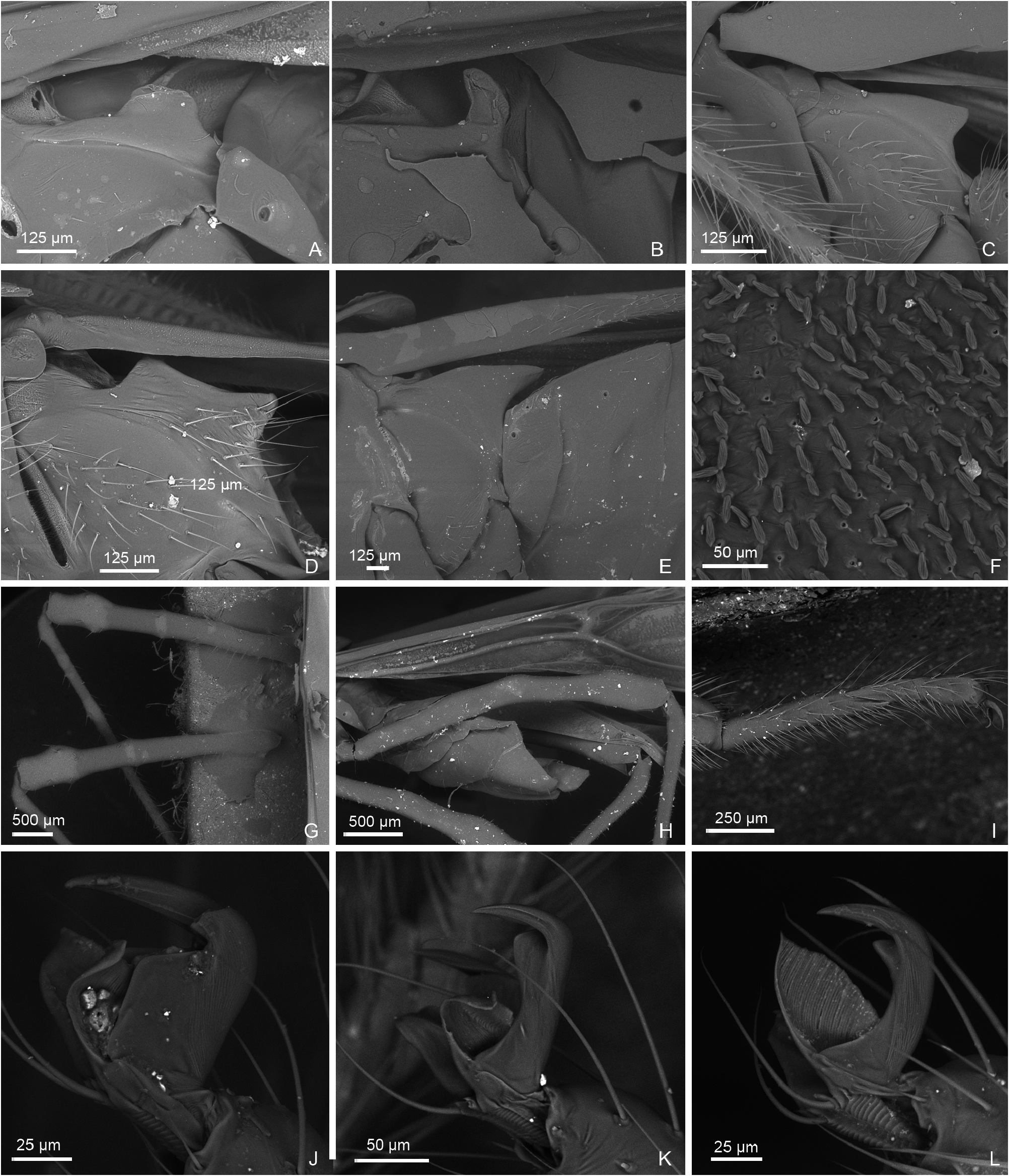
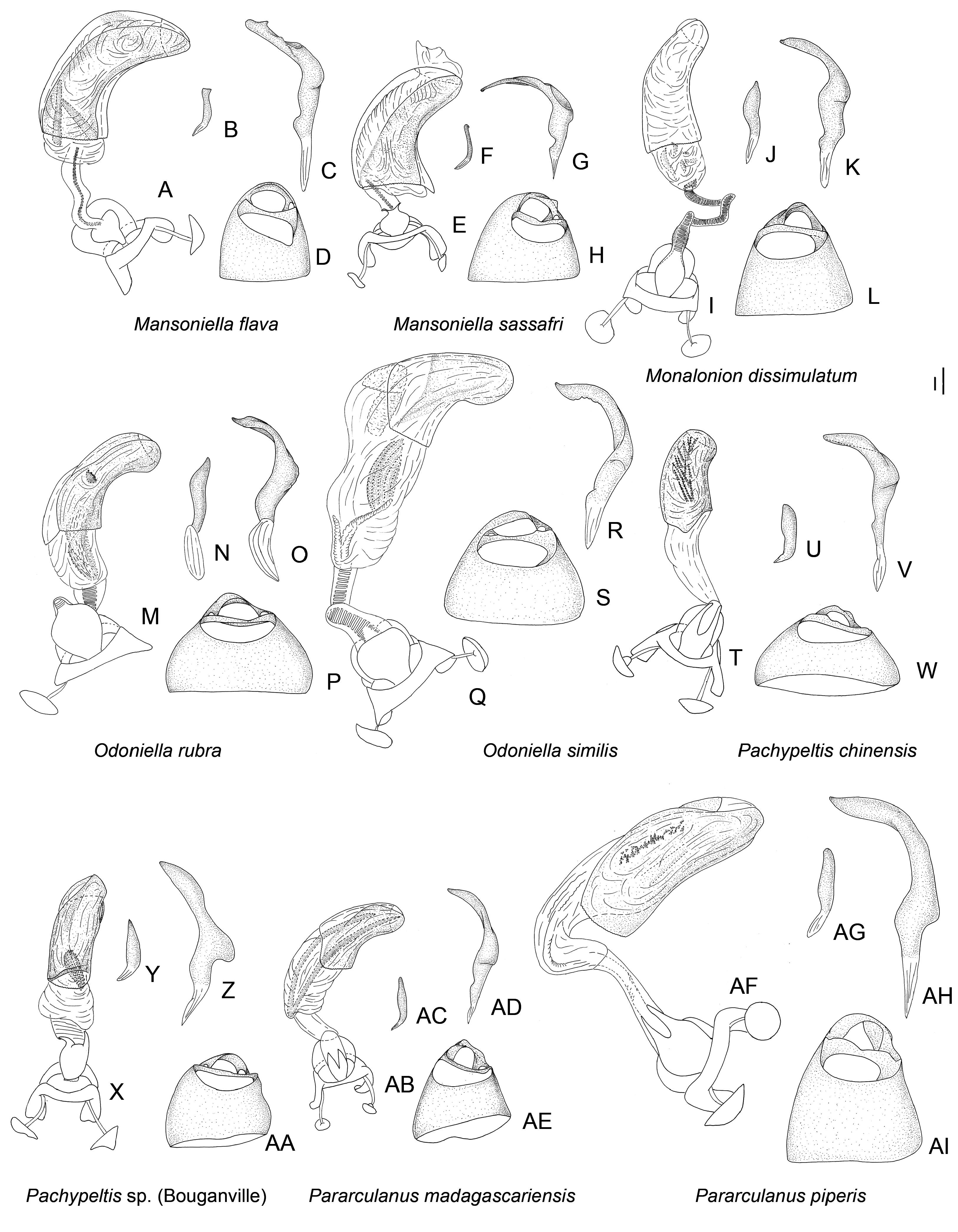
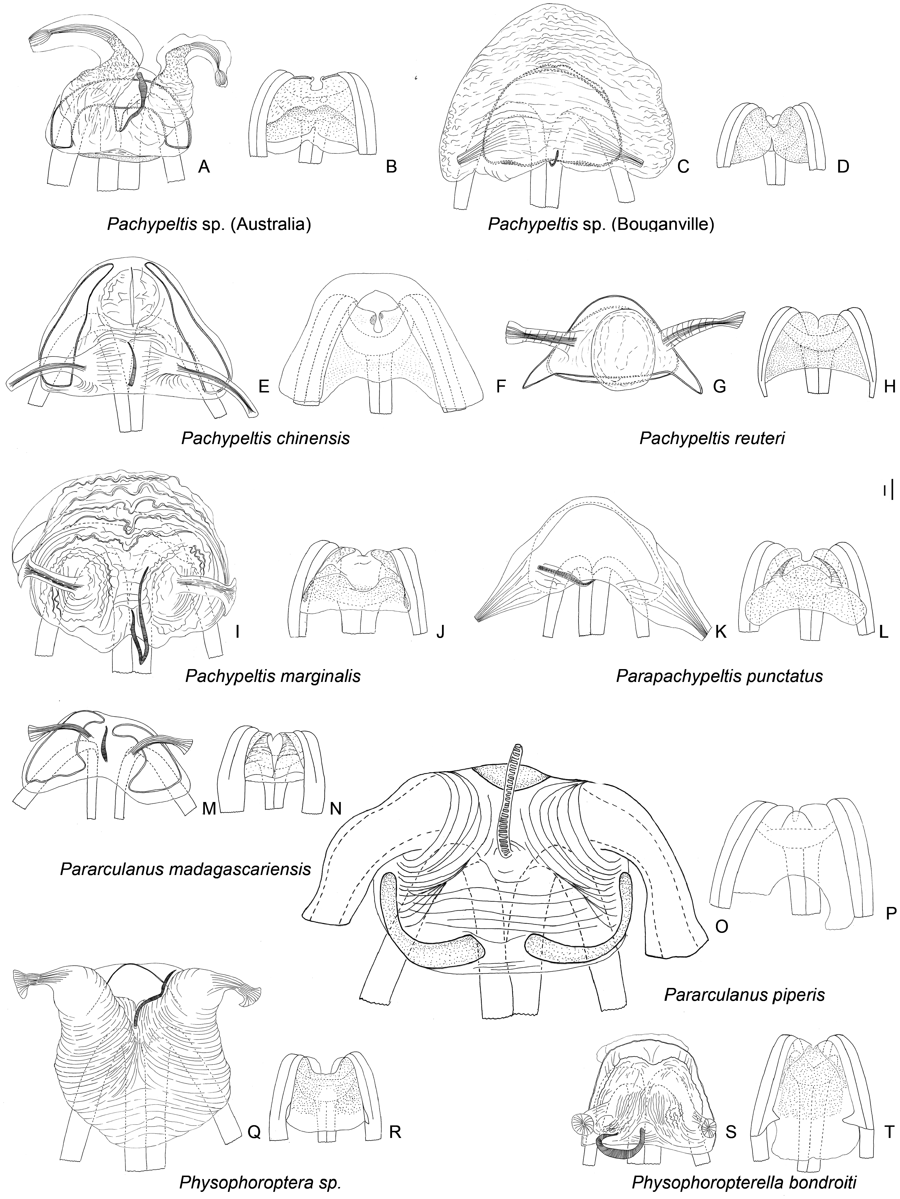
Figures 7 View Figure 7 , 13D, K View Figure 13 , 16T View Figure 16 –AA, 20A–J, 23
Pachypeltis Signoret, 1858: 501 View in CoL (gen. nov.; type species: Pachypeltis chinensis Signoret, 1858 by monotypy); Walker, 1873: 164 (cat.); Atkinson, 1890a: 51 (cat.); Reuter, 1903: 2 (descr.); Kirkaldy, 1906: 134 (list); Kuhlgatz, 1906: 29 (key to gen.); Reuter, 1910: 153 (cat.); Hsiao, 1942: 250 (key to gen., list.); Carvalho, 1952: 59 (cat.); Carvalho, 1955: 40 (key to gen.); Carvalho, 1957: 140 (cat.); Steyskal, 1973: 206 (correction); Carvalho, 1981: 41 (descr., disc., key to spp.); Schuh, 1995: 520 (cat.); Hu and Zheng, 2001: 421 (key to spp.); Schuh, 2002 –2013 (cat.); Namyatova et al., in press (phylogeny).
Disphinctus Stål, 1871: 668 (gen. nov.; type species Disphinctus sahlbergii Stål, 1858 by consequent designation Kirkaldy, 1902: 294); Walker, 1873: 161 (cat.); Atkinson, 1890a: 50 (cat.); Distant, 1904b: 443 (descr.); Kirkaldy, 1906: 134 (list); Reuter, 1910: 166 (syn.); Carvalho, 1957: 140 (cat.); Schuh, 1995: 520 (cat.); Schuh, 2002 –2013 (cat.).
Diagnosis: Pachypeltis is diagnosed by the following characters: row of punctures on clavus and R + M (fig. 11C, D in Namyatova et al., in press); calli merged (fig. 4A in Namyatova et al., in press); apex of scutellum often rounded (fig. 12C, D in Namyatova et al., in press); distance between head and pronotum subequal to half of eye diameter (fig. 4A in Namyatova et al., in press); frons without ridges or spines, without depression medially; LSI–II only twice as long as wide, LSII often more than twice as long as wide; ASII–IV filiform; scutellum often rounded apically; hind femur often distinctly curved (fig. 18C in Namyatova et al., in press); hemelytra with outer margins straight or slightly concave; membrane cell distinctly acute (fig. 13A in Namyatova et al., in press), distinctly longer than pronotum; ductus seminis shorter than phallotheca, without coils; and, outgrowths on phallobase supporting ductus seminis present, long or short ( Fig. 16T View Figure 16 , fig. 22A in Namyatova et al., in press).
Redescription: Male: Body length 7–9 mm. COLORA- TION ( Fig. 7 View Figure 7 ). Usually bright-colored, coloration varies from yellow with dark markings to dark brown to black with paler markings, sometimes orange or red; antennae, or, at least, ASII–IV, usually darker than body, brown to dark brown, corium sometimes with dark marking or darkened posteriorly. TEXTURE. Body smooth, without tubercles; head without wrinkles and flattened areas; pronotum impunctate, without wrinkles; scutellum impunctate, smooth or with transverse wrinkles; pair of punctures between mesoscutum and scutellum, striations on lateral margin of scutellum, and punctures on clavus and R + M present (fig. 11C, D in Namyatova et al., in press), pair of punctures on depression delimiting callosite region andsemicircular depression between scutellum and mesoscutum absent. VESTITURE. Body clothed with dense pale or dark simple setae, usually suberect, adpressed hemelytron and thoracic pleura, setae on abdomen suberect and adpressed; setae usually shorter, sometimes longer than width of hind tibia, tibia regularly setose; black spinules on femora absent, rows of black spinules on tibia present (as in fig. 18D in Namyatova et al., in press). STRUCTURE. Head. Distance between eye and pronotum shorter or subequal to eye length dorsally (fig. 4A in Namyatova et al., in press); occipital region not delimited or distinctly delimited with transverse depression; longitudinal depression on vertex indistinct; eyes not stylate, in line with contour of head, c. 0.2–0.3× as wide as head; distance between antennal fossae slightly longer than antennal fossae diameter; frons from only slightly to distinctly swollen, without ridges, outgrowths or longitudinal depression; anterior view of head c. 1.3– 1.4× as wide as high; eye c. 1.3–2× as high as distance from eye to apex of clypeus; antennal fossae round, diameter of fossa ca.0.25–0.35× as long as eye diameter, only slightly raised (fig. 3A in Namyatova et al., in press), inferior margin placed above of inferior margin of eye; base of clypeus placed near inferior margin of antennal fossa, delimited with more or less distinct or very shallow depression (fig. 3A in Namyatova et al., in press); head from slightly to distinctly swollen dorsally in lateral view; gula straight or slightly convex, c. 1.5–2× as long as buccula length. Labium. Length varying from slightly surpassing anterior margin of mesosternum to almost reaching middle of mesosternum; LSI twice as long as wide, LSII c. 2–3.5× as long as wide, as long as or longer than LSII; LSIII ca.1.5– 3.5× subequal to or slightly shorter than LSII; LSIV c. 2.5–6× as long as wide, c. 1.5–2× as long as LSIII. Antenna. Length varying from reaching base of cuneus to surpassing apex of cuneus; LSI c. 0.5–0.7× as long as head width, widened medially (fig. 8D in Namyatova et al., in press), LSII c. 3.5–5.8 times as long as LSI, c. 1.1–2× as long as head and pronotum combined; LSIII c. 0.5–0.7× as long as LSIII; LSIV c. 0.4–0.7× as long as LSIII; LSII–IV filiform. Thorax. Collar fused with callosite region posteriorly, flat or swollen; calli fused with each other; callosite region delimited posteriorly with depression (fig. 4A in Namyatova et al., in press); humeral angles of pronotum not dilated; posterior margin of pronotum slightly concave or sinuate (fig. 4A in Namyatova et al., in press); scutellum almost flat or moderately swollen (fig. 11C, D in Namyatova et al., in press), often obtuse apically, rarely acute, without outgrowth or ridge, with or without shallow medial depression; metepimeron enlarged, c. 2–4× as long as wide, usually bifurcate ( Fig. 13D View Figure 13 ); metasternum with medial projection on to the abdominal segment II (fig. 17A in Namyatova et al., in press). Hemelytron. Costal margin of hemelytron straight or slightly concave medially; claval commissure c. 1–2× as long as scutellum, its margins straight (fig. 11C, D in Namyatova et al., in press); R + M distinct, reaching posterior margin of corium; medial fracture strongly inclined towards midline; corium not raised posteriorly; cuneus c. 1–4× as long as wide, c. 0.6–1.3× as long as pronotum, its medial margin straight or slightly concave (fig. 13A in Namyatova et al., in press); membrane cell elongate, distinctly surpassing apex of cuneus, forming acute angle (fig. 13A in Namyatova et al., in press), c. 1–2× as long as pronotum; auxiliary vein often present, short; distance from cell to membrane c. 0.3– 0.6× as long as cell length. Legs. Forecoxae contiguous (fig. 17A in Namyatova et al., in press); femora slightly swollen apically (fig. 18C in Namyatova et al., in press); fore- and middle femora straight, hind femur often distinctly curved, sometimes almost straight; tibia without swellings; foretibia shorter than head and pronotum combined; segments of hind tarsus subequal in length (fig. 29A in Namyatova et al., in press), sometimes segment I longer than segments I and II each and segments I and II subequal in length, or segment I slightly longer than segment II and as long as segment III; apical third part of claw curved; basal tooth on claw elongate, concave ( Fig. 13K View Figure 13 ). Genitalia ( Fig. 16T View Figure 16 –AA, fig. 22A–D in Namyatova et al., in press). Genital capsule longer or shorter than wide, without outgrowth(s), its ventral wall not shortened anteriorly; left paramere c. 3.5–4 times as long as right paramere, distinctly r-shaped; phallobase sclerite of primary gonopore bowl-shaped or oval, with long or short outgrowth(s), supporting ductus seminis; ductus seminis not sclerotized basally or apically, distinctly shorter than phallotheca, without coils; sclerotized part of phallotheca broad, occupying entire dorsal part, rounded apically, without outgrowth or ridge; endosoma with fields of small spicules, sometimes with number of elongate spicules, sometimes with both.
Female: Body length 6–12 mm. Coloration, surface, vestiture and structure as in male ( Fig. 7 View Figure 7 ). Genitalia ( Fig. 20A–J View Figure 20 , fig. 23H in Namyatova et al., in press). DLP with sclerotized ring anteriorly, sometimes covered with membrane, or with two sclerotized bands, or with two sclerotized rings; DLP with or without striations, without additional sclerites; lateral oviduct widely separated, placed in posterior half of DLP, sometimes slightly below midline; spermathecal gland placed in posterior half of DLP, medially, sometimes almost at posterior margin; posterior wall covered with small tubercles, sometimes with outgrowths and small sclerites; base of second valvula slightly concave; ventral wall of bursa copulatrix membranous.

No known copyright restrictions apply. See Agosti, D., Egloff, W., 2009. Taxonomic information exchange and copyright: the Plazi approach. BMC Research Notes 2009, 2:53 for further explanation.
|
Kingdom |
|
|
Phylum |
|
|
Class |
|
|
Order |
|
|
Family |
Pachypeltis
| Namyatova, Anna A. & Cassis, Gerasimos 2016 |
Disphinctus Stål, 1871: 668
| Carvalho JCM 1957: 140 |
| Reuter OM 1910: 166 |
| Kirkaldy GW 1906: 134 |
| Distant WL 1904: 443 |
| Kirkaldy GW 1902: 294 |
| Atkinson ET 1890: 50 |
| Walker F 1873: 161 |
| Stal C 1871: 668 |
Pachypeltis
| Hu Q & Zheng LY 2001: 421 |
| Carvalho JCM 1981: 41 |
| Steyskal GC 1973: 206 |
| Carvalho JCM 1957: 140 |
| Carvalho JCM 1955: 40 |
| Carvalho JCM 1952: 59 |
| Hsiao TY 1942: 250 |
| Reuter OM 1910: 153 |
| Kirkaldy GW 1906: 134 |
| Kuhlgatz T 1906: 29 |
| Reuter OM 1903: 2 |
| Atkinson ET 1890: 51 |
| Walker F 1873: 164 |
| Signoret V 1858: 501 |