
Berosus ( Berosus ) japonicus Sharp, 1873
|
publication ID |
https://doi.org/10.5281/zenodo.5303133 |
|
publication LSID |
lsid:zoobank.org:pub:8CDA7151-FBCD-484B-AF8D-C31EB76E9C67 |
|
persistent identifier |
https://treatment.plazi.org/id/1237F952-FFE1-2632-6B1E-FA61FEABFAB0 |
|
treatment provided by |
Marcus |
|
scientific name |
Berosus ( Berosus ) japonicus Sharp, 1873 |
| status |
|
Berosus ( Berosus) japonicus Sharp, 1873 View in CoL
(Figs 1–7, 17A–B, 18A)
Material examined. JAPAN: HONSHÛ: Shimane Prefecture: 6 L1 ( HGF, KMNH), Okinoshima, Sono-chô, Izumo- -shi, paddy field, 29.ii.2008 (rearing adults collected in the field), 17.iii.2008 (date of fixation), M. Hayashi leg. & reared ; 5 L1( KMNH), Okinoshima, Sono-chô, Izumo-shi, paddy field, 20.v.2008 (egg-cases collected in the field) ,
26.v.2008 (date of fixation), M. Hayashi leg.; 20 L3 ( HGF, KMNH), Okinoshima, Sono-chô, Izumo-shi, paddy field, 22.vi.2008, M. Hayashi leg. ; 6 L1, 5 L2 ( HGF, KMNH), Okinoshima, Sono-chô, Izumo-shi, paddy field, 22.v.2009 (egg-cases collected in the field), 31.v.2009 (date of fixation), M. Hayashi leg.
Description. General morphology. Third instar (Figs 1, 5–7). Body moderately thick, widest between second and third abdominal segments. Colour light brown to light greyish brown, with sclerotised parts darker.
Head ( Fig. 5A View Fig ). Head capsule subquadrate; cervical sclerites small. Frontal lines visible only basally; coronal line absent. Surface of head capsule smooth. Six stemmata on each anterolateral corner of head capsule. Clypeolabrum ( Fig. 5B View Fig ) strongly asymmetrical. Nasale with median projection bearing ca. five small teeth. Lateral lobes of epistome present on left side but absent on right side; left lobe very strongly projecting anteriorly, lateral margin of left lobe partially membranous; anterior margin of right epistome almost straight or very weakly rounded.
Antenna ( Figs 6A–B View Fig ) 3-segmented, short, rather slender. Antennomere 1 longer than antennomeres 2 and 3 combined, with one subapical membranous projection on inner face. Antennomere 2 narrower than antennomere 1. Antennomere 3 the narrowest and shortest. Sensorium slightly shorter than antennomere 3.
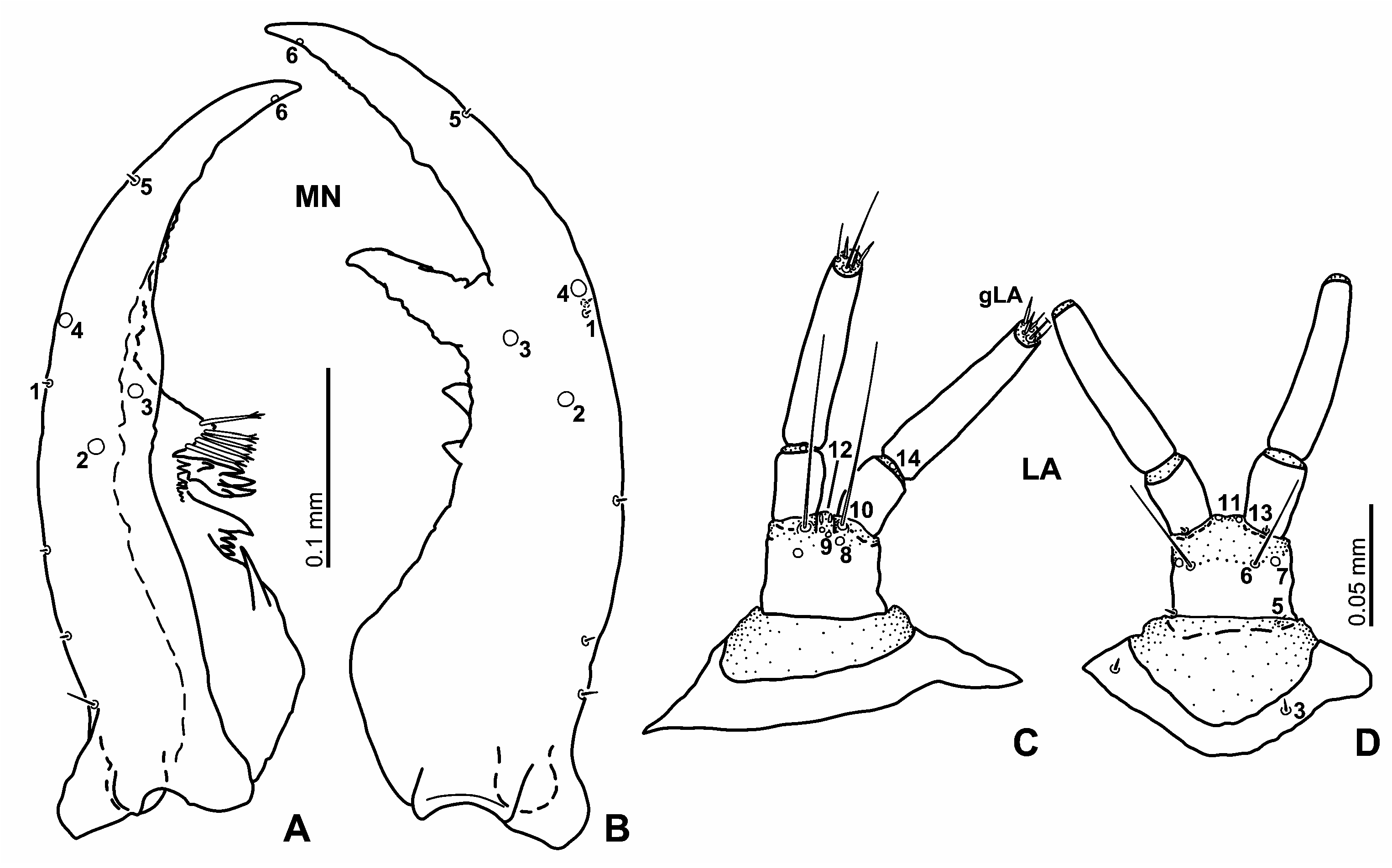
Mandibles ( Figs 7A–B View Fig ) strongly asymmetrical; left mandible shorter than right one. Right mandible with three inner teeth on median part; distal one large, with serrated edge; proximal two small, subequal in size. Left mandible with three inner teeth and one strong inner spine posteriorly to inner teeth on dorsal surface; distal tooth large, with a group (usually five) of seta-like projections, apex of projections trifid; median tooth moderately sized, quadrifurcate, with small cuticular projections basally; basal tooth moderately sized, with at least four projections pointed apically, distal one larger than basal ones.
Maxilla ( Figs 6C–D View Fig ) 6-segmented, longer than antenna. Cardo moderate in size, subtriangular. Stipes the longest and widest, longer than palpomeres 1–4 combined; inner face without spine-like cuticular projections; a small cuticular projection subapically on inner face undetectable. Maxillary palpus 4-segmented, palpomere 1 widest, longer than palpomere 2 and 3, palpomere 3 the longest, palpomere 2 the shortest, palpomere 4 narrowest; palpomere 1 may be completely sclerotised but anterior margin of sclerotised part invisible; inner process sclerotised.
Labium ( Figs 2B View Fig , 7C–D View Fig ) small, partly reduced. Submentum (e.g., Fig. 2B View Fig ) fused to head capsule, large, subpentagonal, wider than mentum. Mentum transverse, narrow, cylindrically sclerotised, wider than prementum; dorsal surface bare. Prementum subquadrate. Ligula strongly reduced, very short, completely membranous. Labial palpus long, straight, without cuticular projections; palpomere 1 as wide as palpomere 2 and slightly shorter than prementum, palpomere 2 distinctly longer than palpomere 1.
Thorax (Fig. 1). Thoracic membrane covered with fine cuticular pubescence. Prothorax wider than head capsule. Proscutum formed by one large plate subdivided by fine sagittal line, anterior part rather weakly sclerotised; whole sclerite bearing densely arranged fine cuticular projections. Prosternal sclerite subpentagonal, large, with long and fine sagittal line. Mesonotum with two sclerites on each side; anterior one small, narrow; posterior one large, subtriangular; one small tubercle behind each posterior mesonotal sclerite; lobe-like lateral projection on each side. Mesonotal spiracles situated anteriorly on dorsolateral face. Metanotal sclerites absent; one pair of membranous tubercles present on median part; lobelike lateral projection on each side. Legs long, slender, visible in dorsal view, 5-segmented; all three pairs similar in shape.
Abdomen (Fig. 1). Ten segmented, tapering posteriad, covered with fine cuticular projections densely arranged; segments 1 to 6 similar in shape and size, segment 7 smaller than others. Lateral sides of segments 1–7 with one long to very long tracheal gill each; dorsal sclerites on segment 1–7 absent. Segments 1–3 with four small tubercles, two on median part, remaining ones laterally behind spiracles; lateral tubercles smaller than median ones. Segments 4–7 with two small tubercles medially. Segment 7 without tubercle.
Spiracular atrium ( Fig. 5D View Fig ) reduced. Segment 8 with oval dorsal plate; segment 9 trilobed, median lobe and each lateral lobe of spiracular atrium very small, hardy visible from dorsal view; procercus, acrocercus, urogomphi and prostyli reduced, undetectable. Ventral surface of spiracular atrium with two bulbous projections.
Second instar. Very similar to third instar larva, slightly more slender than third instar.
Head. Frontal lines clearly visible, nearly straight in median to anterior parts, strongly curved outwards at base.
Antenna proportionally stouter than in third instar; antennomere 1 proportionally shorter than in third instar, slightly longer than or as long as antennomeres 2 and 3 combined.
Maxilla proportionally stouter than third instar.
Mandible. Basal inner tooth on right smaller than median one.
Thorax and abdomen. Arrangements of cuticular projections and pubescence on thorax and abdomen similar to third instar but projections and pubescence finer than in third instar. Lateral tubercles on third abdominal segment indistinct.
First instar ( Figs 2–4 View Fig View Fig View Fig ). Similar to second instar larva; weakly sclerotised than second instar. Spiracles on mesothorax and abdominal segments 1–7 undetectable.
Head. Anterior margin of right epistome almost straight ( Fig. 3A View Fig ). Basal part of frontal line more weakly curved laterally than in second instar ( Fig. 2A View Fig ).
Antenna ( Figs 4A–B View Fig ) proportionally stouter than in second and third instar larvae; antennomere 1 proportionally shorter than in third instar larva, about as long as or slightly shorter than antennomeres 2 and 3 combined.
Maxilla ( Figs 4E–F View Fig ) proportionally stouter than that of second and third instar larva.
Labial palpus ( Figs 4G–H View Fig ) proportionally stouter than third instar; palpomere 1 slightly wider than palpomere 2.
Thorax and abdomen. Arrangements of cuticular projections and pubescence on thorax and abdomen similar to third instar but projections and pubescence finer than in second instar. Prothorax as wide as or slightly wider than head capsule.
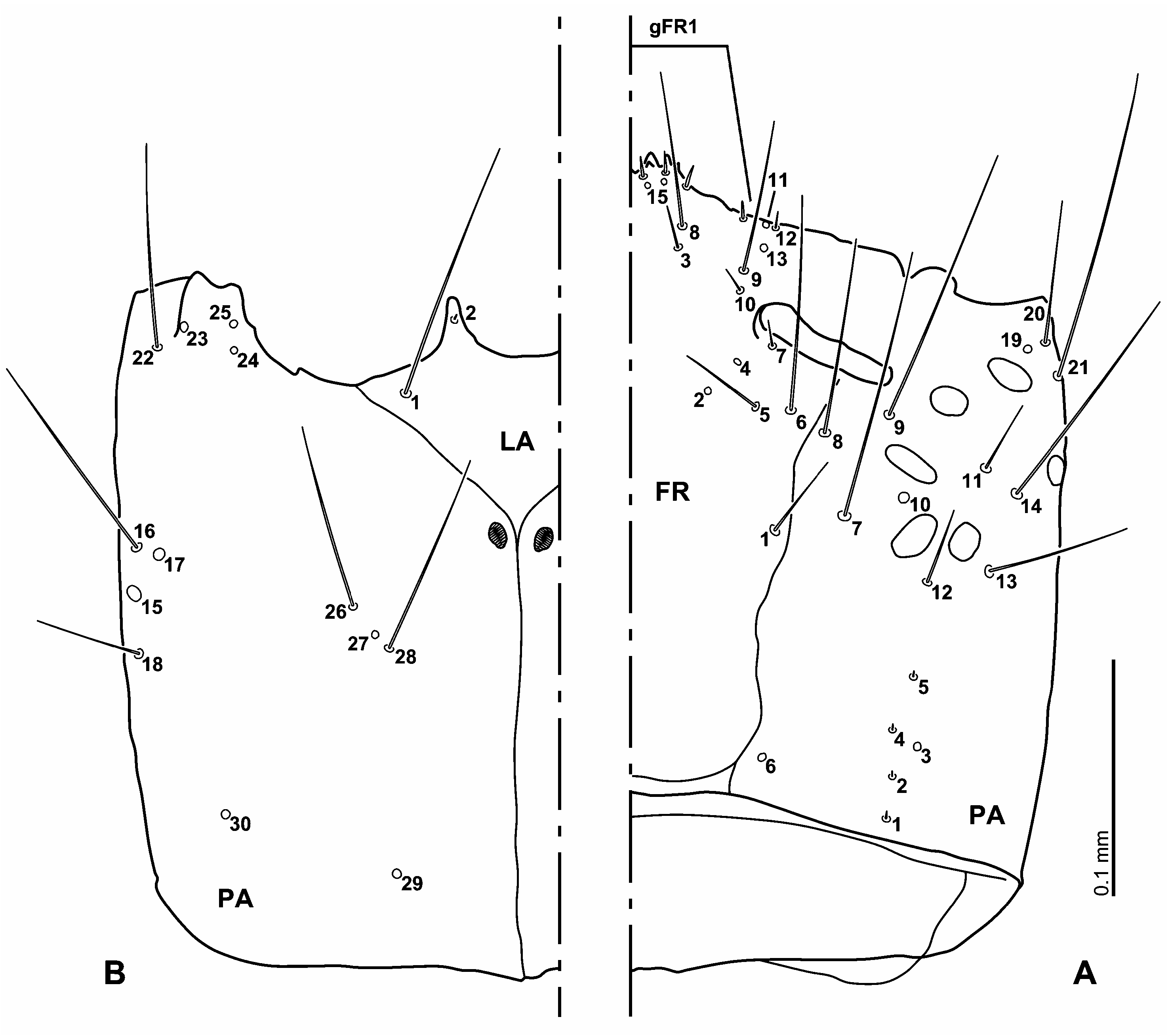
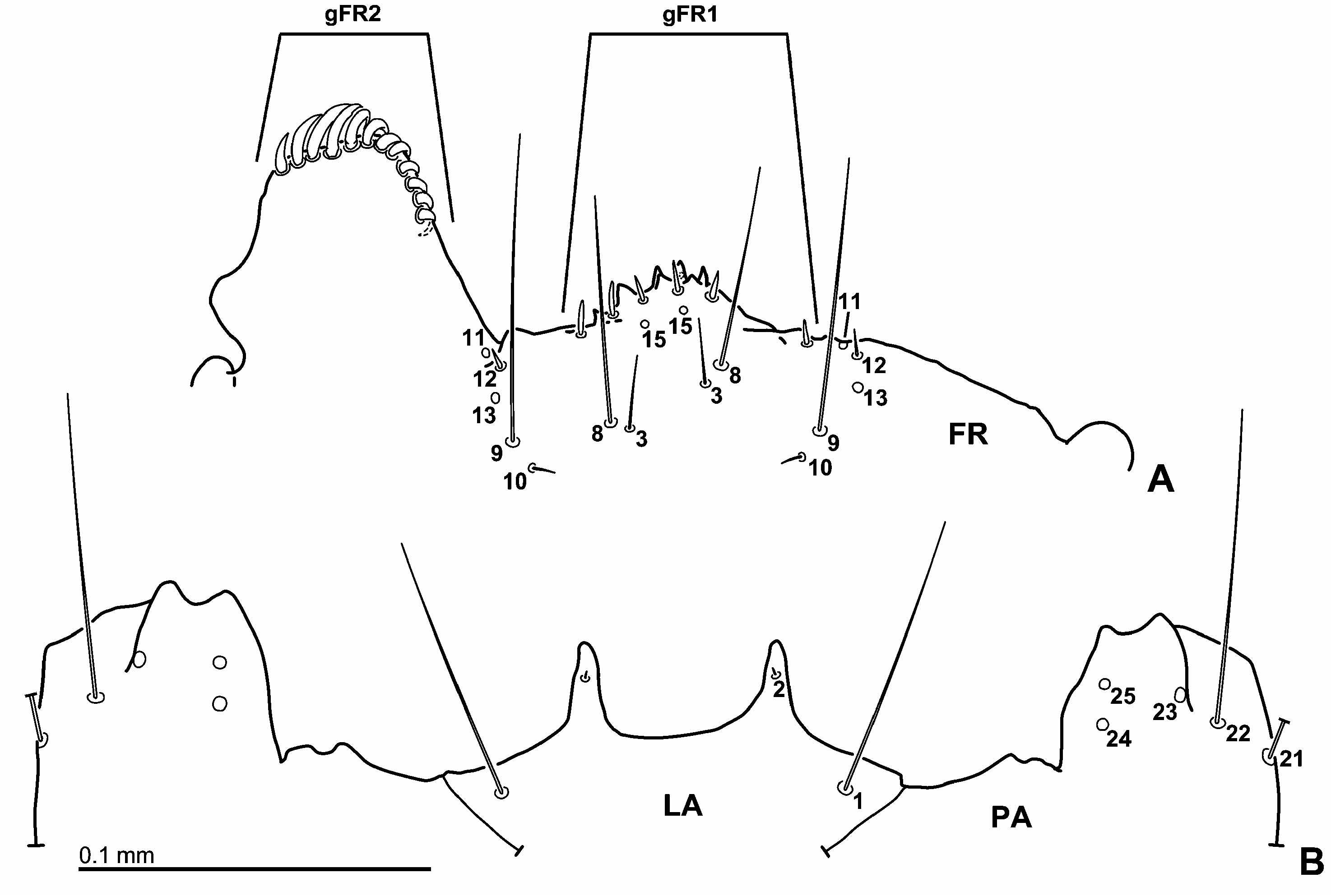
Chaetotaxy of head. Primary chaetotaxy ( Figs 2–4 View Fig View Fig View Fig ). Frontale ( Figs 2A View Fig , 3A View Fig ). Rather long seta FR1 on midlength of frontale close to frontal line. Pore-like sensilla FR2 and FR4 and setae FR5–7 posteromesal to antennal socket, close to anterior end of frontal line; FR5 rather long, FR6 very long (homology of FR5 and FR6 unclear), FR7 short; FR2 and FR6–7 forming a triangular group laterally to FR6–7; FR6 close and lateral to FR5; FR7 anterior to remaining sensilla (FR2, FR4–6), close to inner margin of antennal socket. FR9–13 on epistome, situated anteriorly and slightly mesally to antennal socket, forming irregularly longitudinal row; arrangement of FR9–13 slightly asymmetrical; FR10 and FR12 short setae, FR9 long seta, FR11 and FR13 pore-like sensilla; FR12 on right side stouter than left side. FR10 posterior to FR9 and FR11–13, FR9 anterior to FR10, between FR10 and FR13, FR13 between FR9 and FR12; FR11 on anterior margin of epistome, close to FR12; FR12 slightly posterior to FR11. Rather short seta FR3 and long seta FR8 behind nasale, FR3 close and posterior to FR8. Pore-like sensilla FR15 on median part of nasale. Nasale with a group of six equidistant, stout and short setae and with (at least) one pore-like sensillum (gFR1). Left epistomal lobe with a group of about 12 stout setae densely arranged on anterior margin (gFR2), mesal ones strongly bent towards ventrally; gFR2 absent on right side.
Parietale ( Figs 2A–B View Fig ). Dorsal surface with a group of five sensilla (PA1–5) forming irregularly longitudinal row at midwidth in posterior part of parietale; PA1–2 and 4–5 short setae, PA3 pore-like. PA6 pore-like, located posteromesally, close to posterior end of frontal line, more distant from posterior margin of head than PA1. Pore-like sensilla PA10 between mesal two stemmata. Setae PA7–9 posterior to antennal socket; PA8 long, PA7 and PA9 very long; PA9 close to lateral margin of antennal socket; PA8 posteromesal to PA9, close to frontal line and FR6; PA7 posterior to PA8 and PA9, mesal to PA10, between FR1 and PA10. Rather long seta PA12 and long seta PA13 close to mesal two stemmata of posterior row; PA12 behind mesal-most one, PA13 posterolateral to median one. Setae PA11 and PA 14 in the line connecting lateral four stemmata, PA11 rather long, PA14 very long; PA11 anteromesal to PA14; PA14 between lateral two stemmata of posterior row, anterolateral to PA13. PA19–22 on anterior corner of head capsule, PA19 pore-like, PA20 long seta, PA21 very long seta, PA22 moderately long seta; PA19 dorsal to PA20–22, close to PA20. PA22 ventral to PA19–21, PA20–21 between PA19 and PA22; PA21 behind PA20; PA22 posterolateral to ventral mandibular articulation. Pore-like sensilla PA23–25 close to ventral mandibular articulation; PA23 lateral to PA24–25; PA24 posterior to PA25. PA15–18 situated lateroventrally in midlength of parietale; PA15 and PA17 pore-like, PA16 very long seta, PA18 long seta; PA16 and PA17 anterior to PA15 and PA18, PA17 close and mesal to PA16, PA15 between PA16 and PA18. Two long to very long setae (PA26 and PA28) and pore-like sensillum PA27 situated ventrally on median part of parietale; PA26 posterolateral to PA27 and PA28, PA27 between PA26 and PA28. Two pore-like sensilla (PA29–30) on posterior part of ventral parietale; PA29 mesal to PA30, posterior to PA26–28; PA30 on posterolateral part.
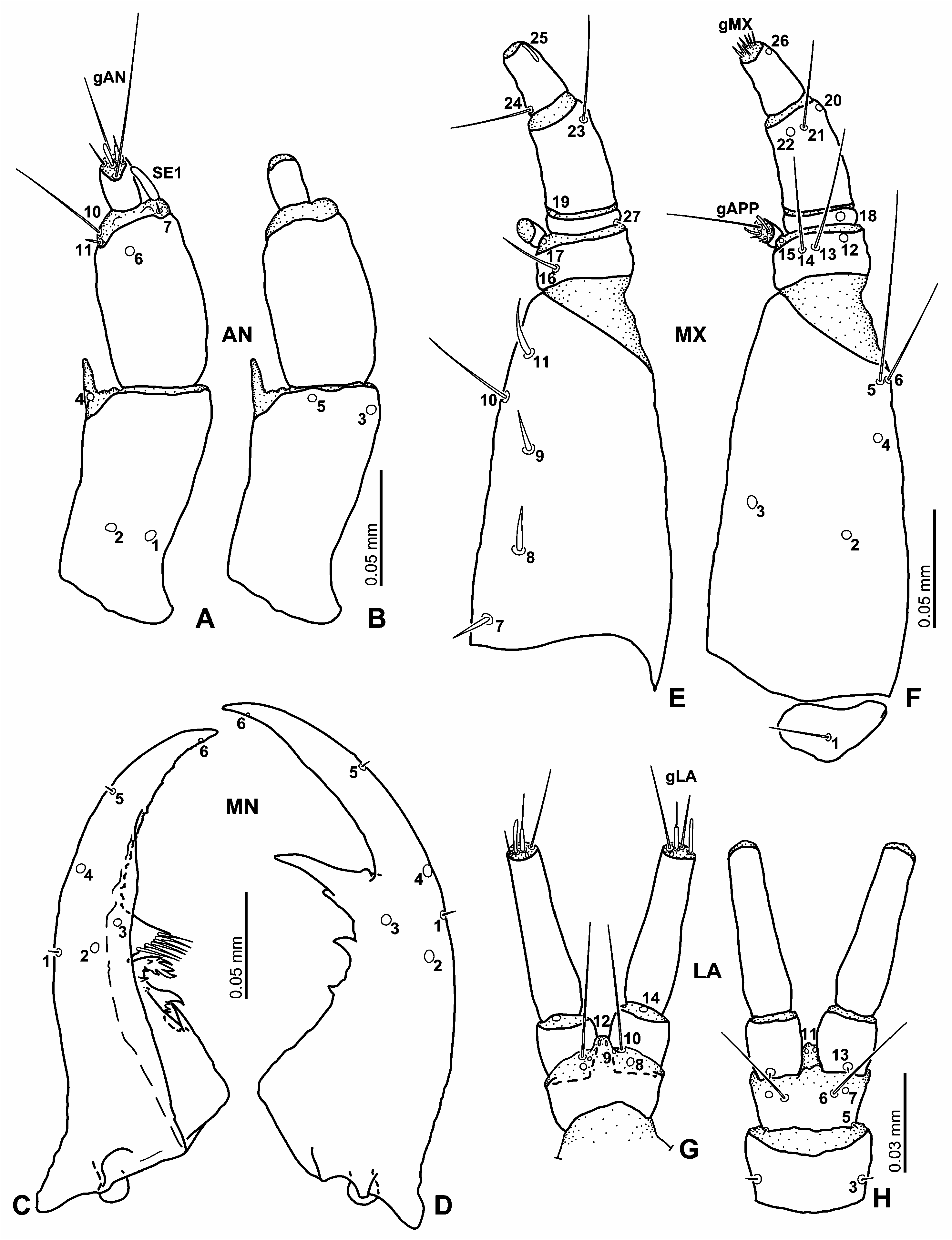
Antenna ( Figs 4A–B View Fig ). Antennomere 1 with five pore-like sensilla (AN1–5); AN1–2 situated dorsally on posterior third, AN1 laterally to AN2, AN3 subapically on lateral face, AN4 on base of inner projection, AN5 ventrally on median portion of anterior margin of sclerite. Antennomere 2 with one pore-like sensillum (AN6) situated dorsally on subapical part of sclerite; minute seta AN7 and sensorium SE1 on lateral face of intersegmental membrane between antennomeres 2 and 3, AN8–9 absent; SE1 slender, about as long as antennomere 3; setae AN10–11 on inner face of intersegmental membrane between antennomeres 2 and 3, AN10 long, AN11 short, both setae close to each other. Antennomere 3 with apical sensilla (gAN) in apical membranous area; gAN with two rather long setae and a few (at least four) short setae of variable shape.
Mandibles ( Figs 4C–D View Fig ). Mandible with two setae (MN1 and MN5) and three pore-like sensilla (MN2–4). Pore-like sensillum MX 6 situated on apical part of incisors area. Very short seta MN1 on midlength of lateral face of mandible. MN2–3 on median part of mandible; MN3 mesal to MN1–2, MN2 posterior to line connecting MN1 and MN3, MN2 on right mandible more distant than left. MN4 and minute seta MN5 on lateral face anteriorly to MN1; MN5 subapical; MN4 at midlength between MN1 and MN5 on left, closer to MN1 on right.
Maxilla ( Figs 4E–F View Fig ). Cardo with one moderately short ventral seta ( MX 1). Stipes with irregular row of five setae ( MX 7–11) situated dorsally along inner face; MX 7–9 and MX 11 stout, moderately short, MX 10 trichoid, long; MX 7–9 and MX 11 almost equidistant from each other; MX 10 between MX 9 and MX 11 but situated more ventrally. Two setae ( MX 5–6) situated apically on outer face of sclerite; MX 5 very long, MX 6 long; MX 5 very close and dorsal to MX 6. Pore-like sensilla MX 2–3 situated ventrally on median part of sclerite; MX 2 on outer part, MX 3 on inner part; pore-like sensillum MX 4 behind MX 5–6, between MX 2 and MX 5. Dorsal surface of palpomere 1 with one rather long, slightly stout seta ( MX 16) on inner face; ventral surface of sclerite with three sensilla ( MX 12–14) close to distal margin of sclerite; MX 12 pore-like on lateral part, MX 13 long seta between MX 12 and MX 14, MX 14 long seta on inner part, close to MX 13. Rather small pore-like sensilla ( MX 15 and MX 17) on membrane behind inner appendage; MX 17 dorsal, MX 15 ventral. Inner appendage with one long seta and a few short setae (gAPP). Palpomere 2 with two pore-like sensilla ( MX 18 and MX 19) and one minute seta ( MX 27); MX 18 situated ventrally on outer part of sclerite; MX 19 on inner face of intersegmental membrane between palpomeres 2 and 3; MX 27 at base of outer face of sclerite. Palpomere 3 with two setae ( MX 21 and MX 23) and two pore-like sensilla ( MX 20 and MX 22); MX 21 rather long, MX 23 long; MX 20 on outer face very close to distal margin of sclerite; MX 21–22 on median part of sclerite close to distal margin of sclerite; MX 21 lateral to MX 22; MX 23 dorsal on outer face close to distal margin. Palpomere 4 with one rather long seta ( MX 24) situated basally on inner face, and with digitiform sensillum ( MX 25) and pore-like sensillum ( MX 26) apically on outer face of sclerite; MX 25 dorsal, MX 26 ventral. Apical membranous area of palpomere 4 with several minute setae (gMX).
Labium ( Figs 2B View Fig , 3B View Fig , 4G–H View Fig ). Submentum with two pairs of setae (LA1–2); LA1 very long, in each lateral corner, LA2 minute, on anterior margin. Ventral surface of mentum with one pair of short setae (LA3) situated on median part of outer face; LA4 absent. Prementum with three pairs of sensilla (LA8–10) on dorsal surface and with three pairs of sensilla (LA5–7) on ventral surface. LA8–10 on anterior membranous area of prementum, close to each other; arrangement of LA8–10 vary, LA8–9 sometimes absent; LA8–9 pore-like, LA10 long seta; LA9–10 at basal part of ligula, LA8 behind LA9–10. Minute seta LA5 at base of outer face; long seta LA6 and pore-like sensillum LA7 on anterior part, close to borderline between sclerite and membrane of prementum; LA6 close and mesally to LA7. Ligula with two pairs of pore-like sensilla (LA11–12) apically; LA12 dorsal, LA11 ventral. One minute seta (LA13) situated ventrally on basal margin of palpomere 1; pore-like sensillum LA14 on dorsal surface of intersegmental membrane between palpomeres 1 and 2. LA15 absent. Apical membranous area of palpomere 2 with several setae of variable length and shape (gLA).
Second instar. Primary sensilla similar to first instar, and secondary chaetotaxy similar to third instar.
Parietale with four rather short secondary setae. Two dorsal setae close to frontal line, one between PA6 and PA7, one between PA8 and PA9; one seta lateroventral, anterior to PA16; one seta on median part of lateral face, between PA13 and PA15–18.
Mandible (e.g., Figs 7A–B View Fig ). Outer face of mandible bearing a few short to minute secondary setae.
Maxilla (e.g., Figs 6C–D View Fig ). Stipes bearing three secondary setae; one moderately short seta situated dorsally on basal part of outer face, one moderately long seta on median portion of outer face, one long seta ventrally on subapical part of sclerite, close to and behind MX 5.
Third instar ( Figs 5B–C View Fig , 6–7 View Fig View Fig ). Similar to second instar.
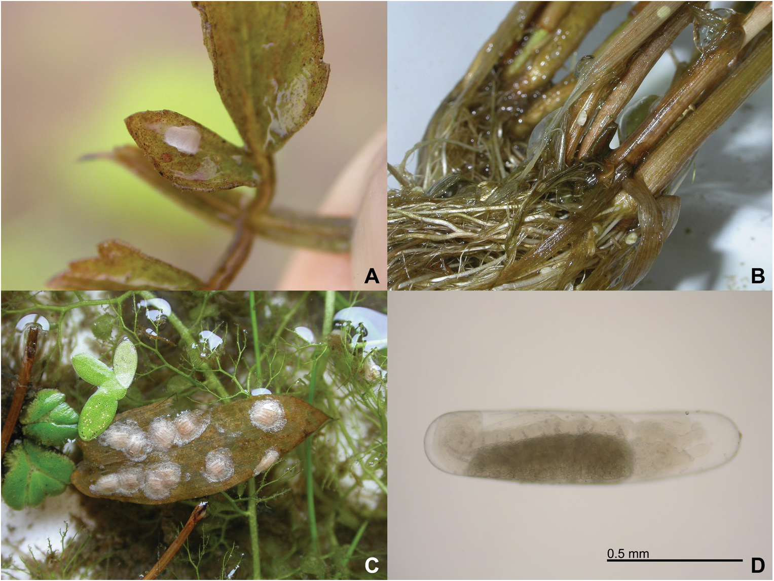
Egg-case. Egg-cases were laid on substrate ( Figs 17A–B; HAYASHI View Fig 2009a).
Biology. Berosus japonicus inhabits paddy fields in the locality of Shimane prefecture; larvae prefer masses of algae, possibly Spirogyra or Zygnema (Zygnemataceae) . Nearly one hundred larvae were occasionally found in such masses of algae, together with larvae of Peltodytes intermedius ( Sharp, 1873) ( Coleoptera : Haliplidae ) (Hayashi, personal observation). This habitat preference might be the reason for confusion about the diet of larval Berosus , which was considered to consist of algae ( WILSON 1923, BØVING & HENRIKSEN 1938, PETERSON 1951). As BØVING & HENRIKSEN (1938) and ARCHANGELSKY (1997, 2008) questioned, Berosus larvae will be carnivorous.


| KMNH |
Kitakyushu Museum and Institute of Natural History |
No known copyright restrictions apply. See Agosti, D., Egloff, W., 2009. Taxonomic information exchange and copyright: the Plazi approach. BMC Research Notes 2009, 2:53 for further explanation.