
Horstia kincaidi, Deland & Cameron & Rao & Ritter & Bullock, 2010
|
publication ID |
https://doi.org/10.11646/zootaxa.2408.1.1 |
|
publication LSID |
lsid:zoobank.org:pub:9BBB84BB-239C-41EA-9CFC-682449F96281 |
|
persistent identifier |
https://treatment.plazi.org/id/101A87CA-FF93-FFB8-DBA6-FC3CF944FBC5 |
|
treatment provided by |
Felipe |
|
scientific name |
Horstia kincaidi |
| status |
n. gen. et n. sp. |
Horstia kincaidi View in CoL n. gen. et n. sp.
( Figs 2A,F View FIGURE 2 ; 9A–F View FIGURE 9 )
Material examined. In 1899 Professor Trevor Kincaid collected a dozen specimens on Whidbey Island in Puget Sound , Washington ( 47°59' N, 122°26' W). The species has not been found again. We have both old Ritter sections and more recent ones made by Bullock of nine of the specimens. Considering the faded condition of the former it seems best to designate one of the latter, accession no. USNM 71439 About USNM as the holotype GoogleMaps ; 58879 becomes the paratype.
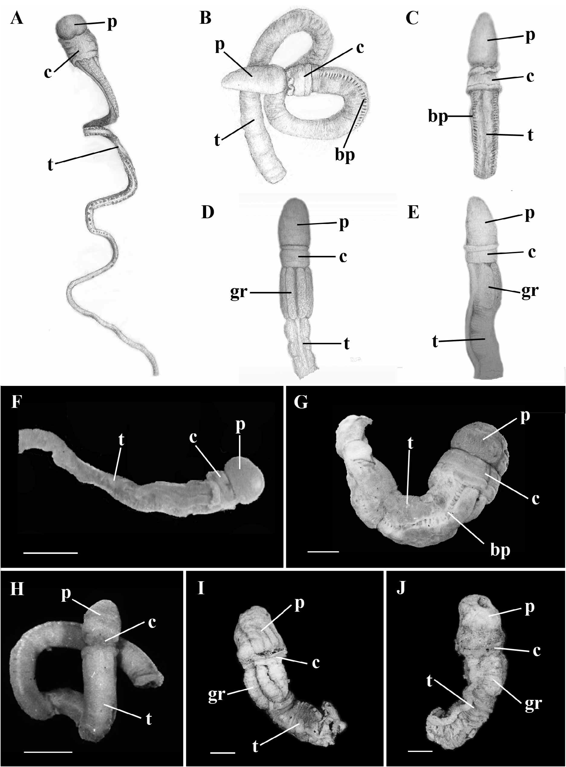
External features ( Fig. 2A,F View FIGURE 2 ). Total length 30–40 mm. Proboscis 2 mm long, as short as wide, collar 1.5– 2 mm long with irregular surface markings. Branchial region 6 mm long, tapering to 1.0–1.5 mm wide, exceptionally narrow, postbranchial trunk even more attenuated. Genital wings and hepatic lobes lacking. Branchial orifices conspicuous, their arrangement distinctive, with left and right rows close together on elevated ridge bounded laterally by groove. Gonads also conspicuous as long series of protruding nodules.
Color in life reported by Kincaid as brown. Proboscis uniform creamy white in material preserved by Perenyi's fixative, collar and anterior portion of thorax only slightly darker; remainder of animal (most of it) greenish brown with gonads yellowish white.
Internal features. Longitudinal muscle fibers of proboscis arranged in well-defined wedge-shaped radial plates ( Fig. 9A View FIGURE 9 ). No conspicuous thickening of nerve-fiber layer in proboscis or any apparent dorsal longitudinal groove. Proboscis coelom extending to tip of organ. Glomerulus poorly developed ( Fig. 9A View FIGURE 9 ), limited to two small bilateral masses (usually well defined in other species), standing out like wings. Pericardial sac quite spacious, covering dorsal and lateral sides of stomochord. Blood vessel also spacious, almost entirely filling inside of pericardial sac ( Fig. 9A View FIGURE 9 ). Ventral and dorsal mesenteries present in proboscis to tip of proboscis complex ( Fig. 9A View FIGURE 9 ).
Proboscis portion of stomochord large, simple, its cavity clearly defined, with slight posteriorly directed ventral blind lumen. A single proboscis pore present, on right or left. In one case, coelomic spaces, canals, and terminal vesicles were so nearly symmetrical apart from the single pore, that Ritter predicted individuals will be found with both pores.
Peribuccal diverticula absent. Perihaemal spaces extending forward of proboscis pore; cornua of proboscis skeleton extending nearly back to posterior edge of collar and notable for steepness and extent of their ventral projection, from their very origin, approaching transverse plane and nearly reaching ventral side of buccal cavity. Keel deep and narrow; almost no chondroid tissue developed in connection with body of skeleton ( Fig. 9B View FIGURE 9 ).
Stomochord continuous through neck, though narrow. Collar nerve cord with very large lacunae scattered throughout its length. A small anterior neuropore; dorsal wall of cord thicker than ventral for some distance behind neuropore ( Fig. 9C View FIGURE 9 inset). No dorsal crest or dorsal roots but dorsal mesentery well developed through collar ( Fig. 9C View FIGURE 9 ). Collar canals large, vertically orientated ( Fig. 9D View FIGURE 9 and inset). Collar longitudinal muscles well developed, forming wing-shaped bundles on either side of collar lumen ( Fig. 9D View FIGURE 9 ).
Ventral portion of pharynx about a third of size of branchial portion. Branchial pores on each side numbering about 30. Gonads beginning somewhat posterior to middle of branchial region ( Fig. 9F View FIGURE 9 ). Individual gonads well separated from each other, standing out prominently from surface of body. Intestinal pores and hepatic caeca absent.
Remarks. We here carry out Ritter's intention to name the species for Professor Trevor Kincaid, long deceased of the University of Washington and a pioneer northwestern naturalist.
The defining characters of Horstia kincaidi are listed below:
A. Body very tapered, proboscis round.
B. Short proboscis and collar.
C. Longitudinal musculature of the proboscis in radial plates.
D. Deep and narrow skeletal keel with the cornua steeply bent ventralwards at their posterior end.
E. The dorsal and ventral mesenteries are present in the proboscis and collar.
F. Left or right proboscis pore.
G. Anterior neuropore.
H. Large vertical collar canals.
I. Two rows of lateral gonads forming small protuberances starting in the middle of the branchial region (no genital ridges or wings).
J. Gonopores on an elevated ridge.

No known copyright restrictions apply. See Agosti, D., Egloff, W., 2009. Taxonomic information exchange and copyright: the Plazi approach. BMC Research Notes 2009, 2:53 for further explanation.
|
Kingdom |
|
|
Phylum |
|
|
Class |
|
|
Family |
|
|
Genus |