
Metapolystoma theroni, Landman & Verneau & Raharivololoniaina & Preez, 2021
|
publication ID |
https://doi.org/ 10.1016/j.ijppaw.2021.01.012 |
|
DOI |
https://doi.org/10.5281/zenodo.10914311 |
|
persistent identifier |
https://treatment.plazi.org/id/0F7C4239-994F-FFDB-B56F-FB59FBE0DA96 |
|
treatment provided by |
Felipe |
|
scientific name |
Metapolystoma theroni |
| status |
sp. nov. |
3.1.4. Metapolystoma theroni View in CoL n. sp. ( Fig. 10‒11 View Fig View Fig ; Table 2 View Table 2 )
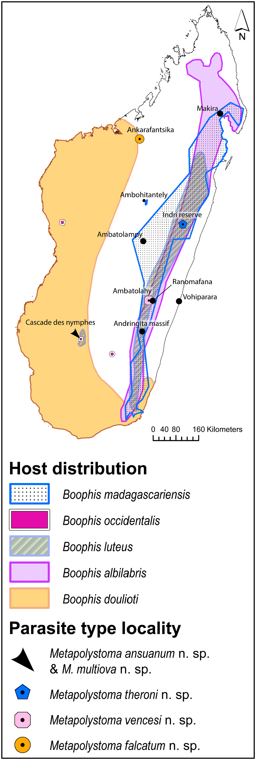
3.1.4.1. Type host. Boophis madagascariensis ( Mantellidae ).
new name Metapolystoma ansuanum n. sp. Landman et al. is: urn:lsid:zoobank.org:act:4761BF6E-F309-4625-A7D7-CA64F3A3F6F8 .
3.1.3.7. Etymology. This species is named for Mrs Anna-Susan van der Linde, known as Ansu, in acknowledgement of her teaching and inspiration of many secondary school pupils in the field of biology.
3.1.4.2. Type locality. Indri Reserve in Andasibe , Madagascar ( Fig. 1 View Fig ), (18.930856S; 48.413611E) GoogleMaps .
3.1.4.3. Site in host. Urinary bladder.
3.1.4.4. Level of infection. Three of 30 frogs collected were infected with a total of one mature and 71 juvenile parasites, while as many as 40 parasites were infecting a single host (prevalence of 10%, mean intensity 24).
3.1.4.5. Type material. The morphological descriptions are based on one mature and 27 juvenile parasites. One sexually mature specimen (Holotype 573) and four immature ones (Paratypes 574–577), all from the type locality, were deposited in the parasitic worm collection, National Museum, Aliwal Street, Bloemfontein 9301.
3.1.4.6. Voucher material. Remaining specimens in polystome collection, North-West University, Potchefstroom, South Africa.
3.1.4.7. Zoobank registration. The Life Science Identifier ( LSID) of the article is: 59F6A99A-C667-48 EB-9EA4-881D43956065. The LSID of the new name Metapolystoma theroni n. sp. Landman et al. is: urn:lsid:
170
zoobank.org:act: 55203AE0-217D-45D0-B3C8-07DDE53FA9A3.
3.1.4.8. Etymology. This species is named in honour of emeritus Professor Pieter Daniel Theron at the North-West University, South Africa,
in recognition of 54 years of inspiring teaching and dedication to the field of zoology.
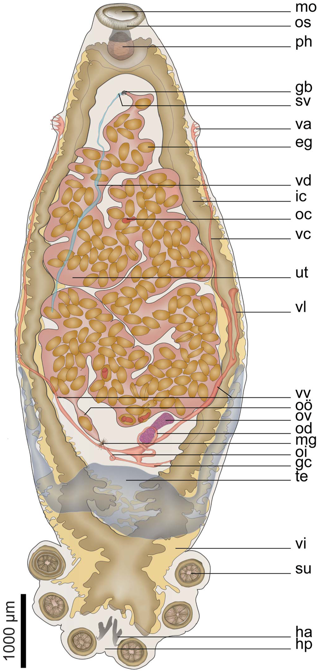
3.1.4.9. Description. Measurements reflected in Table 2 View Table 2 . Body pyriform
171
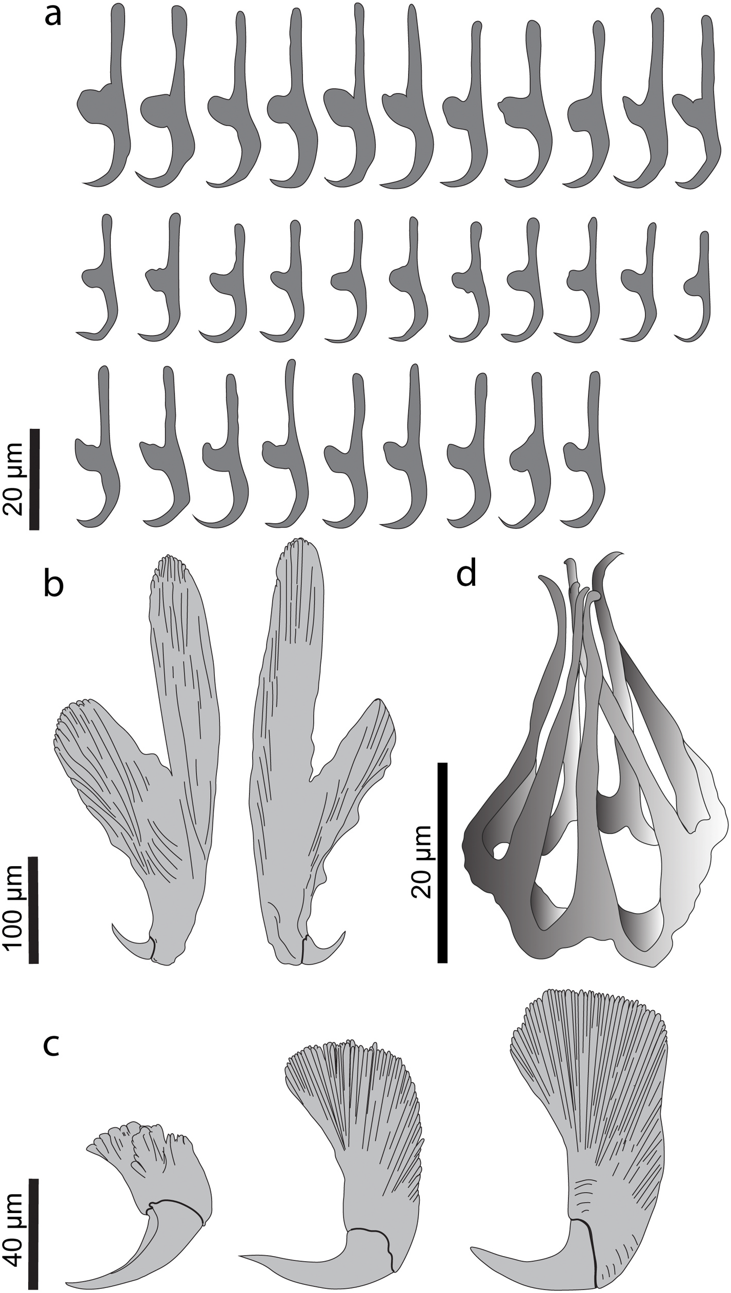
( Fig. 10 View Fig ) dorsoventrally flat, widest section at 51% from anterior end, body length 2.6 times greater than width, mouth sub-ventral, surrounded by false oral sucker. Posterior haptor occupying 18% of total body length, bearing three pairs of haptoral suckers, equal in size. Marginal hooklets placed as for other polystomes, pairs one and two between hamuli, pairs three to five embedded in suckers, pairs six to eight in area between anterior-most suckers, pairs one and eight larger than pairs two to seven ( Fig. 11a View Fig ). Well-developed hamuli positioned between posterior-most haptoral suckers with deep cut between handle and guard ( Fig. 11b View Fig ). Hamuli development presented in Fig. 11c View Fig . Medial pharynx length greater than width, positioned immediately posterior to or at margin of false oral sucker. Intestine bifurcates immediately posterior to pharynx at 10% from anterior, converging posteriorly at 80% from anterior; no prehaptoral anastomoses. Lateral intestinal diverticula in first three quarters length equal to width, in last quarter length greater than width. Medial diverticula only posterior to ovary, length greater than width.
Testis follicular, u-shaped, mainly positioned posterior to the ovary with two lateral processes extending forward along the lateral line past the ovary up to one-third of the body proper, ventral to intestine. Vas deferens widens anteriorly to form sinuous semen vesicle 23–65 (46 ± 18; 1) wide, 122 long, measuring 1% of body length, narrowing towards genital bulb, opening in common genital opening. Genital pore opening mid-ventral, posterior to intestinal ceca bifurcation, positioned 13% from anterior, genital bulb muscular, surrounded by glandular cells, armed with genital crown bearing seven genital spines ( Fig. 11d View Fig ).
Ovary elongate, not lobed, positioned posterior to midbody, length 1.2 times greater than width, measuring 5% of body length. Oviduct 1167 long, 20–51 (33 ± 9; 1) wide. Uterus massive, occupying 50% of body proper, tubiform, serpentines between posterior connection at ootype ¨and anterior connection at genital bulb, containing 176 ovoid, operculate eggs, some contain fully developed oncomiracidia. Hatched intrauterine oncomiracidia present. Mehlis’ gland distinct. Two parallel vaginae 270–304 long, 132–177 wide, on lateral margins, with multiple marginal openings, vaginal vestibule cup-shaped at 18% from anterior. Vitellaria dorsal to intestinal tract, extended throughout most of body and haptor, except areas occupied by female reproductive organs. Genito-intestinal canal prominent 457 long, 30–77 (52 ± 16; 1) wide, situated posterior to ovary.
No known copyright restrictions apply. See Agosti, D., Egloff, W., 2009. Taxonomic information exchange and copyright: the Plazi approach. BMC Research Notes 2009, 2:53 for further explanation.
|
Kingdom |
|
|
Phylum |
|
|
Class |
|
|
Order |
|
|
Family |
|
|
Genus |