
Dendronotus noahi, Pola & Stout, 2008
|
publication ID |
https://doi.org/ 10.11646/zootaxa.1960.1.2 |
|
persistent identifier |
https://treatment.plazi.org/id/087D7947-FF88-FFF2-1A86-204B8EE0F86A |
|
treatment provided by |
Felipe |
|
scientific name |
Dendronotus noahi |
| status |
sp. nov. |
Dendronotus noahi View in CoL sp. nov.
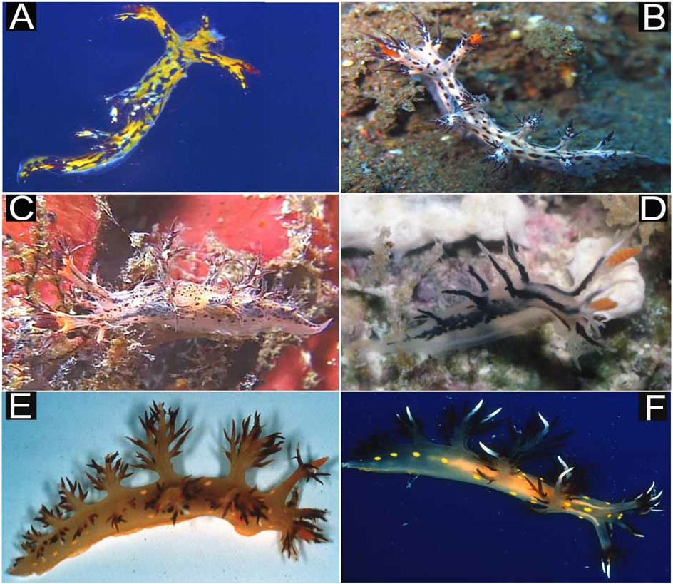
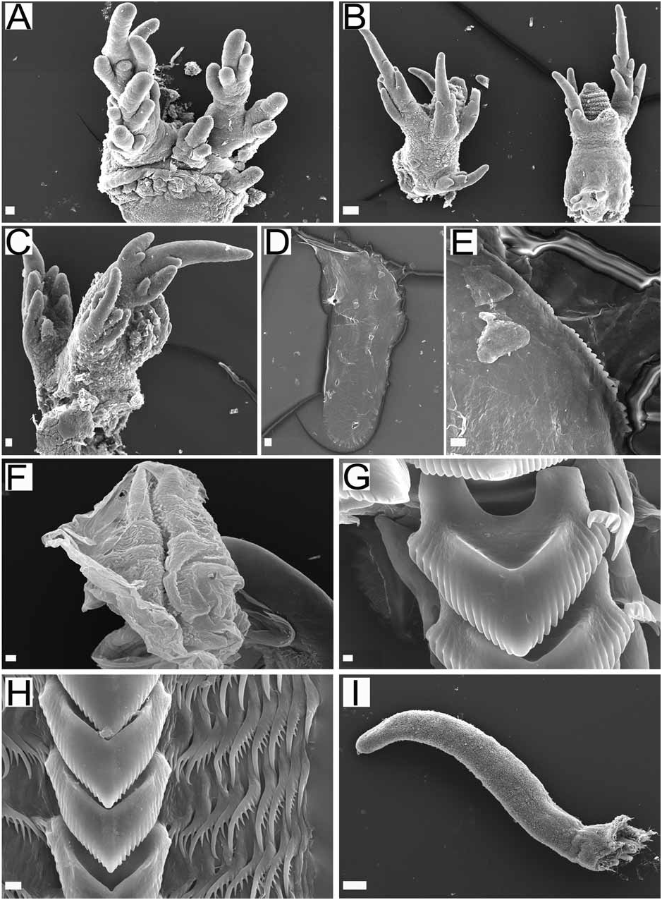
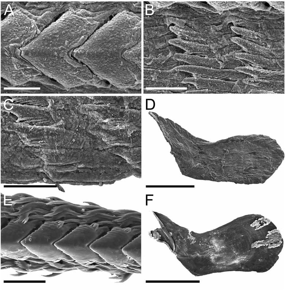
( Figures 1D View FIGURE 1 , 6A–B View FIGURE 6 )
Material Examined: Holotype: CASIZ 075085 , Papua New Guinea, North coast, outer barrier reef, Bagabag Island, off New Year’s Bay, 4 mm preserved, immature, 30.5 m depth, 26 November 1990, collected by T. M. Gosliner.
Etymology: Named after junior author’s nephew.
Distribution: Thus far, known from the type locality, Bagabag Island ( Papua New Guinea).
External morphology ( Fig. 1D View FIGURE 1 ): The general body shape is limaciform, tall and laterally compressed with a short, pointed posterior end of the foot. The foot is narrow, rounded in front, not sharply marked off from the body, tapering rapidly to the posterior end of the foot. The lateral margin is not prominent other than the line of processes. The body surface has a few low tubercles, some in the mid-dorsal line and some on both sides of the body. The cardiac elevation is prominent. The head is rounded, its velar margin marked by a series of three paired, stout, branched processes oriented anteriorly. The two nearest the mouth on either side are smaller with three short blunt branches and the next outermost is larger with a longer central branch and three short branches. The outermost pair is the largest with six branches of irregular length. The rhinophores consist of tall stalks inclined anteriorly. The sheath margin is expanded into four simple processes; the two most anterior processes are simple and about the same length and the two most posterior processes are the longest. Of these latter two processes, the inner one is longer and with a small branch about half way to the tip. On the outer side of each stalk, at its base, there is also a small papilla. The conical clavus is perfoliate with 9 robust lamellae. The lamellae of the rhinophores are orange. There are four processes on either side, arranged in pairs along the dorsolateral margins of the back. The distance between of each pair of dorsolateral processes is about the same. The first pair is the largest, the remaining decreasing in size towards the tail. The last pair is very small and simple. The first and second pairs of dorsolateral processes have a main central branch with two very small processes close to the tip. Close to the base on the outer side there are two elongate papillae and a slightly more distally on the inner side there are two more elongate papillae. The third pair of dorsolateral processes is similar to the first and second pairs but the outer and inner papillae are very small. The anal opening is along the dorsolateral line about midway between the first and second dorsolateral processes on the right side. The small renal pore lies just dorsal to the anal elevation. The reproductive openings are not visible in this specimen.
The background color of the animal is transparent white. Wide, black branches of the digestive gland can be seen through the transparent tissue ( Fig. 1D View FIGURE 1 ). Once dissected, the digestive gland shows a brown pigmentation with darker brown spots on it.
Alimentary Canal: The mouth opening is T-shaped surrounded by thick, muscular lips. There are two close sets of six relatively long tubular labial glands extending deep into the sub-epithelial tissue. The pharyngeal bulb is large. The lip disk is small and covered by a thin and delicate cuticle with small rodlets on it. The inner side of this cuticle is continuous with the masticatory process of the mandibles. The mandibles are concave and oval, resembling a mussel shell in overall shape. They are thin and arched with a masticatory margin armed with a series of plates. The oesophagus is short and narrow. There is a pair of small and branching salivary glands attached to the exit of the oesophagus. The oesophagus emerges from the pharyngeal bulb, bends to the right side and dilates into the anterior part of the wide stomach. The stomach bears a series of folds. In its posterior bend the stomach receives the ducts of the digestive gland. The digestive gland is divided into three separate lobes; paired right and left anterior lobes and a single posterior one, which extends backwards ventrally below the ovotestis to the posterior part of the body cavity. The posterior portion of the digestive gland sends off branches to the tip of the second, third and fourth pairs of dorsolateral processes. The right and left anterior portions of the digestive gland each have two very long branches, one branch going to the tip of the main papillae of the first dorsolateral process and one going to the largest posterior branch of the rhinophore sheath. The radular formula is 18 x 4.1.4. The median tooth is robust and at least twice as long as it is wide. The lateral margins of the thick strong cusp bear about fifteen elongate denticles that decrease in size toward the cusp ( Fig. 6A View FIGURE 6 ). There are four lateral teeth, each consisting of a thin, flattened plate with a thickened posterior margin that is elevated and prolonged into a strong pointed curve toward the mid-line of the radula. The outer border of the cusps bears a series of seven or eight sharp denticles. The innermost lateral has a diminished cusp with seven to eight denticles that are nearly equal size. The outermost lateral is quite narrow, with a small apex that is nearly straight and smooth ( Fig. 6B View FIGURE 6 ).
Reproductive system: The lobules of the ovotestis are not fully developed, but there are traces of about twelve very small and irregular hermaphroditic acini lying above the digestive gland in the posterior part of the body. The rest of the reproductive system remains immature.
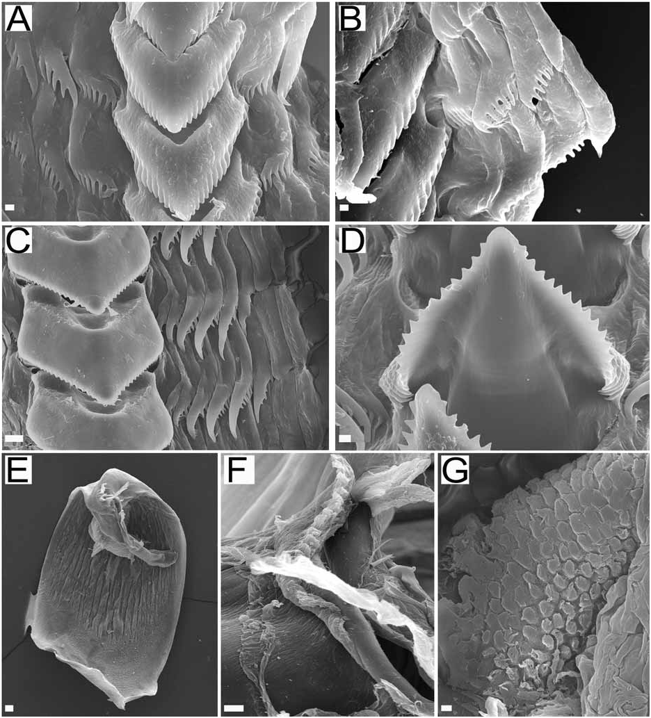
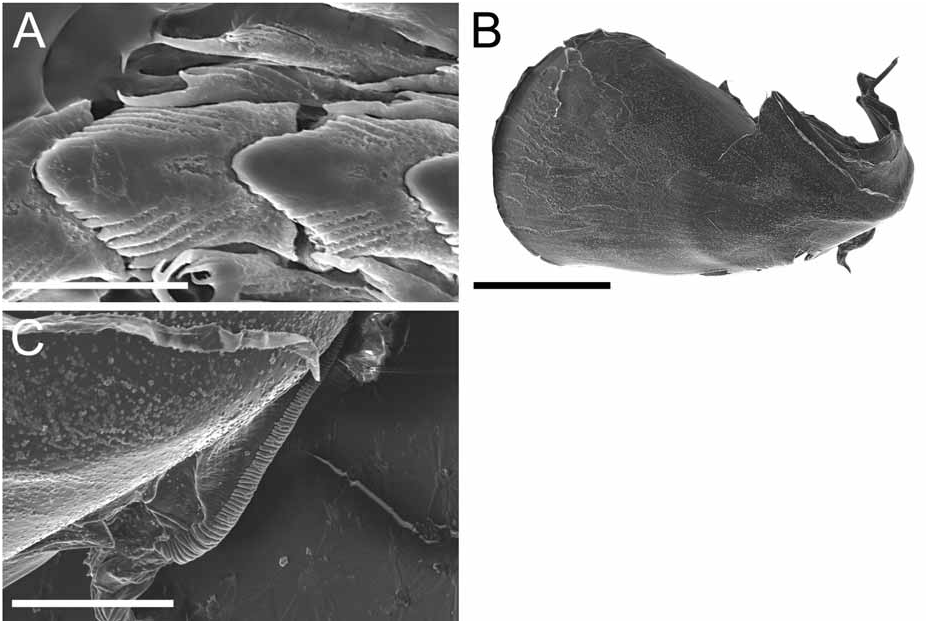
Remarks: Only Dendronotus frondosus , D. subramosus , and a few specimens of D. albus have been reported as having tubercles ( Robilliard 1970). Of these, this specimen of D. noahi most closely resembles D. frondosus in radula morphology ( Figure 11 View FIGURE 11 ). There are differences in the number of rows but it has been suggested that the number of rows in D. frondosus may be correlated with the length of the organism ( Robilliard 1970). In this specimen, the radular formula (18 × 4.1.4) differs from the accepted range of D. frondosus (29–49 × 7–14.1.7–14). The median tooth however has a slightly different shape that is also present in D. regius , although not to the same degree as in D. regius ( Fig. 5 View FIGURE 5 ). All other major radula characteristics are the same with D. frondosus , including number of denticles on median tooth, degree of denticulation (length of furrow), presence of denticulation and slight curvature on all lateral teeth except the outer one or two ( Robilliard 1970). In external morphology, however, D. frondosus and this new species are very different, especially in the degree of branching of the dorsolateral processes and papillae. Dendronotus frondosus has a much more arborescent appearance with three main stalks per fan-shaped dorsolateral process that end with transparent tips on the secondary and tertiary branches ( Robilliard 1970), unlike the single main stalk described in this specimen with two sets of papillae extending laterally and medially at different levels along each dorsolateral process. Like D. noahi , the crown papillae in D. frondosus typically have little or no secondary branching, but sheath papillae are found at the base of this new species instead of midway up the sheath as in D. frondosus (see Robilliard 1970). These papillae are also very small, unlike in D. frondosus where they may branch and be just as long and arborescent as the dorsolateral processes ( Robilliard 1970). Another difference between D. frondosus and this specimen is in the veil papillae. Unlike D. noahi , in which the most distal of the three veil papillae are longest, the innermost (medial) of four veil papillae is longest in D. frondosus (see Robilliard 1970). The background coloration of this specimen is very distinct, especially with the bright orange rhinophores. In D. frondosus the rhinophores match the variable ground color of the body (Table 1). Also, the tubercles are missing the yellow or white pigmentation that is normally found in D. frondosus (see Robilliard 1970). For external morphology, this new specimen most closely resembles D. albus , but only because of its delicate and translucent appearance. They share the extension of dark digestive glands into the dorsolateral processes and rhinophore sheaths, but the similarities end there. In D. albus , the digestive glands blend into a yellow-copper or white color at the tips of the dorsolateral processes and papillae ( MacFarland 1966, Robilliard 1970). In this specimen the tips remain translucent except in the longest crown papilla of each sheath where they also turn white. The radula of D. albus is markedly different in formula (32–38 × 6–8.1.6–8, see Fig. 8A–C View FIGURE 8 and Table 1) and in median tooth morphology ( MacFarland 1966, Robilliard 1970). The only other species with which this specimen shares characteristics that are not commonly found within the genus is D. subramosus , but the only similarities are the body tubercles and the large cardiac prominence. Differences in radulae and external morphology are numerous. The median teeth of D. subramosus are more squared towards the base and the lateral teeth do not curve towards the midline as in D. noahi ( Figs 16A View FIGURE 16 and 6A View FIGURE 6 ). In D. subramosus , only the first pair of dorsolateral processes and the rhinophores receives extensions of the digestive glands, whereas in D. noahi the rhinophores and all of the dorsolateral processes receive these extensions. Also, the pattern of bifurcations in the dorsolateral processes is different, with D. subramosus exhibiting more of a bushy-shaped appearance while D. noahi has more of the fan-shaped appearance (see Fig. 1D View FIGURE 1 ).
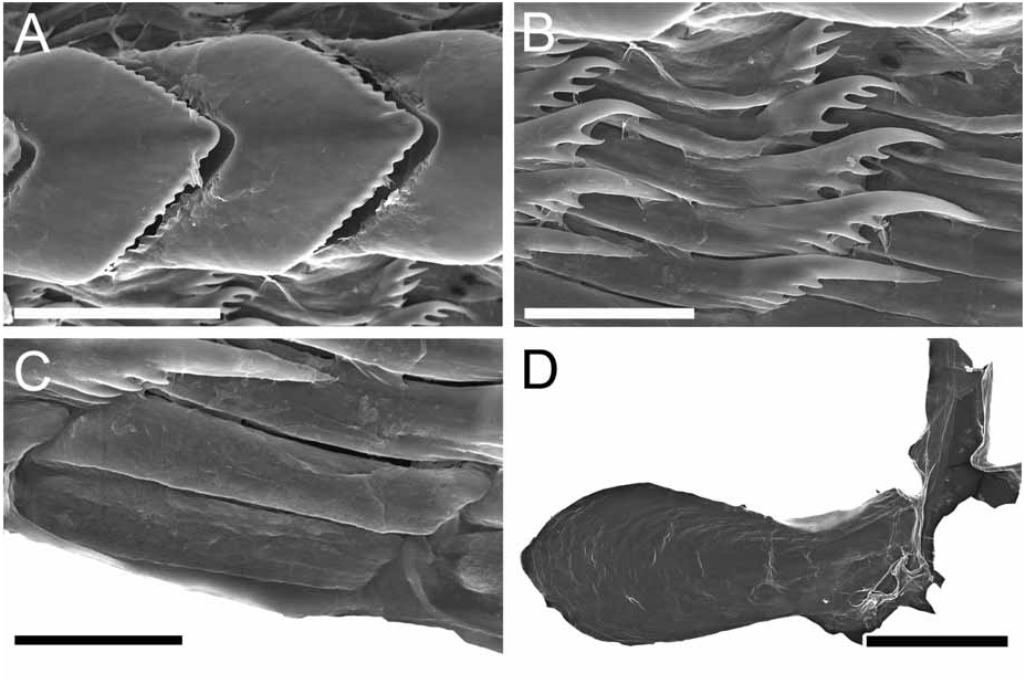
Regarding the new tropical species, D. noahi and D. regius can be distinguished from each other in a number of ways. Besides possessing distinct color patterns, differences are seen in the radulae, tertiary branch lengths, branching of the crown papillae, and branching of the lateral papillae of the rhinophoral sheath. The outermost lateral teeth of D. noahi are thin while those of D. regius are broader and plate-like ( Figs 2G–H View FIGURE 2 , 6A–B View FIGURE 6 ). Dendronotus noahi has short tertiary branch lengths while in D. regius they are long. Branching of the crown papillae along the rhinophoral sheath margin and in the sheath papillae is only present in D. regius . While both species possess a fan-shaped branching pattern of the dorsolateral appendages, the main stalk in D. regius is much more robust and secondary branches do not occur until about halfway up these appendages. In D. noahi , the secondary branches can be found starting very low and near to the base of the dorsolateral appendages. These differences appear to be unrelated to the small size and immature nature of the single specimen of D. noahi and thus the two species are clearly distinct. Based on its tropical distribution and its distinct characteristics we think is more important to describe this new tropical Indo-Pacific species than keep it on the dark. As soon as new specimens come available the description will be improved.

| T |
Tavera, Department of Geology and Geophysics |
No known copyright restrictions apply. See Agosti, D., Egloff, W., 2009. Taxonomic information exchange and copyright: the Plazi approach. BMC Research Notes 2009, 2:53 for further explanation.
|
Kingdom |
|
|
Phylum |
|
|
Class |
|
|
Order |
|
|
Family |
|
|
Genus |