
Micrathena, SUNDEVALL, 1833
|
publication ID |
https://doi.org/10.1111/j.1096-3642.2012.00831.x |
|
persistent identifier |
https://treatment.plazi.org/id/07032673-D103-E65D-FF34-52F857FBFD9A |
|
treatment provided by |
Marcus |
|
scientific name |
Micrathena |
| status |
|
GENUS MICRATHENA SUNDEVALL, 1833 View in CoL View at ENA
Micrathena Sundevall, 1833: 14 View in CoL . Read in April 1833 (F.O.P.- Cambridge, 1904: 525); publication date uncertain [F.O.P.- Cambridge (1904) states that the work was publicly read in April 1833, therefore the actual publication date must be earlier. Thus, the name Micrathena Sundevall, 1833 View in CoL has priority over Acrosoma Perty, 1833 .]. Type species Epeira clypeata , only species listed in section one of the genus, designated by Simon (1895: 848).
Acrosoma Perty, 1833: 193 . Published in December 1833 (F.O.P.- Cambridge, 1904: 525). Type species Acrosoma swainsoni , designated by F.O.P.- Cambridge, 1904: 525. First synonymized with Micrathena View in CoL by F.O.P.- Cambridge (1904).
Meganopla Simon, 1864: 292 . Type species Meganopla cyanospina , designated by Bonnet, 1957: 2752. First synonymized with Acrosoma by Butler (1873).
Keyserlingia O.P.- Cambridge, 1890. Type species by monotypy Keyserlingia cornigera O.P.- Cambridge, 1890 (= M. sexspinosa View in CoL ); preoccupied by Pander, 1861 (Brachiopoda) ( Levi, 1985). First synonymized with Micrathena View in CoL by Simon (1895).
Ildibaha Keyserling, 1892: 31 . Type species by monotypy I. albomaculata Keyserling, 1892 (= M. flaveola (C.L. Koch, 1839)) View in CoL . First synonymized with Micrathena View in CoL by Levi (1985).
Chaetacis Simon, 1895: 863 . Type species by original designation Acrosoma affinis C. L. Koch, 1839 (= C. aureola ). syn. nov.
Thaumastobella Mello-Leitão, 1945 . Type species by monotypy Thaumastobella mourei Mello-Leitão, 1945 [= M. saccata (C.L. Koch, 1836) View in CoL ]. First synonymized with Micrathena View in CoL by Scharff (1991).
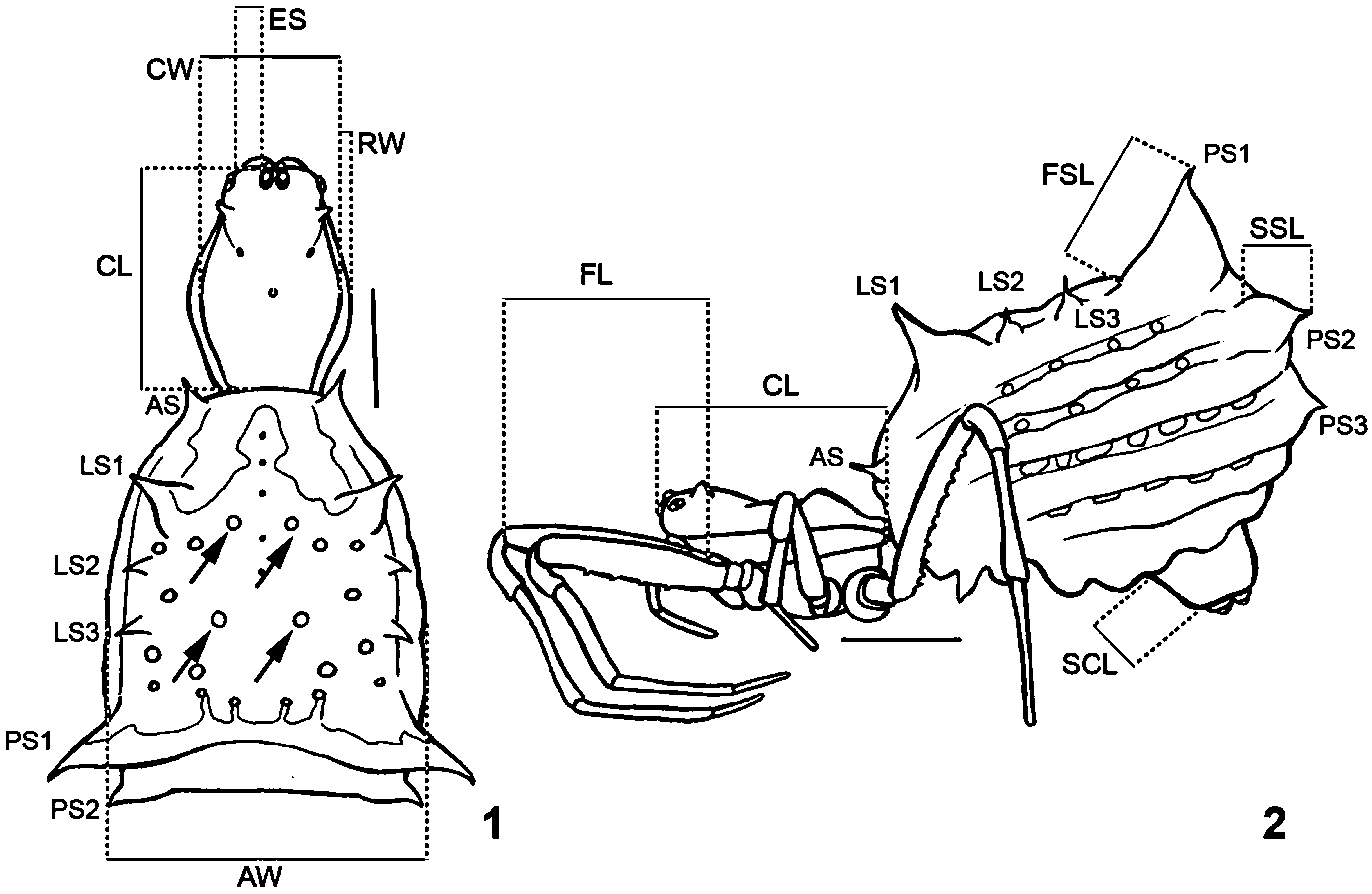
Diagnosis: Micrathena females have a circular, excavated thoracic fovea ( Magalhães & Santos, 2011: figs 15, 18, 21, 33) and a ventral, median line of up to three apodemes in the abdomen (Fig. 12, LMA, SpA). The carapace is glabrous or has reduced setae (but is hirsute in the M. plana and M. furcula groups) and is modified in shape, having a high thoracic region and often a pair of lateral rims and up to three pairs of dimples. The abdomen is usually longer than wide, heavily sclerotized with a ring around the spinnerets (which is found on the tip of a ventrally projected cone; Fig. 2 View Figures 1–2 , SCL), often has the dorsum glabrous (hirsute in the M. plana group) and has from one to nine pairs of spines (usually three to seven), but no median posterior tubercle ( Figs 1, 2 View Figures 1–2 ).
Micrathena View in CoL males also present a circular thoracic fovea. They have an elongated abdomen that is longer than wide and rectangular to subtrapezoidal in form ( Levi, 1985: figs 15, 487, 561; Magalhães & Santos, 2011: figs 15, 18, 21). The paracymbium of the palpus is large (Fig. 13, P) and often has extra lobes or is modified otherwise ( Levi, 1985: figs 541, 711; Magalhães & Santos, 2011: fig. 40). The median apophysis has a basal projection that is partially fused to the frontal base of the radix (Figs 14–16, BP; Magalhães & Santos, 2011: figs 16, 22, 39, BP; frequently small and not fused to the radix in the M. plana View in CoL group, Fig. 17; Magalhães & Santos, 2011: fig. 19).
Description: See descriptions of Micrathena and Chaetacis provided by Levi (1985: 441, 600).
Natural history: See Robinson & Robinson (1980), Uetz & Biere (1980), Biere & Uetz (1981), Shelly (1984), Levi (1985), Hodge (1987a, b), Uetz & Hartsock (1987), Carvalho Jr. (1992), Bukowski & Christenson (1997a, b, 2000), Díaz-Fleischer (2005), Meling-López et al. (2008), Vanderhoff et al. (2008), Moya et al. (2010), Opell et al. (2011), and Gálvez (2011).
Genus subdivisions
We have rediagnosed all of Levi’s (1985) original Micrathena species groups, proposing new groups and species transfers whenever necessary. We have proposed taxonomic rearrangements for both species in the phylogenetic analysis and species not included in it. For these species not included in the analysis, we examined specimens whenever possible to assure correct assignment to a group. When specimens were unavailable, we assigned specimens based on Levi’s (1985) illustrations. However, several species could not be unambiguously assigned to any group and remain as Micrathena incertae sedis (see below).
Micrathena cornuta group
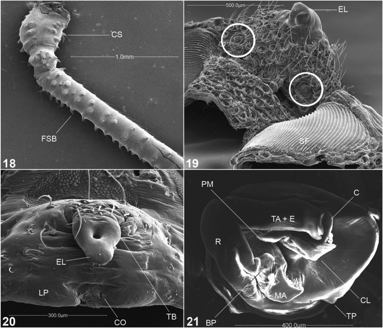
Diagnosis: Females can be diagnosed by the presence of a pair of tubercles or spines in the carapace ( Magalhães & Santos, 2011: figs 33, 34; except M. pungens ), spiny fourth coxae ( Fig. 18 View Figures 18–21 ), reniform spermathecae ( Levi, 1985: figs 805, 823, 855; Magalhães & Santos, 2011: fig. 38; except M. osa , in which it is rounded), epigynal lateral keels and epigynum without lobe, frequently conical and ventrally projected ( Magalhães & Santos, 2011: figs 36, 37). Males can be diagnosed by the presence of spiny setal bases at the margins of the carapace (except M. pungens ) and by the palpus lacking a terminal apophysis, with a long, distal tegular projection, a large conductor that projects beyond the margin of the tegulum ( Magalhães & Santos, 2011: fig. 39), and a large and modified paracymbium with a dorsal lobe ( Magalhães & Santos, 2011: fig. 40, DL).
Composition: This group corresponds to species previously allocated in Chaetacis , plus M. pungens . Eleven species are included: M. abrahami comb. nov., M. aureola comb. nov., M. bandeirante comb. nov., M. carimagua ( Levi, 1985) comb. nov., M. cornuta comb. nov., M. cucharas ( Levi, 1985) comb. nov., M. necopinata Chickering, 1960 comb. nov., M. osa comb. nov., M. picta comb. nov., M. pungens , M. woytkowsii ( Levi, 1985) comb. nov.
Micrathena funebris group
Diagnosis: As for the species (see Levi, 1985: 588).
Composition: Monotypic.
Micrathena furcula group
Diagnosis: Females can be distinguished by the wide carapace with a straight anterior margin and a hairy thoracic region ( Levi, 1985: fig. 170), and by the abdomen, which is wide, short, and has no spines except for one or two pairs of short posterior spines ( Levi, 1985: fig. 170) (except M. digitata and M. furva , which have lateral spines). The epigynum has a transverse bar and looks like a bird’s head laterally ( Levi, 1985: figs 173, 180). Males can be distinguished from those of other groups whose members have a coxal hook by the following combination of characters: rounded thoracic region, abdomen short and rectangular and with a pattern of large, dorsal, paired guanine spots ( Levi, 1985: fig. 174). The terminal apophysis always covers the embolus and has a rounded tip ( Levi, 1985: figs 128, 163). There is no macroseta in the palpal patella (except in M. digitata and M. furva ).
Composition: Ten species: Micrathena bimucronata , M. cubana , M. digitata , M. furcula , M. furva ( Keyserling, 1892) , M. mitrata , M. patruelis , M. rufopunctata , M. saccata , M. similis .
Micrathena gracilis group
Diagnosis: Females vary greatly in abdomen and carapace shape. Nevertheless, all can be distinguished by having a ventrally projected epigynal lobe that is laterally excavated ( Fig. 19 View Figures 18–21 , EL). Most males are unknown; the ones described are minute in comparison to females and can be diagnosed by the palpus with a membranous digitiform projection in the basal projection of the median apophysis and a dorsally pointing conductor (Fig. 15, DP, CL).
·
Figures 12–17. Scanning electron microscopy (SEM) images of species included in the current study. Fig. 12. Micrathena nigrichelis , female abdomen, ventral view, showing median line of apodemes. Fig. 13. Micrathena evansi , cymbium, retrolateral. Fig. 14. Micrathena spinosa , male copulatory bulb, submesal. Fig. 15. Micrathena horrida , male copulatory bulb, mesal. Fig. 16. Micrathena evansi , male copulatory bulb, submesal. Terminal apophysis accidently torn off during the process of preparation for SEM. Fig. 17. Micrathena plana , male copulatory bulb, mesal. Abbreviations: BP, basal projection of the median apophysis; C, conductor; CL, conductor lobe; Cy, cymbium; DP, digitiform projection of the median apophysis; E, embolus; EF, epigastric furrow; LMA, large median apodeme; MA, median apophysis rim; MAL, median apophysis lobe; P, paracymbium; R, radix; S, spiracle; SpA, spinnerets apodeme; SR, spinnerets ring; ST, spinnerets tubercle; TA, terminal apophysis; TAP, terminal apophysis projection; TP, tegular projection.
Composition: Seven species: M. forcipata , M. gracilis , M. horrida , M. margerita Levi, 1985 , M. glyptogonoides Levi, 1985 , M. spinulata F.P.- Cambridge, 1904, M. striata F.P.- Cambridge, 1904.
Micrathena guerini group
Diagnosis: Females of this group often lack the anterior pair of spines (except M. crassispina , M. guanabara , and M. lindenbergi ), having a pair of lateral and two large pairs of posterior spines. There is a transverse bar in the epigynum that is frequently, but not always, elongated, forming a scape-like structure ( Fig. 20 View Figures 18–21 , TB). The epigynum lateral plates are swollen, forming distinct sclerotized cheeks ( Fig. 20 View Figures 18–21 , LP). Males can be distinguished from other Micrathena males with a coxal hook by the elongated thoracic region and the abdomen dorsum with a median dark band with guanine spots by its sides ( Magalhães & Santos, 2011: fig. 15). The terminal apophysis may be entirely fused to the embolus (e.g. M. nigrichelis , M. fissispina ; Fig. 21 View Figures 18–21 , TA + E) or have a membranous projection (e.g. M. gaujoni , M. bifida ; Fig. 17, TAP).
Composition: Eighteen species: Micrathena atuncela Levi, 1985 , M. bifida , M. crassispina (C.L. Koch, 1836) , M. fissispina , M. gaujoni , M. guanabara , M. guerini , M. gurupi Levi, 1985 , M. kochalkai Levi, 1985 , M. lindenbergi Mello-Leitão, 1940 , M. miles Simon, 1895 , M. nigrichelis , M. pilaton Levi, 1985 , M. raimondi (Taczanowski, 1879) , M. reali Levi, 1985 , M. rubicundula (Keyserling, 1864) , M. shealsi Chickering, 1960 , M. teresopolis Levi, 1985 .
Micrathena kirbyi group
Diagnosis: Females can be diagnosed by having ten pairs of abdominal spines, the fourth the longest ( Levi, 1985: figs 262, 267). The epigynum has a transverse bar that is narrow laterally ( Levi, 1985: figs 254, 268). Males have a coxal hook and a rounded thoracic region. The palpus has a sharp basal projection at the median apophysis that points towards the apex of the embolus and a basal membrane in the conductor ( Levi, 1985: fig. 259).
Composition: Three species: Micrathena armigera (C.L. Koch, 1837) , M. kirbyi , M. macfarlanei Chickering, 1961 .
Micrathena lepidoptera group
Diagnosis: Females can be diagnosed by the unique trifid lateral spines in the abdomen ( Levi, 1985: figs 526, 535) and by the pair of ventral, blunt spines beside the spinnerets ( Levi, 1985: figs 525, 534). There is no stridulatory surface on the booklung covers. The genitalia of the males are similar to those of the M. triangularispinosa group: the basal projection of the median apophysis is large and covers the rim, and the paracymbium is laterally projected ( Levi, 1985: figs 532, 541). The terminal apophysis, however, is large and wide, rather than thin, and covers the embolus in mesal view ( Levi, 1985: figs 531, 540).
Composition: Two species: Micrathena decorata , M. lepidoptera .
Micrathena militaris group
Diagnosis: This corresponds to Levi’s (1985) M. militaris and M. spinosa groups. Females have a sculptured sternum and at most four pairs of abdominal spines, none of which are anterior ( Levi, 1985: figs 638, 712, 726). The median posterior eyes lack a tapetum altogether and the lateral eyes may not be juxtaposed. There is no stridulatory surface on the booklung covers. The epigynum lobe is laterally excavated, forming two pockets to which the male’s embolus and terminal apophysis may remain attached after mating (e.g. M. militaris , M. spinosa , M. sexspinosa ; Levi, 1985: figs 605, 729). The male genitalia are quite varied, but all have a simple or bifid digitiform projection in the palpal tibia that is diagnostic (but also present in M. swainsoni ) ( Levi, 1985: figs 681, 718). There is no palpal patella macrosetae (except M. lata ) and the tibiae and femora lack strong spines and macrosetae, having only normal setae.
Composition: Eighteen species: Micrathena anchicaya Levi, 1985 , M. banksi Levi, 1985 , M. brevipes , M. coca Levi, 1985 , M. cyanospina , M. donaldi Chickering, 1961 , M. furcata , M. guayas Levi, 1985 , M. hamifera Simon, 1897 , M. lata , M. militaris , M. petrunkevitchi Levi, 1985 , M. pichincha , M. reimoseri Mello-Leitão, 1935 , M. sagittata , M. sexspinosa (Hahn, 1822) , M. soaresi Levi, 1985 , M. spinosa .
Micrathena plana group
Diagnosis: Females have a hairy carapace and hairs on the abdomen dorsum, which is flattened. There are often several pairs of lateral spines (usually four; five in M. ruschii ; Levi, 1985: fig. 328; Gonzaga & Santos, 2004: fig. 1). There is no stridulatory surface in the booklung covers. The genitalia are quite similar to that of species in the M. furcula group, although some species have spermathecae with two compartments (see char. 86). Males differ from other Micrathena males with a coxal hook by having a rounded thoracic region and by the abdomen, which is wider anteriorly and has two dark bands in the lateral margins ( Magalhães & Santos, 2011: figs 17, 18). The basal projection of the median apophysis is faint and small, and frequently is not fused to the radix, and the rim is only slightly bent when compared to species in the M. furcula , M. guerini , and M. kirbyi groups (Fig. 17; Magalhães & Santos, 2011: fig. 19). The terminal apophysis may have a membranous projection (Fig. 17, TAP).
Composition: Seventeen species: Micrathena alvarengai Levi, 1985 , M. bananal Levi, 1985 , M. brevispina (Keyserling, 1864) , M. duodecimspinosa (O.P.- Cambridge, 1890), M. excavata , M. exlinae Levi, 1985 , M. huanuco Levi, 1985 , M. lenca Levi, 1985 , M. marta Levi, 1985 , M. molesta Chickering, 1960 , M. parallela (O.P.- Cambridge, 1890), M. plana , M. quadriserrata F.O.P.- Cambridge, 1904, M. ruschii , M. triangularis (C.L. Koch, 1836) , M. triserrata , M. tziscao Levi, 1985 .
Micrathena schreibersi group
Diagnosis: Females can be diagnosed by the glabrous thoracic region of the carapace, by the constricted sternum (except M. clypeata , in which it is shieldshaped; see char. 37), lack of the small median ventral apodeme, epigynum with lateral keels ( Levi, 1985: figs 569, 575), and spermathecae with two compartments. The epigynum may ( Levi, 1985: fig. 577) or may not ( Levi, 1985: fig. 547) have a transverse bar. Males may present a median constriction in the abdomen (not present in M. clypeata and M. balzapamba ) ( Levi, 1985: figs 548, 579) and have a distinct palpus structure, lacking a terminal apophysis, having a subsquarish tegular margin ( Levi, 1985: fig. 571) and a large, flattened retrolateral lobe of the paracymbium ( Levi, 1985: fig. 563).
Composition: Six species: M. clypeata , M. balzapamba , M. embira Levi, 1985 , M. schreibersi , M. spitzi , M. vigorsi .
Micrathena swainsoni group
Diagnosis: As for the species. See Levi (1985: 571). Additionally, females lack a tapetum in the median posterior eyes and have an epigynum without lobe. Males have a very large conductor, which holds the embolus and occupies most of the mesal face of the tegulum.
Composition: Monotypic.
Micrathena triangularispinosa group
Diagnosis: All species in this group are small-sized Micrathena , with a dome-shaped carapace (not so in M. schenkeli and M. ucayali ) and at most four pairs of abdominal spines ( Levi, 1985: fig. 490). The median posterior eyes lack a tapetum. The epigynum has a very rounded bulge and a small, frequently pointed lobe ( Magalhães & Santos, 2011: fig. 11). Males have a carapace darker than that of females and a large, median guanine spot in the otherwise dark abdomen ( Levi, 1985: fig. 487). The palpus structure is distinct, with a very sclerotized conductor apex that is notched, a drop-shaped, sclerotized conductor lobe, a thin and membranous terminal apophysis that is parallel to the embolus, and a large, sclerotized basal projection of the median apophysis that covers the rim (Fig. 16; Levi, 1985: fig. 467).
Composition: Eleven species: Micrathena acuta , M. annulata Reimoser, 1917 , M. bicolor (Keyserling, 1864) , M. evansi , M. flaveola (Perty, 1839) , M. jundiai , M. peregrinatorum (Holmberg, 1883) , M. schenkeli , M. triangularispinosa (De Geer, 1778) , M. ucayali Levi, 1985 , M. yanomami Magalhães & Santos, 2011 .
Micrathena incertae sedis
The following species were assigned by Levi (1985) to the M. kirbyi and M. guerini groups. We have not examined specimens of them and thus we are not certain where they belong, although all of them should be within the distal part of the M. clypeata clade. These are M. agriliformis (Taczanowski, 1879) , M. bogota Levi, 1985 , M. coroico Levi, 1985 , M. crassa (Keyserling, 1864) , M. elongata (Keyserling, 1864) , M. fidelis (Banks, 1909) , M. lucasi (Keyserling, 1864) , M. pupa Simon, 1897 , M. stuebeli (Karsch, 1886) , and M. zilchi Kraus, 1955 . Micrathena agriliformis , M. bogota , M. elongata , and M. pupa possibly form a monophyletic group (characterized by a long and narrow abdomen with short spines) nested within the M. guerini group; M. crassa possibly belongs in the M. furcula group; and M. coroico and M. zilchi possibly belong in the M. guerini group. However, these are only tentative suggestions of placement and should be considered carefully only after examination of specimens.
No known copyright restrictions apply. See Agosti, D., Egloff, W., 2009. Taxonomic information exchange and copyright: the Plazi approach. BMC Research Notes 2009, 2:53 for further explanation.
|
Kingdom |
|
|
Phylum |
|
|
Class |
|
|
Order |
|
|
Family |
Micrathena
| Magalhães, Ivan L. F. & Santos, Adalberto J. 2012 |
Chaetacis
| Simon E 1895: 863 |
Ildibaha
| Keyserling E 1892: 31 |
Meganopla
| Bonnet P 1957: 2752 |
| Simon E 1864: 292 |
Micrathena
| Cambridge FOP 1904: 525 |
| Simon E 1895: 848 |
| Sundevall JC 1833: 14 |
Acrosoma
| Cambridge FOP 1904: 525 |
| Cambridge FOP 1904: 525 |
| Perty M 1833: 193 |