
Unaspis lansivora, Watson, Gillian W., 2015
|
publication ID |
https://doi.org/10.11646/zootaxa.3905.3.9 |
|
publication LSID |
lsid:zoobank.org:pub:35AC1A3B-72F5-482F-AB0E-353CA74E017D |
|
DOI |
https://doi.org/10.5281/zenodo.6097983 |
|
persistent identifier |
https://treatment.plazi.org/id/03FF87F9-FFEA-8246-FF38-742A97B1FEE4 |
|
treatment provided by |
Plazi |
|
scientific name |
Unaspis lansivora |
| status |
sp. nov. |
Unaspis lansivora sp. n.
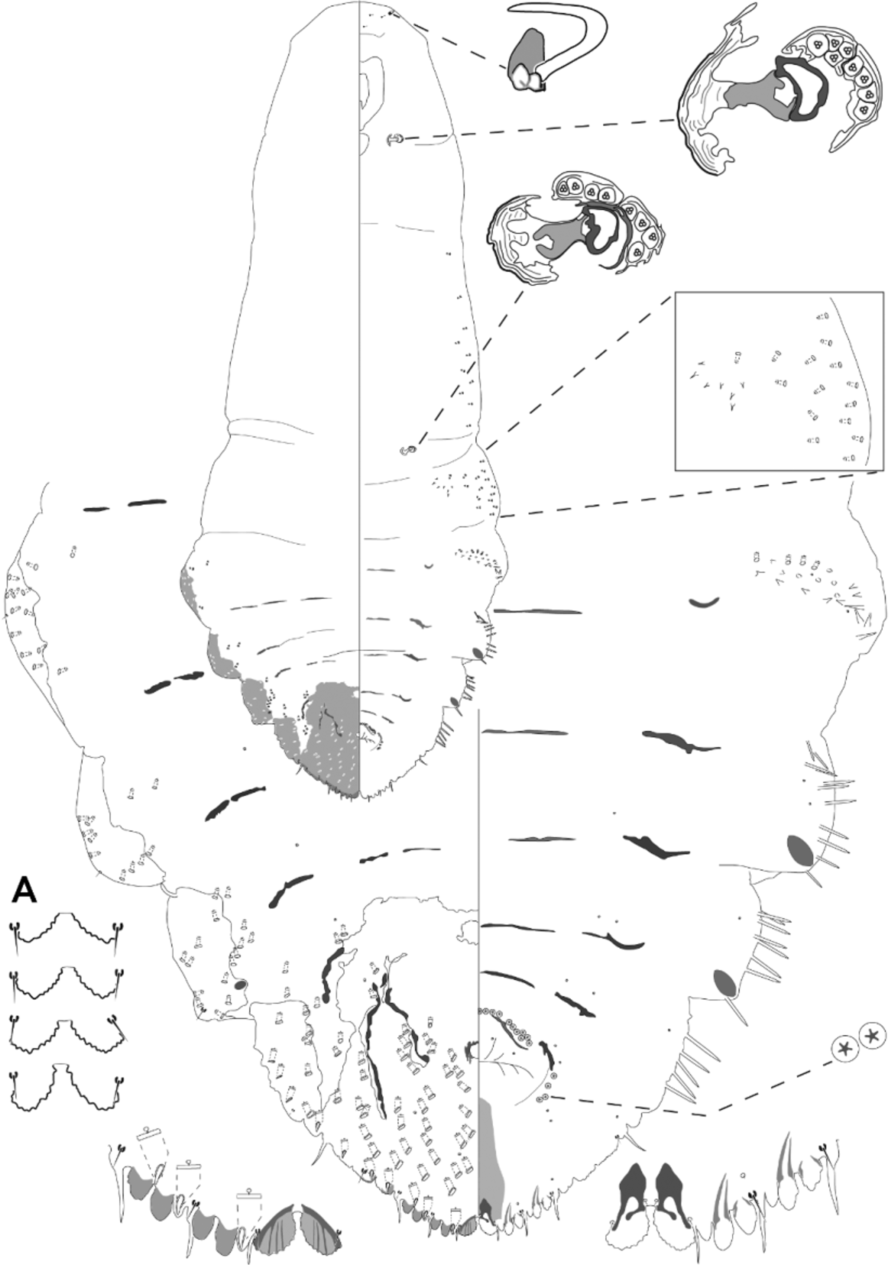
( Fig. 1 View FIGURE 1 )
Suggested common name: Lanzones scale.
Material studied. Holotype female: PHILIPPINES, Luzon Island, Laguna, San Pablo City, on Lansium domesticum leaves, coll. M.V. Navasero, 31.iii.2014 (at CSCA, 2nd specimen from the right end of a row of 4 adult females, clearly mapped on the slide label).
Paratypes female: PHILIPPINES, same data as holotype (at BMNH, 2 slides each containing 4 adult females, 1 slide containing 1 adult female and one second instar exuviae, and 3 slides each containing 1 adult female; at CSCA, same slide as holotype, 3 adult females; 1 slide containing 2 adult females; 1 slide containing 1 adult female and one second instar exuviae; and 2 slides each containing one adult female; at UPLBM, 1 slide containing 4 adult females, 2 slides each containing 1 adult female and one second instar exuviae, and 2 slides each containing a single adult female; at USNM, 3 slides each containing 1 adult female and one second instar exuviae, and 1 slide containing one adult female). PHILIPPINES, Luzon Island, Batangas prov., Malvar, on Lansium domesticum leaves, coll. B.M. Shepard, G. Carner, C.B. Adalla 1, 18.1 2014 (at CSCA, 1 slide containing 3 adult females; at USNM, 1 slide containing 4 adult females).
Description. In life: found feeding on the leaves of Lansium domesticum , mainly on the ventral surface. Scale cover of adult female elongate, slender mussel-shaped, moderately convex, not very thick but fairly rigid, rippled, with a rough, approximately median longitudinal ridge, mid-brown and very slightly translucent, with yellowbrown terminal exuviae. Scale cover of immature male much smaller than that of adult female, white, felted, tricarinate, with yellow terminal exuviae.
Adult female in slide mount: the figures provided below are for the holotype specimen, and ( holotype + 39 paratype specimens, range and mean value). Body elongate, up to 3 times as long as wide; widest at abdominal segment I, then tapering to apex of pygidium but abdominal segments I–III each with moderately produced lateral lobes; segment 3 with a dorsal submarginal boss or cicatrix on either side; marginal sclerotized spurs absent. In young adults, head outline smoothly convex and prosomal derm membranous but with increasing maturity, margin of head becomes more prominently convex and develops a pair of lateral tubercles ( Fig. 1 View FIGURE 1 ), becoming more sclerotized with maturity. A few microducts present on venter of head, anterior to clypeus. Maturation also results in slightly increased sclerotization of the rest of the prosoma and the free abdominal segments, particularly the lateral lobes of abdominal segments I–III ( Fig. 1 View FIGURE 1 ).
Pygidium slightly rounded, with a small apical notch formed by divergent median lobes. Median lobes well developed, not zygotic, slightly recessed into the pygidial margin, set quite close together basally, with a pair of minute marginal setae between the bases, often difficult to see; these lobes slightly (to considerably) divergent ( Fig. 1 View FIGURE 1 A), with inner margins longer than outer margins, forming a slight notch at apex of pygidium; margins of median lobes serrate. Second and third lobes smaller than median lobes, well developed, deeply bilobed, the lobules rounded; second lobes sometimes slightly smaller than third lobes. Slender ventral paraphyses arising from the angles of the inner lobules of second and sometimes third lobes, converging anteriorly. Anus approximately circular, situated approximately at centre of pygidium; a median, sclerotized furrow extending from anus to median lobes, textured with longitudinal striae. Anteromedial area of sclerotized shield textured like tree bark. Vulva situated approximately at centre of pygidium.
Marginal macroducts mostly larger than dorsal ducts; each orifice sclerotized, with the long axis perpendicular to margin; some situated in pore prominences; numbering 7 on each side of the pygidium (absent from between median lobes; 1 between median and second lobe, 2 by third lobe, 2 on segment V and 2 on segment IV). Dorsal pygidial macroducts smaller than the smaller marginal ducts, present on all pygidial segments including segments VII and VIII, becoming smaller towards anterior of pygidium, a few sometimes present immediately anterior to anus. Dorsal macroducts on segments V–VIII numbering 75 (61–102, mean 81.53), loosely arranged in segmental series submarginally and submedially ( Fig. 1 View FIGURE 1 ). Perivulvar pores, each pore containing 5 loculi, present in 5 groups, with 2–4 (0–5, mean 3.31) pores in each posterolateral group, 7 (3–11, mean 7.24) in each anterolateral group, and 5 (3–8, mean 5.75) in anteromedial group, total 25 pores (19–33, mean 26.85).
Marginal gland spines well developed, absent from between the median lobes; present singly between pygidial lobes, more numerous on margins of abdominal segments II–IV. On abdominal segment I, gland spines reduced to conical duct tubercles, forming a series on ventral surface extending inwards from margin, together with a few macro- and microducts and duct tubercles ( Fig. 1 View FIGURE 1 ). Metathorax with a similar group of macro- and microducts extending inwards from the margin, sometimes including a few very small gland spines and duct tubercles. A small to moderate number of small macroducts and microducts scattered along margin of mesothorax. Stigmatic disc pores, each with 3 loculi, present in a compact cluster of 7 or 8 (4–15, mean 9.65) by each anterior spiracle; a cluster of 8 (0–14, mean 8.20) pores present by each posterior spiracle. Antennae small, heavily sclerotized, situated near or on anterior margin, each bearing 1 curved, robust seta.
Etymology. The species name lansivora means ‘feeding on Lansium’.
Comments. Unaspis lansivora sp. n. was initially misidentified as U. citri . It resembles U. citri in possessing perivulvar pores and often having 60–70 pygidial macroducts. It differs from U. citri in having more than 18 perivulvar pores (no more than 8 in U. citri ); a series of small gland spines, duct tubercles and ducts extending in from the margin on the venter of abdominal segment I and on the metathorax (both absent in U. citri ); and the lateral lobes of the prepygidial abdominal segments becoming sclerotized with maturity (they remain membranous in mature U. citri ).
Unaspis lansivora sp. n. also resembles U. pseudaesculus in possessing perivulvar pores in 5 groups; lacking gland spines on the mesothorax; and in having median lobes asymmetric and slightly to moderately divergent. It differs from U. pseudaesculus in having a series of small gland spines, duct tubercles and ducts extending inwards from the margin on the venter of abdominal segment I (they are confined to the margin in U. pseudaesculus ), posterior spiracle with about the same number of spiracular pores as those by the anterior spiracle (fewer by the posterior spiracle than by the anterior spiracle in U. pseudaesculus ), and the lateral lobes of the prepygidial abdominal segments becoming sclerotized with maturity (they remain membranous in mature U. pseudaesculus ).
No known copyright restrictions apply. See Agosti, D., Egloff, W., 2009. Taxonomic information exchange and copyright: the Plazi approach. BMC Research Notes 2009, 2:53 for further explanation.
|
Kingdom |
|
|
Phylum |
|
|
Class |
|
|
Order |
|
|
Family |
|
|
Genus |