
Homogryllacris platycis Liu & Bian, 2021
|
publication ID |
https://doi.org/ 10.11646/zootaxa.5067.2.11 |
|
publication LSID |
lsid:zoobank.org:pub:43680906-B4A0-451B-9E4A-7BB80FCD03B6 |
|
DOI |
https://doi.org/10.5281/zenodo.5698877 |
|
persistent identifier |
https://treatment.plazi.org/id/03FD87B7-6250-FFD9-5FBB-23EAFE8AFD5F |
|
treatment provided by |
Plazi |
|
scientific name |
Homogryllacris platycis Liu & Bian |
| status |
sp. nov. |
Homogryllacris platycis Liu & Bian View in CoL sp. nov.
ĒḦ同ďễ
Figures 3–6 View FIGURE 3 View FIGURE 4 View FIGURE 5 View FIGURE 6
Material examined. Holotype: male, Gulinjing, Maguan, Yunnan, June 30, 2021, coll. by Jinxiang Liu ; 1 female, Gulinjing, Maguan, Yunnan, July 28, coll. by Wei Bin, Xiaoyu Peng and Xun Bian. Other specimen: 1 female, other information as holotype.
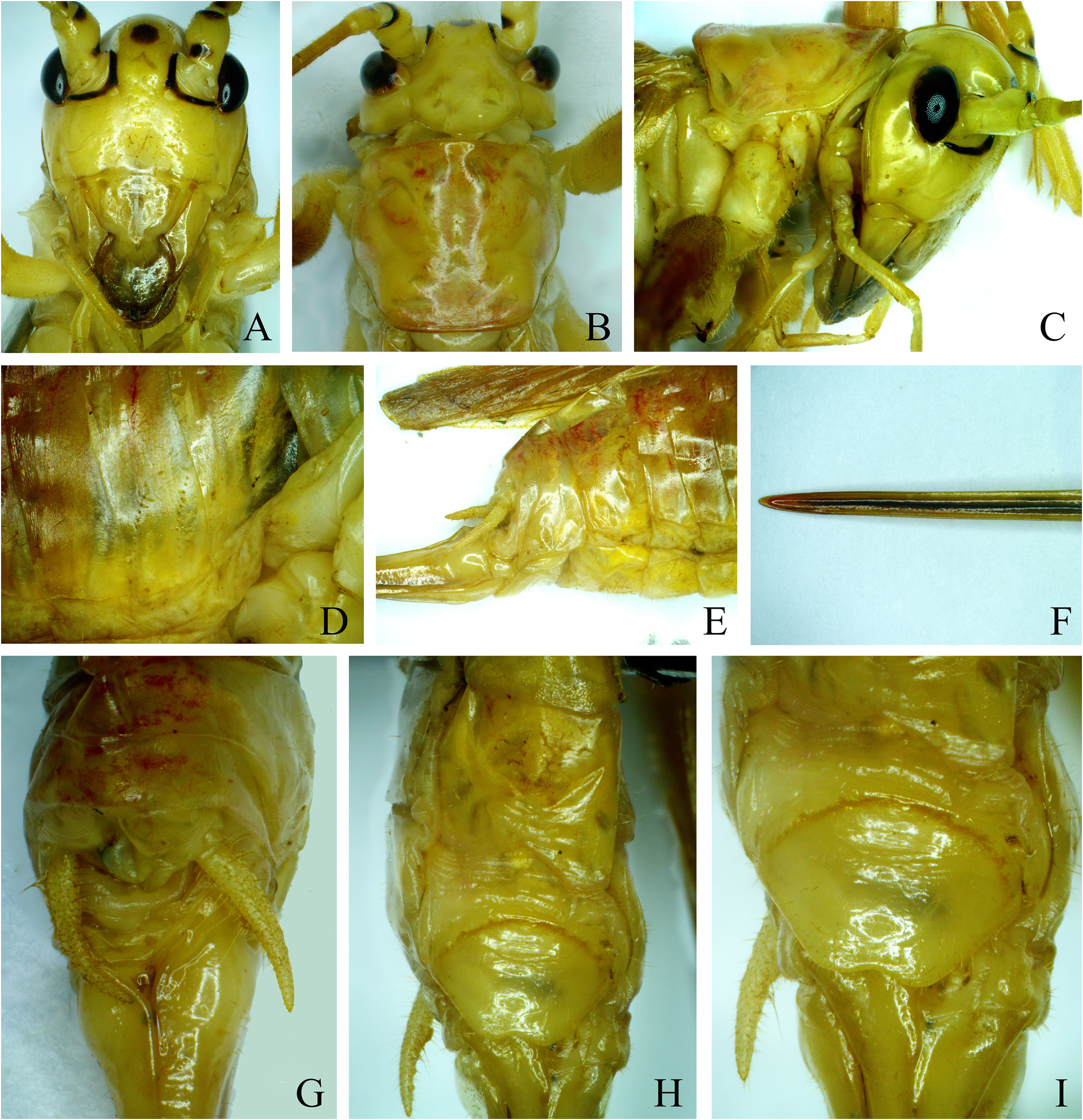
Diagnosis. The new species is close to Homogryllacris brevispina Shi, Guo & Bian, 2012 , but differs from the latter in: processes of male tenth abdominal tergite not constriction in the middle area, the apical spine wider, shorter and straight ( Fig. 3H View FIGURE 3 ); ventral surface of female seventh abdominal sternite without stiffened flap which may be copulatory depressions, subgenital plate triangular in ventral view, posterior margin with 1 median concavity. The female of the new species differs from Homogryllacris yunnana Shi, Guo & Bian, 2012 by subgenital plate triangular with medially concave on posterior margin ( Fig. 5H View FIGURE 5 ), while the latter has nearly semicircular subgenital plate and no concavity on posterior margin.
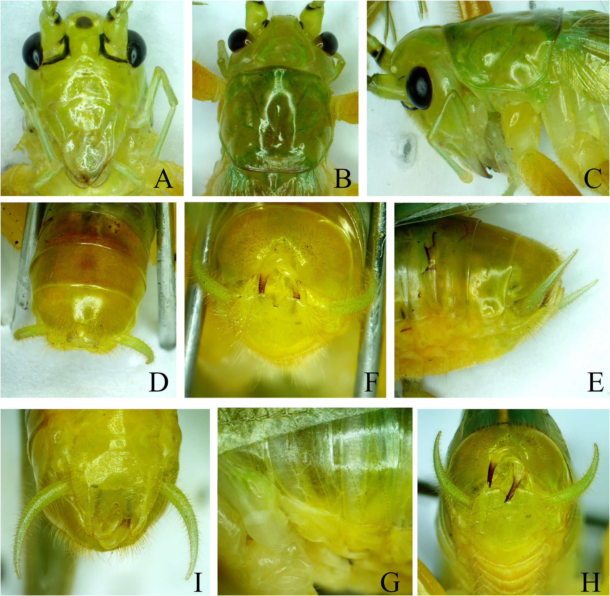
Description. Male. Body medium. Fastigium verticis slightly wider than scape. Face with scattered impressed dots ( Fig. 3A View FIGURE 3 ). Eyes long ovoid, ocelli distinct.
Anterior margin of pronotum slightly projecting, posterior margin nearly straight ( Fig. 3B View FIGURE 3 ), lateral lobes longer than high.
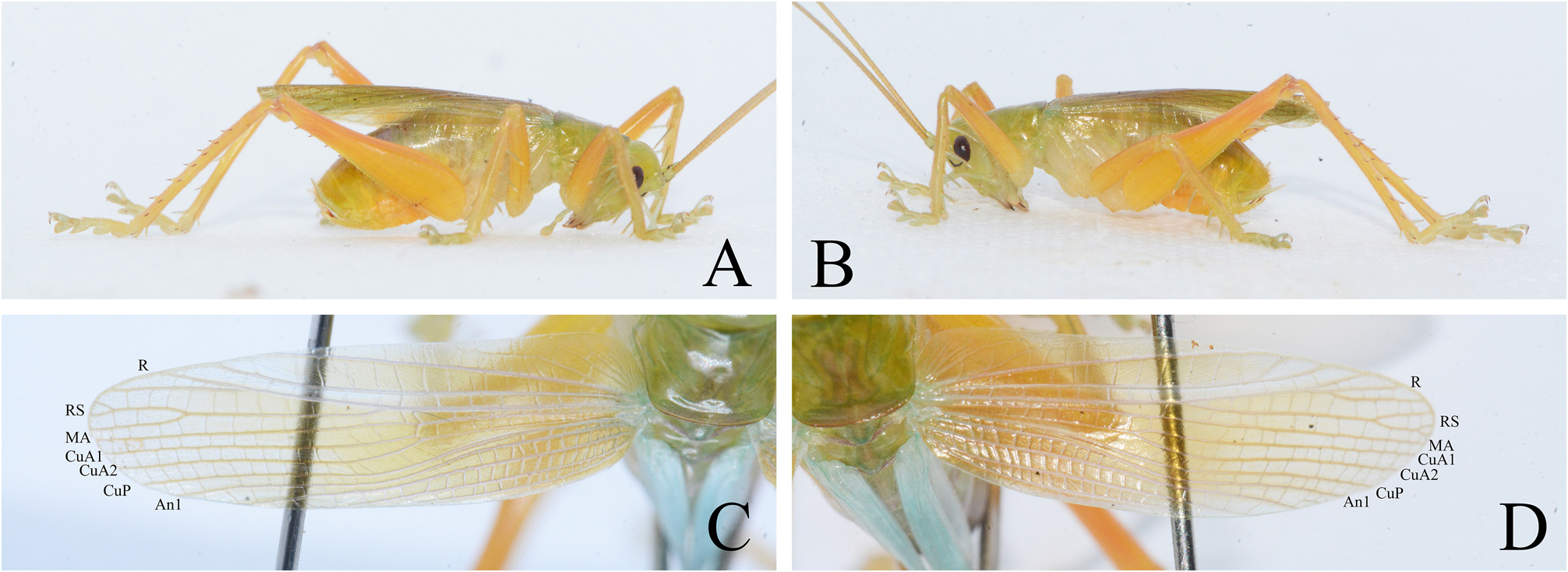
Wings developed well, slightly surpassing apex of abdomen and reaching the basal third of hind tibiae ( Fig. 4A–B View FIGURE 4 ). Tegmina ( Fig. 4C–D View FIGURE 4 ): radius with RS branching little before middle of tegmen, both forked near tip; MA undivided; cubitus anterior forks after basal third into two branches, CuA1 and CuA2; cubitus posterior undivided, free throughout; with 4 anal veins, the last two with common base.
Fore coxae with 1 small spine. Fore and middle tibiae with 4 pairs of long spurs and a pair of short apical spurs on ventral surfaces separately; middle tibiae with an internal apical spine on dorsal surface. Hind femora with 11 internal and 6–8 external spines on ventral surface; tibiae with 6 internal and 7 external spines on dorsal surface, 1 pair of dorsal apical spurs, 2 pairs of ventral apical spurs, and 1 pair of ventral subapical spurs.
Two and three abdominal tergites each with two rows of minute stridulatory pegs ( Fig. 3G View FIGURE 3 ). Ninth abdominal tergite semiglobular in dorsal view, posterior margin with a semicircular concavity ( Fig. 3D, F View FIGURE 3 ). Tenth abdominal tergite very short, with a pair of long projections, in basal half crossing each other and largely hidden under the ninth abdominal tergite, the apical half terminating into a compressed spine pointing to ventrad and slightly forward and with subacute tip ( Fig. 3G, H View FIGURE 3 ). Subgenital plate longer than wide, the lateral margins convex, posterior margin with V-shaped concavity in the middle ( Fig. 3I View FIGURE 3 ). Styli located at apico-lateral angles of subgenital plate ( Fig. 3I View FIGURE 3 ).
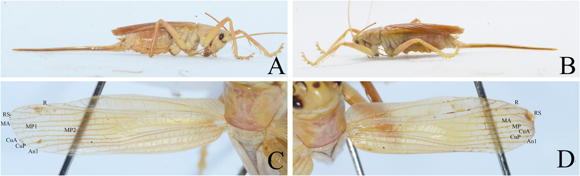
Female. Body longer than male. Tegmina ( Fig. 6C–D View FIGURE 6 ): radius with RS branching about apical third area of tegmina, both forked in subapical area; media of left tegmen forks behind basal third into two branches, MA and MP, shortly after the second branch divides into MP1 and MP2, while MP of right tegmen not divide till tip of tegmen; cubitus anterior and cubitus posterior undivided, free throughout; with 4 anal veins, the last two with common base. Seventh abdominal sternite obviously longer than preceding sternites; apical half membranous with tansverse folds ( Fig. 5H View FIGURE 5 ). Subgenital plate nearly triangular, posterior margin concave in the middle, the lateral lobes obtuse ( Fig. 5I View FIGURE 5 ). Ovipositor very long and straight, margins slightly converging ( Fig. 6A–B View FIGURE 6 ); apices narrowly obtuse ( Fig. 5F View FIGURE 5 ).
Coloration. Body yellowish green ( Figs. 4A–B View FIGURE 4 , 6A–B View FIGURE 6 ). Fastigium verticis with a transverse black spot ( Figs. 3A View FIGURE 3 , 5A View FIGURE 5 ); margins of antennal sockets black, internal margin of scape with one black spot on basal and apical areas separately, basal area of pedicel internally with a black spot ( Figs. 3A View FIGURE 3 , 5A View FIGURE 5 ). Processes of male tenth abdominal tergite with brown apical half ( Fig. 3H View FIGURE 3 ).
Measurements (mm). Male: BL 18.4, PL 5.4, TL 18.9, HFL 15.2; female (paratype): BL 27.5, PL 5.3, TL 18.1, HFL 13.9, OvL 25.8.
Distribution. China (Yunnan).
Etymology. The new species name is derived from Greek word “ platy ” referring the spines of tenth abdominal tergite wider.
No known copyright restrictions apply. See Agosti, D., Egloff, W., 2009. Taxonomic information exchange and copyright: the Plazi approach. BMC Research Notes 2009, 2:53 for further explanation.