
Limnophyes viribus Namayandeh, Eiseman, Palmer & van der Linden, 2023
|
publication ID |
https://doi.org/10.11646/zootaxa.5249.1.3 |
|
publication LSID |
lsid:zoobank.org:pub:6AD83534-E480-4CA2-99AE-785E25133F76 |
|
DOI |
https://doi.org/10.5281/zenodo.7688348 |
|
persistent identifier |
https://treatment.plazi.org/id/53EF97E1-5C99-4E60-9847-D3E1E61AAE0C |
|
taxon LSID |
lsid:zoobank.org:act:53EF97E1-5C99-4E60-9847-D3E1E61AAE0C |
|
treatment provided by |
Plazi |
|
scientific name |
Limnophyes viribus Namayandeh, Eiseman, Palmer & van der Linden |
| status |
sp. nov. |
Limnophyes viribus Namayandeh, Eiseman, Palmer & van der Linden View in CoL sp. nov.
( Figs. 9–11 View FIGURE 9 View FIGURE 10 View FIGURE 11 )
LSID: urn:lsid:zoobank.org:act:53ef97e1-5c99-4e60-9847-d3e1e61aae0c
Holotype. USA: OREGON: Lane Co., Blue River , 44.1535, -122.328, 5.iv.2022, em. 10–14.iv.2022, leg. M. W. Palmer, ex Erythranthe guttata complex (1Ô, USNM). GoogleMaps
Paratypes. USA: IOWA: Winneshiek Co., Decorah, Van Peenan Spring, Van Peenan Park, 43.312834, - 91.776010, 17.v.2022, em. 21.v.2022, leg. J. van der Linden, ex. Impatiens sp. (1Ô, USNM) GoogleMaps ; OREGON: same data as holotype (5ÔÔ, 4♀♀, 1 larva, USNM) GoogleMaps .
Other material examined. USA: OREGON: same data as holotype (9ÔÔ, 2♀♀, ANC) GoogleMaps .
Etymology. The new species is named after a phoenix sculpture named “ Viribus,” which represents resiliency and rebirth, made by sculptor Jud Turner. The monument stands in the McKenzie River Corridor town of Blue River, the type locality, which was ravaged by the Holiday Farm Fire of 2020. The word is Latin and means “strength.”
Description.
Male (n = 9).
Total length 1.8–2.0, 1.9 mm. Wing 1.3–1.4 mm long and 0.3–0.4 mm wide.
Coloration. Head, thorax, legs, tergites, sternites, and hypopygium black. Wings and halters grey.
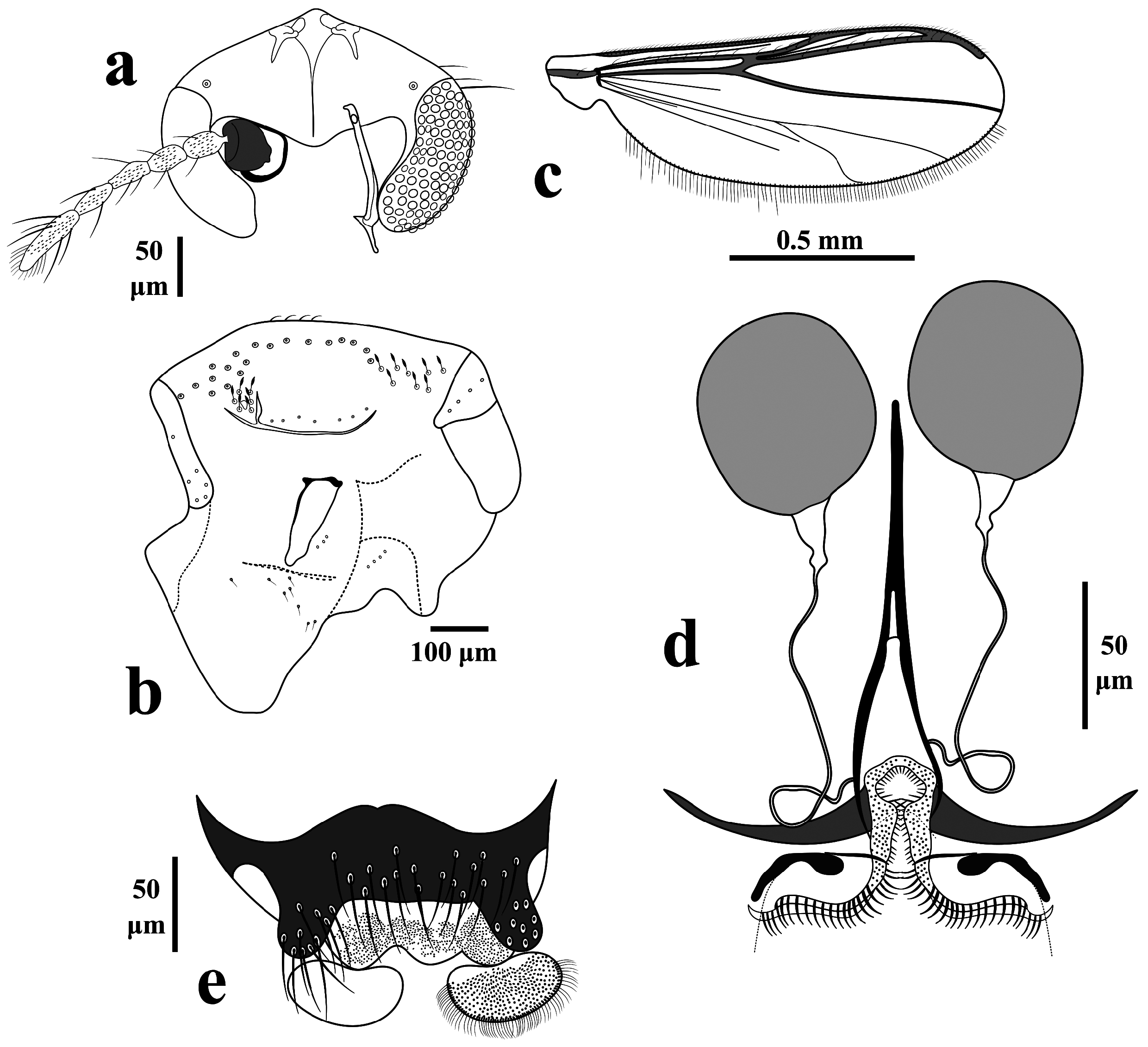
Head. Antenna with 13 flagellomeres, last flagellomere with 6 sensilla chaetica, groove starts at third segment, AR 0.3–0.6, 0.5. Eyes bare, without dorsomedial extension. Temporal setae few, 2 outer verticals and 1 frontal. Tentorium 132–135, 133 μm long ( Fig. 9a View FIGURE 9 ). Clypeus rectangular, 60 μm long and 114 μm wide, bearing 15 setae, setae 57–70, 63 μm long. Palpal segment lengths (in μm): 27–29, 28; 32–39, 36; 65–68, 66; 49–57, 53; 70–108, 89. Third palpomere with 1 sensilla clavata.
Thorax ( Fig. 9b View FIGURE 9 ). Acrostichals 4; dorsocentrals 20–25, around 23 in a single row and remainder in post humeral region double rows; prealars 6–7; scutellars 6–7 in single row; 3–6 lanceolate humerals; 6–8 lanceolate prescutellars; 10–14, 12 antepronotals; 5 posterior anepisternals II; 3 median anepisternals II; 2 epimeron II; 8 preepisternals, 6 anteriorly clustered and diagonal, separated from 2 vertical.
Wing ( Fig. 9c View FIGURE 9 ). Brachiolum with 1 seta. Squama bare. R with 11 setae, R 1 with 4 setae, other veins bare. Costa extension 45 μm. Anal lobe not projecting. Microtrichia visible at 10 ×.
Legs. Fore tibia spur 37–45, 41 μm long, mid tibia spurs 20–24, 22 and 19 μm long, hind tibia spurs 42–50, 46 and 14–18, 16 μm long, hind tibia comb with around 12 spines. Lengths and proportions of legs as in Table 3 View TABLE 3 .
Hypopygium ( Fig. 9d View FIGURE 9 ). Tergite IX with around 4 setae close to the base of anal point. Anal point extremely short, almost receded, wide with apex rounded; anal point 7–12, 10 μm long and 17–26, 21 μm wide. Virga consists of single long mid-spine with around 4 shorter lateral spines, the main spine 25–32, 28 μm long 7. Sternapodeme transverse with well-developed oral projections; sternapodeme 74–91, 81 μm long. Phallapodeme 33–42, 38 μm long. Inferior volsella a large triangular lobe with narrow apex; covered in numerous simple setae. Gonostylus large, expanded apically and with small spine-like distal outer projection, 60–77, 70 μm long; crista dorsalis large, overarching the apex of gonostylus. Gonocoxite 110–123, 116 μm long. HR 1.5–1.8, 1.7, HV 2.5–2.6.
Female (n = 2).
Total length 1.7–1.9, 1.8 mm. Wing 1.2 mm long and 0.37–0.43, 0.40 mm wide.
Coloration. Same as the male.
Head ( Fig. 10a View FIGURE 10 ). Antenna with 5 flagellomeres, last flagellomere with 8 sensilla chaetica, 1 st –4 th segments each with 2 sensilla chaetica, AR 0.4–0.7, 0.5. Eyes bare. Temporal setae 3–4 including 2–3 outer vertical and 1 frontal. Tentorium 120–123, 122 μm long. Clypeus rectangular, 65–82, 74 μm long and 96–110, 104 μm wide, bearing 20 setae, setae 53–63, 59 μm long. Palpal segment lengths (in μm): 27–31, 29; 31–34, 32; 57–71, 64; 61; 72–90, 81.
Thorax ( Fig. 10b View FIGURE 10 ). Acrostichals 4; dorsocentrals 26, around 23 in a single row and remainder in post humeral region double rows; prealars 5; scutellars 7 in single row; 8 lanceolate humerals; 8 lanceolate prescutellars; 6 antepronotals; 4 posterior anepisternals II; 3 epimeron II; 8 preepisternals, 6 anteriorly clustered and diagonal, separated from 2 vertical.
Wing ( Fig. 10c View FIGURE 10 ). Brachiolum with 1 seta. Squama bare. R with 9–13, 11 setae; R 1 with 5–7 setae; R 4+5 12–15 setae; other veins without setae. Costa extension 27–33 μm. Microtrichia visible at 10 ×.
Legs. Hind and mid femur with keel. Fore tibia spur 24 μm long, mid tibia spurs 18–20, 19 and 13–17, 15 μm long, hind tibia spurs 40–47, 45 and 16–17 μm long; hind tibia comb with around 12 spines. Lengths and proportions of legs as in Table 4 View TABLE 4 .
Genitalia ( Figs. 10d–e View FIGURE 10 ). Seminal capsules comparatively large, 66–70, 68 μm long, and 49–57, 53 μm wide, semi-circular, spermathecal ducts with loops, with well-developed bulb ( Fig. 10d View FIGURE 10 ). Notum 154–174, 158 µm long. Gonapophysis VIII divided into ventrolateral and thin dorsomesal lobe ( Fig. 10d View FIGURE 10 ).Apodeme lobe distinct. Gonocoxite developed with around 8–9 setae ( Fig. 10e View FIGURE 10 ). Tergite IX undivided. Cercus small, crescent-shaped, 60–61 µm long, and 31–41, 36 µm wide ( Fig. 10e View FIGURE 10 ).
Pupa. At present not known. Pupal exuviae could not be retrieved from the leaves.
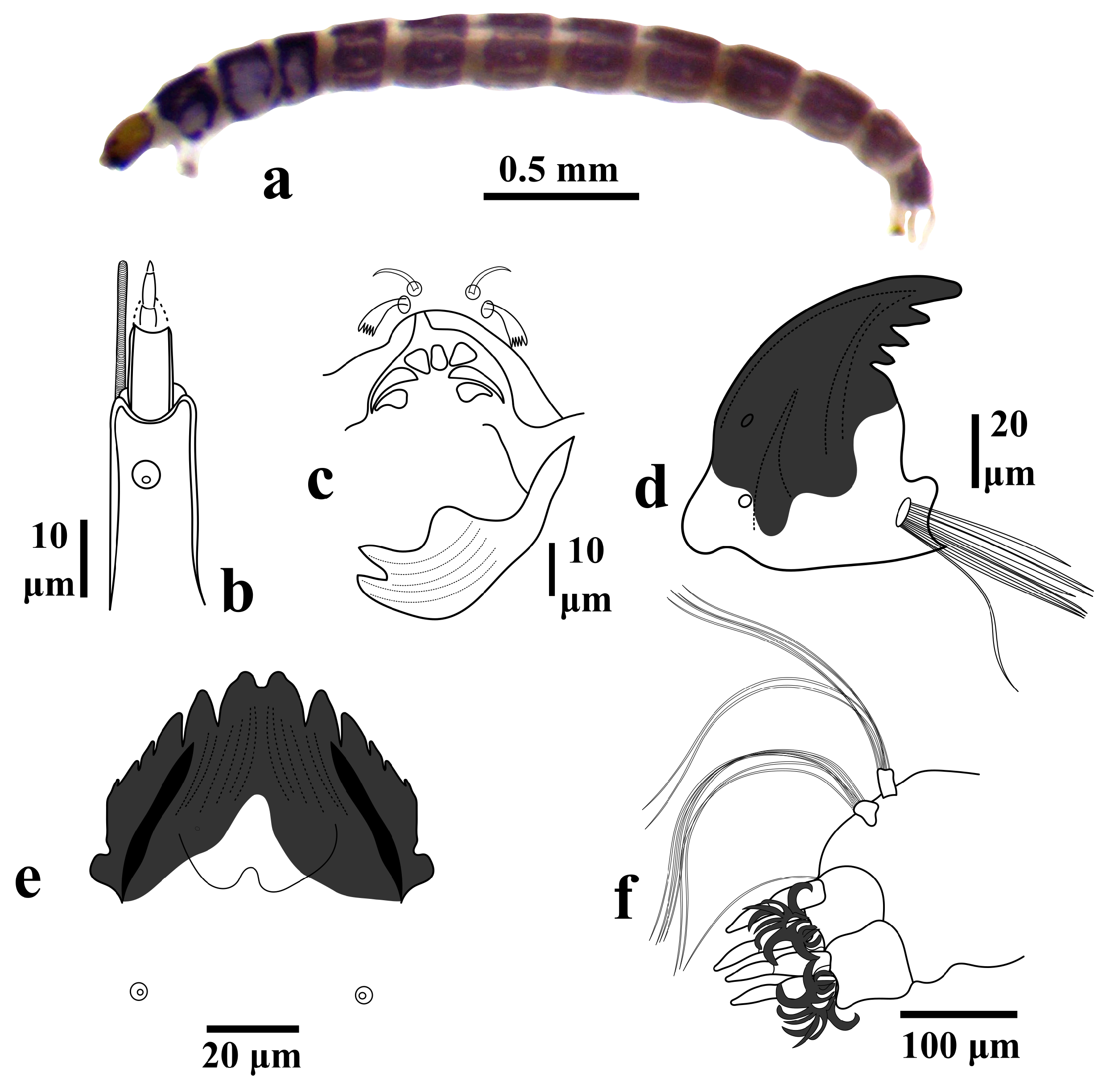
Larva (n = 1).
Total length 3.0 mm. Head 231 μm long and 267 μm wide.
Coloration ( Fig. 11a View FIGURE 11 ). Head capsule yellow with postmentum region darker. Occipital margin much darker in contrast to the remainder of the head. Abdomen bluish grey with patches of white.
Head. Antenna 5 segmented; segments length in μm: 30, 11, 2, 5, 3; ring organ closer to the apex of the basal segment; blade as long as the flagellum, blade 21 μm long ( Fig. 11b View FIGURE 11 ); AR 1.4. Labral SI serrate, SII–SIII simple ( Fig. 11c View FIGURE 11 ). Premandible wide and bifid, dark apically, 49 μm long ( Fig. 11c View FIGURE 11 ). Mandible dark, apical tooth shorter than combined width of 3 inner teeth; seta subdentalis very small; setae interna with several very long branches ( Fig. 11d View FIGURE 11 ), mandible 86 μm long. Mentum dark, with large bifid median tooth and 5 pairs of lateral teeth, median tooth 2.8 × the 1 st lateral teeth; seta submenti posteriad to mentum aligned with 2 nd lateral tooth ( Fig. 11e View FIGURE 11 ); mentum 57 μm long and 68 μm wide; ventromental plate 39 μm long, and 14 μm wide, large, slightly reaching beyond the margin of mentum.
Abdomen. Posterior parapods 65 μm long and 40 μm long, bearing around 12 simple dark claws ( Fig. 11f View FIGURE 11 ). Procercus 23 μm long and 17 μm wide, bearing 6 apical setae, apical setae 217 μm long; supraanal setae 144 μm long; four anal tubules with constriction, anal tubules 91 μm long ( Fig. 11f View FIGURE 11 ).
Diagnostic characters. Limnophyes viribus can be separated from other related species by the combination of the following characteristics: The adult male is characterized by AR of 0.3–0.6; 3–6 lanceolate humerals; 6– 8 lanceolate prescutellars; 8 preepisternals, 6 anteriorly clustered and diagonal, separated from 2 vertical; anal point extremely short, almost receded; virga consists of single long mid-spine with around 4 shorter lateral spines; inferior volsella a large triangular lobe with narrow apex; gonostylus large, expanded apically with small spine-like distal outer projection. The adult female is characterized by AR of 0.4–0.7; 8 lanceolate humerals; 8 lanceolate prescutellars; 8 preepisternals, 6 anteriorly clustered and diagonal, separated from 2 vertical; seminal capsules comparatively large. The larva is characterized by AR 1.4; premandible wide and bifid; setae interna of the mandible with several very long branches; mentum with large bifid median tooth and 5 pairs of lateral teeth; procercus bearing 6 apical setae; supraanal setae long; anal tubules longer than posterior parapods.
Taxonomic remarks. The adults of L. viribus resemble Limnophyes pilicistulus Saether, 1975 . The two species are probably related and form a sister group. Adults of L. viribus can be separated from L. pilicistulus by the lower number of temporal setae, higher number of lanceolate humerals and prescutellars. Additionally, the adult male of L. viribus has a virga with more branches, and a gonostylus with more overarching crista dorsalis, and a small spinelike distal outer projection. The adult female of L. viribus has longer cercus and notum and shorter seminal capsules compared to L. pilicistulus .
Biological notes. The larvae are apparently secondary inhabitants in leaf mines of Metriocnemus species, as with the undetermined Limnophyes species discussed below. The single Iowa specimen emerged in a batch rearing of M. eurynotus larvae feeding on Impatiens cotyledons. Four days earlier, in another rearing container, a mine with a larva of M. eurynotus had been found to also contain a much smaller larva that appeared uniformly dark and may have been an individual of L. viribus (photos at https://bugguide.net/node/view/2126834). Also in Iowa, a female very likely representing L. viribus was reared from a collection of M. erythranthei larvae mining Veronica leaves (see below under “ Limnophyes spp. ”). The Oregon specimens were reared along with M. erythranthei from plants of the Erythranthe guttata complex, although it was not entirely clear whether they emerged from leaves or the muck surrounding the roots.

| USNM |
Smithsonian Institution, National Museum of Natural History |
| ANC |
Dipartimento di Scienze Agrarie, Alimentari ed Ambientali, Università Politecnica delle Marche |
No known copyright restrictions apply. See Agosti, D., Egloff, W., 2009. Taxonomic information exchange and copyright: the Plazi approach. BMC Research Notes 2009, 2:53 for further explanation.
|
Kingdom |
|
|
Phylum |
|
|
Class |
|
|
Order |
|
|
Family |
|
|
Genus |