
Hypolagus balearicus, Quintana, Bover, Alcover, Agusti & Bailon, 2010
|
publication ID |
https://doi.org/ 10.5252/g2014n2a4 |
|
DOI |
https://doi.org/10.5281/zenodo.4822237 |
|
persistent identifier |
https://treatment.plazi.org/id/03F83642-021F-FFBF-FED3-FB3357B3209D |
|
treatment provided by |
Felipe |
|
scientific name |
Hypolagus balearicus |
| status |
|
Hypolagus balearicus Quintana, Bover, Alcover, Agustí & Bailon, 2010
DESCRIPTION
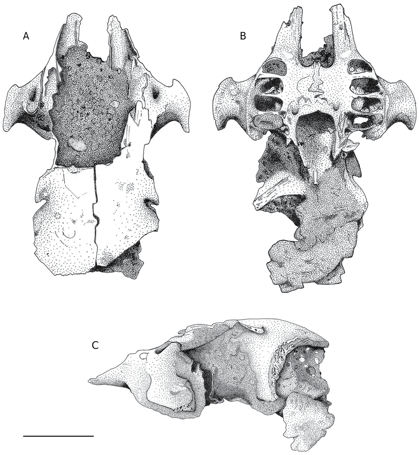
Cranium (IPS-41726; Fig. 2 View FIG ; Tables 1 View TABLE , 2 View TABLE ) Relatively small, badly preserved, without teeth. The body of maxilla is partially preserved. The two maxillas show the alveolus of P2, P3, P4 and M1. The M2 alveolus is only partially preserved. Only the facial tubercles of the two zygomatic archs are preserved. The maximum distance between the ventral surface of the facial tubercle and the dental alveolus is very small. The two major palatine foramina are situated very close to the anterior side of
the choana. The posterior side of the palatine fissure is situated at the same level as the mean length of P2. The frontal bones are partially preserved. The supraorbital caudal notch appears shallow.
The posterior zone of the zygomatic process (and the supraorbital caudal notch) is only little developed, which is likely due to the poor preservation of this part of the cranium.
Though both orbits are badly preserved, the distance between the zygomatic process of the frontal bone and the dorsal side of the facial tubercle suggests that the orbits of H. balearicus are proportionally smaller than those of Oryctolagus cuniculus (Linnaeus, 1758) .
Left incisor I1 (IPS-61674; Fig. 3H View FIG )
The linguoventral and medioventral margins show a well rounded outline. The central part of the ventral margin is slightly concave. The dorsal face is crossed by a rather wide V-shaped groove.It shows a depth of 18% in relation to the dorso-ventral width of tooth. Labial lobule with a well rounded outline, wider than the lingual lobule, which is acuter and narrower. The enamel is only present on the dorsal half of the tooth.
Left premolar P2 (IPS-61601; Fig. 3D View FIG )
The paraflexus has a depth equivalent to the 36% of the anteroposterior tooth length. The lagiocone is formed by two lobes with similar contour, separated by a mesoflexus that shows a depth that equals 1/3 of the paraflexus length and 12% of the anteroposterior length. The metaflexus forms a very slight concavity. The hypercone antero-lingual contour is genuinely rounded, without hypoflexus.
P3-M2 (IPS-61661, 61662, 61665 [ Fig. 3E View FIG ]; IPS-61663 [ Fig. 3F View FIG ]; IPS-61664, 61666) Variable hypoflexus morphology, but with no more than three lobules on the anterior margin. The hypoflexus enters up to half of the tooth approximately. Posterior side of the hypoflexus only slightly undulated.
Mandible
– IPS-26592A: mandibular fragment with p4, m1 and m2 ( Fig. 3C View FIG );
– IPS-26592B: left/right mandibular fragment;
– IPS-41727: incomplete right mandible ( Fig. 4 View FIG ); – IPS-26590: incomplete left mandible;
– IPS-26591: left mandibular fragment ( Table 4).
The retroalveolar foramen is small with an elliptic outline. The mandibular ramus is 134° inclined in relation to the dorsal side of the mandibular body ( Table 3 View TABLE ). The posterior side of the mental foramen is in line with the anterior side of p3.
Remarks. The inclination of the mandibular ramus appears to be related to the cranium length. The inclination is similar in H. balearicus and other leporids with a proportionally short skull ( Table 3 View TABLE ).
Left incisor i1 (IPS-61675; Fig. 3G View FIG )
Tooth with a rounded trapezoidal outline. The enamel only covers the ventral side of the tooth.
Right premolar p3 (IPS-61602; Fig. 3A View FIG )
and left premolar p3 (IPS-61603; Fig. 3B View FIG )
Tooth with trapezoidal outline. Little pronounced or shallow anteroflexid and absent paraflexid. The anteroconid shows a rounded, slightly sharpened outline. V-shaped protoflexid, with a depth equal to 13.5%-16% of the total width of the tooth. The hypoflexid relative depth is about 48%-52%; undulation of the flexid is not observed; the mesial side of the hypoflexid shows, sometimes, a marked convexity; distal and mesial walls run almost parallel. Lingual side with rounded outline, slightly wider (anteroposterior direction) than the medial zone of the hypoflexid; labial anteroconid with trapezoidal outline. Lingual anteroconid with straight or convex contour.
Remarks. The two p3 from Eivissa differ in size and in the morphology of the hypoflexid. There are considerable differences in size between the Eivissa p3 and that of Caló d’en Rafelino (Mallorca) ( Quintana et al. 2010: fig. 3). However, the morphological differences between the p3 of the two island populations are little significant, and consist basically in the absence of undulation on the hypoflexids of the p3 from Eivissa.
p4-m2 (IPS-61667-61673; Fig. 3C View FIG )
Hypoflexid with smooth or slightly undulated anterior and posterior margins. The lingual extreme of the hypoflexid is curved towards the anterior margin of the tooth.This curvature increases progressively from p4 to m2. The enamel of the posterior margin of the hypoflexid is extremely thin, similar to that on the anterior margin of the trigonid. The enamel disappears on the lingual face on both trigonid and talonid. Humerus (IPS-41728, left incomplete humerus,
see Table 5 View TABLE ; IPS-61604, distal epiphysis fragment
of left humerus, see Fig. 5 View FIG )
The head of the humerus shows, from a lateral view, a very rounded outline. The lateral and medial faces of the proximal epiphysis are largely worn. The crests of the trochlea show a slightly sharp outline ( Fig. 5 View FIG ) and are quite separated.
Remarks. The trochlea of the distal epiphysis is relatively wide ( Fig. 6B View FIG ) and the crests show a light acute outline in comparison to the other species included in the genus Hypolagus (Dawson 1958: fig. 30C; Fladerer 1984; Fladerer & Fiore 2003: pl. 2, fig. 2; Fostowicz-Frelik 2007b: figs 24-30). We exclude the possibility that the particular morphology of the IPS-61604 distal epiphysis is caused by abrasion due to taphonomic processes.
In leporids, the development and separation of the crests and pits on the elbow articulation are likely related to the speed attained during running and leaping. Species better adapted to high speeds are those with more acute crests, whereas the non-running species have lower crests and the distal epiphysis is wider transversally, as it happens in N. rex (Quintana 2005: fig. 56) or, to a lesser extent, in Pentalagus furnessi (Stone, 1900) .
Radius (IPS-61605, left proximal epiphysis)
The fovea is shallow and the surface portion of the articulation situated on the lateral side is bended 42° in relation to the diaphysis.
Remarks. The craniocaudal diameter of the proximal radius from Eivissa is larger than that of its Mallorcan homologue, and the lateral margin is substantially lower ( Quintana et al. 2011: fig. 8) ( Table 6). On H. balearicus the fovea is less concave than H. beremendensis (Petényi, 1864) (Fladerer 1984: abb. 5, figs 1, 2) and Hypolagus petenyii Čermák & Fladerer in Čermák, 2009 (Fostowicz-Frelik 2007a: figs 32, 34). The poor depth of the fovea is related to the low sharp outline of the lateral crest of the distal humeral epiphysis.
Ulna (IPS-41733, right ulna without distal epiphysis, see Fig. 7 View FIG ; IPS-61606, left proximal epiphysis)
From an anterior view, the anconeal process shows a well-rounded outline. The diaphysis is wide, both transversally and anteroposteriorly. ( Fig. 8 View FIG ; Table 7 View TABLE ).
Remarks. The rounded outline of the anconeal process suggests that the trochlea on the distal epiphysis of the humerus forms an arch with an opened outline, even more than in N. rex (Quintana 2005: fig.56a). Both, this trait and the morphology of the proximal radius in H. balearicus from Mallorca ( Quintana et al. 2010: fig. 8), suggest that the crests and the elbow articulation pits in H. balearicus have a low acute contour.
The ulna proximal epiphysis of H. balearicus shows an aspect considerably more robust than H. beremendensis (Fladerer 1984: abb. 6, fig. 1). In lateral view, the trochlear incisure of H. balearicus forms a similar arch than N. rex (Quintana 2005: fig. 72) and slight more open than H. beremendensis .
Second metacarpal (IPS-61607, right proximal epiphysis, see Fig. 9B View FIG and Table 8 View TABLE ; IPS-61608, right proximal epiphysis)
From cranial view, the epiphysis shows a quadrangular outline. The trapezoid fossa forms an arch slightly open in mediolateral direction. The lateral crest (dorsal view) forms an angle of 75°. The medial crest is proportionally lower and rounded. The capitatum facet is bended more or less 40° in relation to the longitudinal axis of the diaphysis. From a lateral view, this facet shows a V-form outline, rather open and symmetric. The crest, which separates the facets for the capitatum and the third metacarpal, as well as the pit for the third meta- carpal form a gentle arch. On the first metacarpal facet, the ventral extreme is missed. It presents an elongated surface in dorsoventral direction, slightly irregular and concave in anteroposterior direction.
Remarks. The proximal epiphysis of the second metacarpal of H. balearicus differs from O. cuniculus and Lepus granatensis Rosenhauer,1856 by a trapezoid fossa that is more open and wider in mediolateral direction and proportionally shorter in dorsoventral direction.The lateral crest is wider and less acute in H.balearicus .Cranial view, the proximal epiphysis of H. balearicus shows a quadrangular outline, while in N. rex and H. beremendensis is more elongated in dorsoventral direction (Quintana 2005: fig. 84; Fladerer 1984: abb. 19e).
Femur (IPS-26589, left proximal epiphysis, see Fig. 9C View FIG ; IPS-41730, left proximal left epiphysis;
IPS-41729, right diaphysis from a juvenile individual) From a dorsal view, the transversal section of the cranial face of the greater trochanter shows a rounded outline. The lesser trochanter is little elevated in mediolateral direction and in relation to the femoral neck. The dorsal margin of the femoral neck is short, so that the femoral head and the greater trochanter are close to each other. On the cranial face, the margin that separates the femoral head from the neck shows a low sharpened outline. The transversal section of the diaphysis has a circular outline.
Remarks. The lateral margin of the femoral head of H. balearicus is proportionally closer to the greater trochanter than in O. cuniculus and H. petenyii (Fostowicz-Frelik 2007b: fig. 4). From a cranial view, the femoral neck length is proportionally shorter than in O. cuniculus but longer than H. petenyii .
The minor trochanter is proportionally less developed in H. balearicus . The anteroposterior diameter of femoral head ( Table 9 View TABLE ) is intermediate between Pronolagus rupestris (A. Smith, 1834) and H. petenyii . Tibia (IPS-61609, right distal epiphysis; IPS-61658, left distal epiphysis;
IPS-61659, right distal epiphysis, see Fig. 9A View FIG )
The facet where the lateral margin of the astragalus is articulated shows a U-shaped outline. A groove, separating the lateral and medial face from this facet, is not observed. The cranial half of the calcaneus facet is wider than the caudal half and shows, in craniocaudal direction, a slightly concave outline. The area that separates the calcaneus and astaragalus facets shows an elliptic outline, with the principal axis pointing in craniocaudal direction.
Remarks. The lateral fossa that articulates with the astragalus shows a mediolateral outline similar to that of L. granatensis and slightly less acute and more open than the one of O. cuniculus . However, the facet for articulation with calcaneus of H. balearicus shows a concavity (in craniocaudal direction) similar to O. cuniculus and clearly smaller than that in L. granatensis . The transversal diameter of H. balearicus tibia is slightly larger than that of O. cuniculus but smaller than that of Bunolagus monticularis (Thomas, 1903) ( Table 10).
Navicular (IPS-61660, right incomplete bone, see Fig. 9D View FIG ) The astragalus pit shows a subquadrangular outline and a little marked concavity.On the plantar process, the ventral surface is slightly convex without any groove. Remarks. Due to its special traits, the recovered navicular of H. balearicus is interpreted as belonging to a juvenil individual. The astragalus fossa is notably more opened than in H. petenyii (Fostowicz-Frelik 2007: fig. 13), O. cuniculus and L. granatensis . The size of the navicular of H. balearicus is smaller in comparison to P.furnessi , Sylvilagus floridanus (J. A. Allen, 1890) or Lepus europaeus Pallas, 1778 (Fostowicz-Frelik 2007: pl. 3, p. 474).
Calcaneus (IPS-41732, right incomplete bone, see Fig. 10)
The proximal facet of the astragalus shows a very acute anterior extreme and a concave surface, while the distal facet presents an elliptic outline (with the major axis slightly oblique in relation to the calcaneus length) and a slightly convex surface. The boundary between both facets shows a gentle outline. From medial view, the posterior half of the facet for the tibia shows a circular outline. The cuboid facet shows a piriform outline with a rather enlarged and slightly concave surface.
Remarks. The caudal half of calcaneus is proportionally robuster in H. balearicus than in O. cuniculus and L. granatensis because the caudal side of the cuboid facet is less pronounced. The lateroproximal astragalus facet is concave in H. balearicus , while it shows a marked convexity in O. cuniculus and L. granatensis . The form of the lateral outline of the tibial facet of H. balearicus shows an intermediate curvature, different from those in L. granatensis (clearly circular) and O. cuniculus (eliptico-oval).The mediolateral diameter of calcaneus of H. balearicus is larger than in O. cuniculus but smaller than in B. monticularis ( Table 11).


No known copyright restrictions apply. See Agosti, D., Egloff, W., 2009. Taxonomic information exchange and copyright: the Plazi approach. BMC Research Notes 2009, 2:53 for further explanation.