
Varanasifaustula, Dronen & Blend & Mohammed & Bannai, 2021
|
publication ID |
https://doi.org/10.11646/zootaxa.5027.2.5 |
|
publication LSID |
lsid:zoobank.org:pub:041275C5-9611-4218-8D72-2BF0AA584C5F |
|
DOI |
https://doi.org/10.5281/zenodo.5492772 |
|
persistent identifier |
https://treatment.plazi.org/id/03F787B6-AC02-D62C-F8F6-F359FDE11B6D |
|
treatment provided by |
Plazi |
|
scientific name |
Varanasifaustula |
| status |
gen. nov. |
Varanasifaustula View in CoL n. gen.
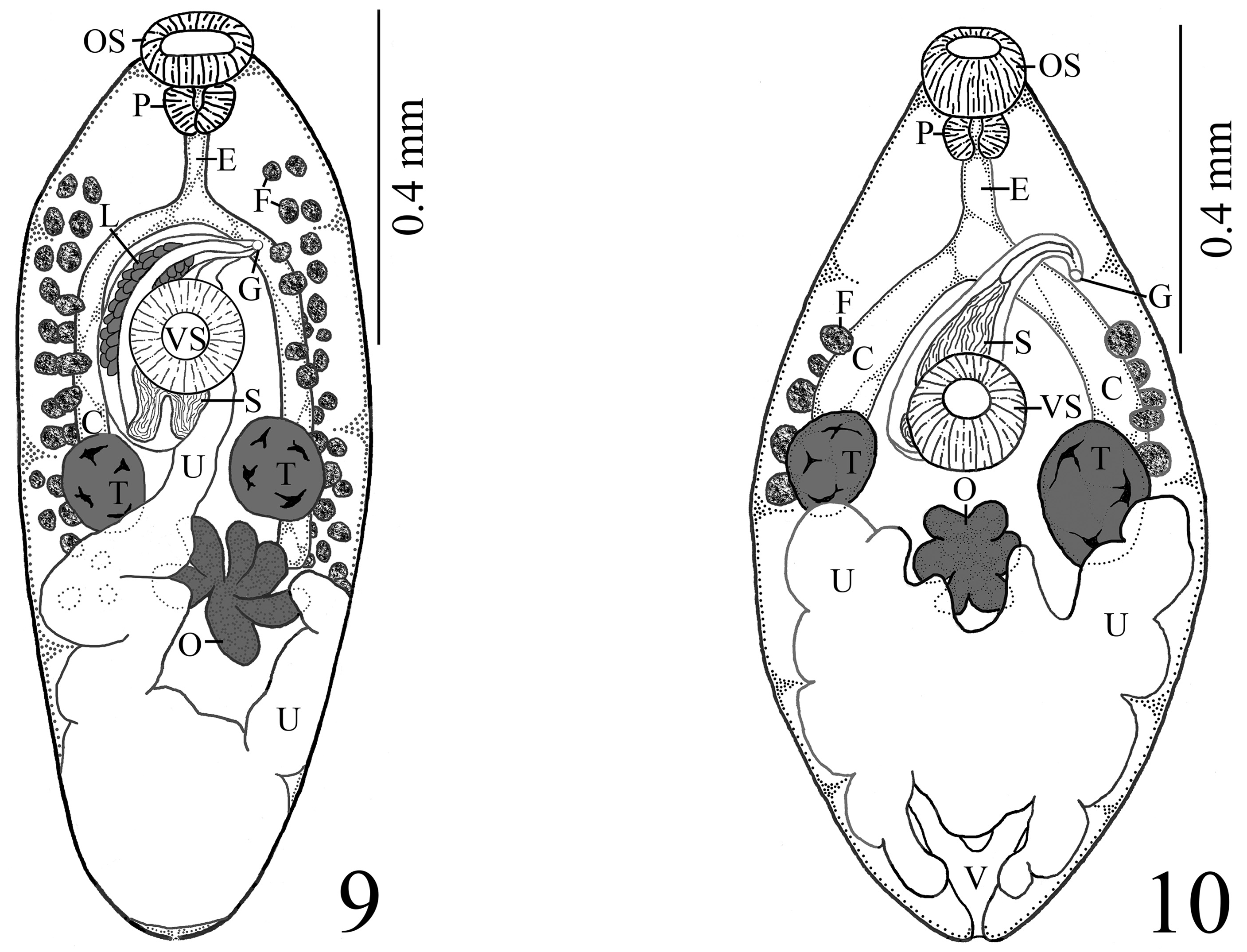
( Fig. 10 View FIGURES 9–10 )
Type species: Varanasifaustula indica ( Agarwal & Verma, 1981) View in CoL n. comb. Type and only species.
(Syn. Faustula indica Agarwal & Verma, 1981 View in CoL )
Diagnosis: Body small, somewhat elliptical, extremities tapered to form bluntly pointed ends; tegumental spines not reported. Oral sucker, globose, slightly subterminal. Ventral sucker muscular, globose, anterior to midlevel of body, larger than oral sucker. Prepharynx absent. Pharynx oval, nearly circular, muscular. Esophagus relatively short. Intestinal bifurcation about half way between suckers. Ceca relatively short, terminate slightly anterior to midlevel of body. Testes 2, entire, symmetrical, near midlevel of body; anterior extent may overlap posterior margin of ventral sucker dorsally almost to its midlevel. Cirrus sac clavate, overlaps ventral sucker dorsally, may surpass it posteriorly by short distance; sac encloses cirrus, narrow ejaculatory duct, short tubular pars prostatica, relatively long, saccate, naked seminal vesicle. Genital pore at about level of posterior margin of intestinal bifurcation, distinctly submedian, extracecal, immediately left of left cecum. Ovary with 6 lobes, immediately posttesticular or slightly more anterior, near midline of body to slightly submedian. Seminal receptacle described as absent. Uterus largely posterior to gonads, filling most of posttesticular space. Vitellarium composed of few, relatively large follicles (approximately 5–7/side); follicles linearly organized near lateral margins of body from about midlevel of ceca to level of posterior margin of testes or slightly more posterior. Eggs small, operculate. Excretory vesicle V-shaped, extent of excretory arms unknown; excretory pore slightly subterminal. Reported as intestinal parasite of species of clupeid fishes in the Ganges River, India.
Etymology: The genus is named from the location where the type species was collected in India ( Varanasi) and its probable assignment within the faustulid trematodes ( Faustula ).
Remarks: Varanasifaustula indica (Syn. F. indica ) differs from species of Faustula by having a simple cirrus; a narrow ejaculatory duct; a tubular pars prostatica and a simple, saccate, unipartite, naked seminal vesicle vs a winding tubular seminal vesicle that is at least partially embedded in large glandular cells; a distinctly submedian, extracecal genital pore; few (5–7/side), relatively large vitelline follicles somewhat longitudinally arranged along the lateral fields of the body from about the midlevel of the ceca to the level of the posterior margin of the testes vs smaller more numerous follicles that range from some distance posterior to the level of the intestinal bifurcation, posteriorly to about the level of the ovary or slightly more anterior; the gonads located near the midlevel of the body and a median ovary with 6 lobes vs 8 or more lobes as seen in species in Faustula . Note the illustration representing F. indica (= V. indica ) in the original description by Agarwal & Verma (1981) (see fig. 2) is identified as a ventral view, but the location of the cirrus sac and vitelline follicles relative to the ceca, and the ventral sucker relative to the testes and cirrus sac suggest it is a dorsal view. It appears that the specimen illustrated in fig. 2 was rolled so that the structures more closely associated with the dorsal aspect of the body (e.g., posterior aspect of the cirrus sac, gonads) were displaced to the right while structures more closely associated with the ventral surface (e.g., ventral sucker, genital pore) may have been displaced to the left. It is our opinion that the cirrus sac in this species likely more extensively overlaps the ventral sucker, but that the position of the genital pore was not appreciably altered. Varanasifaustul a indica (Syn. F. indica ) is somewhat similar to L. hilsai discussed above, most notably by having 6 ovarian lobes vs 5–6 ovarian lobes in L. hilsai (see description and Fig. 2A View FIGURES 1–2 of Kumar &Agarwal 1984). Lingulitrema hilsai also differs from V. indica by having longer ceca that surpass the testes for some distance posteriorly, reaching about the midlevel of the ovary; an S-shaped seminal vesicle that lacks large glandular cells; a distinctly submedian genital pore that approaches the cecum and small, more numerous vitelline follicles forming more extensive vitelline fields that extend from the midlevel of the esophagus posteriorly to about the level of the ovary.
The following key to genera within the Faustulidae was developed to accomomodate both previous genera and the 5 new genera proposed herein.

No known copyright restrictions apply. See Agosti, D., Egloff, W., 2009. Taxonomic information exchange and copyright: the Plazi approach. BMC Research Notes 2009, 2:53 for further explanation.