
Cunninghamella saisamornae N. Suwannarach & P. Wongputtisin, 2021
|
publication ID |
https://doi.org/ 10.11646/phytotaxa.509.3.4 |
|
persistent identifier |
https://treatment.plazi.org/id/03F6879B-E224-FFBD-FF52-FDB52134FA09 |
|
treatment provided by |
Marcus |
|
scientific name |
Cunninghamella saisamornae N. Suwannarach & P. Wongputtisin |
| status |
sp. nov. |
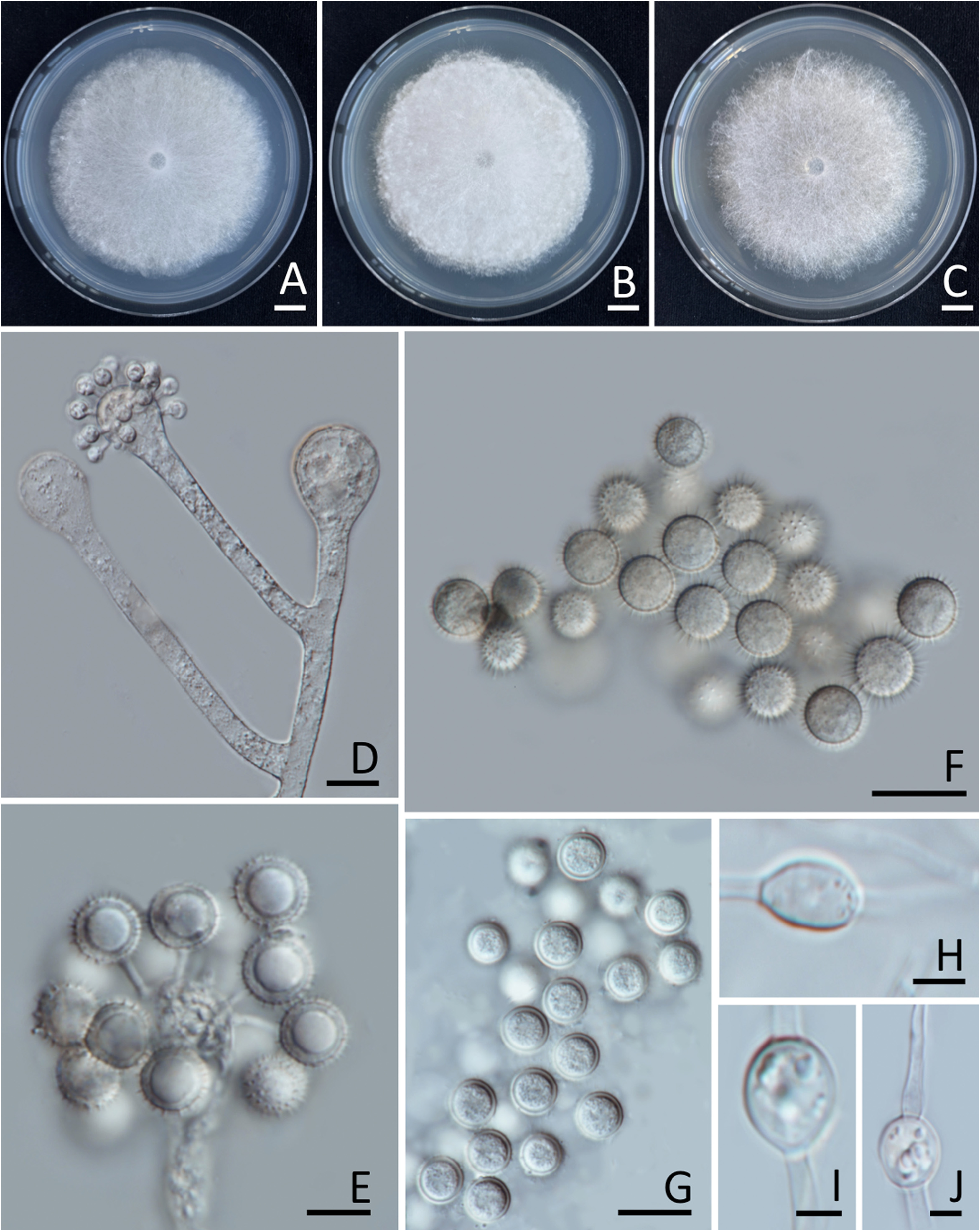
Cunninghamella saisamornae N. Suwannarach & P. Wongputtisin , sp. nov. Figure 2 View FIGURE 2 .
MycoBank number: MB 839021
Diagnosis: —Distinguishable from other members of the Cunninghamella species by a lack of the zygosporic stage as well as by a chlamydospore formation and its maximum growth temperature at 45°C.
Etymology: —refers to the name of Emeritus Prof. Dr. Saisamorn Lumyong in honour of her 70th birthday and her over 30 years’ contribution to Thai mycology.
Holotype: — THAILAND, Chiang Mai Province, San Sai District , 19°15’42”N 101°0’16”E, isolated from soil in paddy field, March 2016, P. Wongputtisin & N. Suwannarach, dried culture: SDBR-CMU291 , ex-holotype TBRC 14914 View Materials . GoogleMaps
Culture characteristics: —Colonies growing on CZA, PDA and SMA were 7.00–7.15, 6.90–7.10 and 6.80–7.00 mm in diameter after two days at 25°C. Colonies on CZA, PDA and MEA floccose, initially white and became yellowish white to pale yellow, reverse pale yellow after two weeks of incubation at 25°C ( Fig. 2 View FIGURE 2 A−C). On all media the hyphae are branched, hyaline, smooth-walled, non-septate when young, irregularly septate when aging, 5–20 μm wide. Sporophores hyaline, erect to recumbent, smooth-walled, opposite or verticillate branches, main axes of the branches predominantly terminated with a vesicle. Vesicles subglobose to pyriform; terminal vesicles, smooth-walled, 25–50 μm in diameter (average 37.5 μm in diameter, n = 50); lateral vesicles smaller, smooth-walled, 20–37 μm in diameter (average 28.5 μm in diameter, n = 50) ( Fig. 2D View FIGURE 2 ). Pedicels 4−10 μm long. Sporangioles borne on pedicles on vesicles, globose to subglobose, 13–18 μm in diameter (n = 50), hyaline, thick-walled, echinulate with short spines; spines 2–3 μm long ( Fig. 2E–G View FIGURE 2 ). Zygosporangia not observed. Chlamydospores, subglobose, spherical to ellipsoid, 10–25 × 8–20 μm (n = 50) ( Fig. 2H–J View FIGURE 2 ).
Additional specimens examined: — THAILAND, Chiang Mai Province, San Sai District , 19°15’42”N 101°0’16”E, isolated from soil in paddy field, March 2016, isolated by P. Wongputtisin & C. Supo: SDBR-CMUPFCM-9 GoogleMaps .
Known distribution: — Chiang Mai Province, Thailand.
| PDA |
Royal Botanic Gardens |
No known copyright restrictions apply. See Agosti, D., Egloff, W., 2009. Taxonomic information exchange and copyright: the Plazi approach. BMC Research Notes 2009, 2:53 for further explanation.
|
Kingdom |
|
|
Phylum |
|
|
Class |
|
|
Order |
|
|
Family |
|
|
Genus |