
Progonatocerus Huber
|
publication ID |
https://doi.org/ 10.11646/zootaxa.3967.1.1 |
|
publication LSID |
lsid:zoobank.org:pub:809A05D1-3BAD-4A32-8D56-C91A6B609D00 |
|
DOI |
https://doi.org/10.5281/zenodo.6112343 |
|
persistent identifier |
https://treatment.plazi.org/id/03F587E3-355A-FFF2-41CB-FD98CCDDF66D |
|
treatment provided by |
Plazi |
|
scientific name |
Progonatocerus Huber |
| status |
gen. nov. |
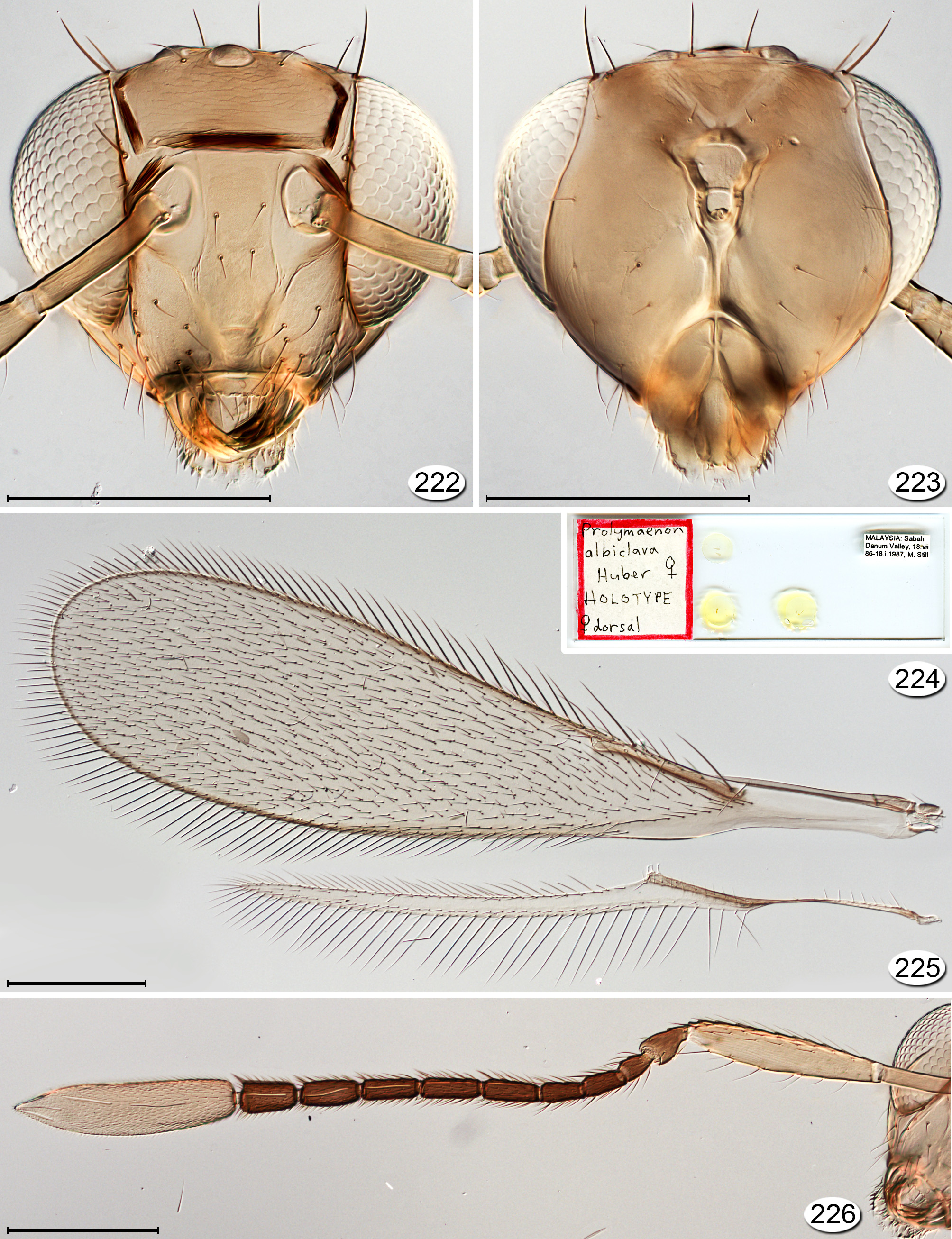
Progonatocerus Huber , gen. n.
( Figs 221–247 View FIGURE 221 View FIGURES 222 – 226 View FIGURES 227, 228 View FIGURES 229 – 231 View FIGURES 232 – 237 View FIGURES 238 – 243 View FIGURES 244 – 247 )
Type species. Progonatocerus albiclava , sp. n., by present designation.
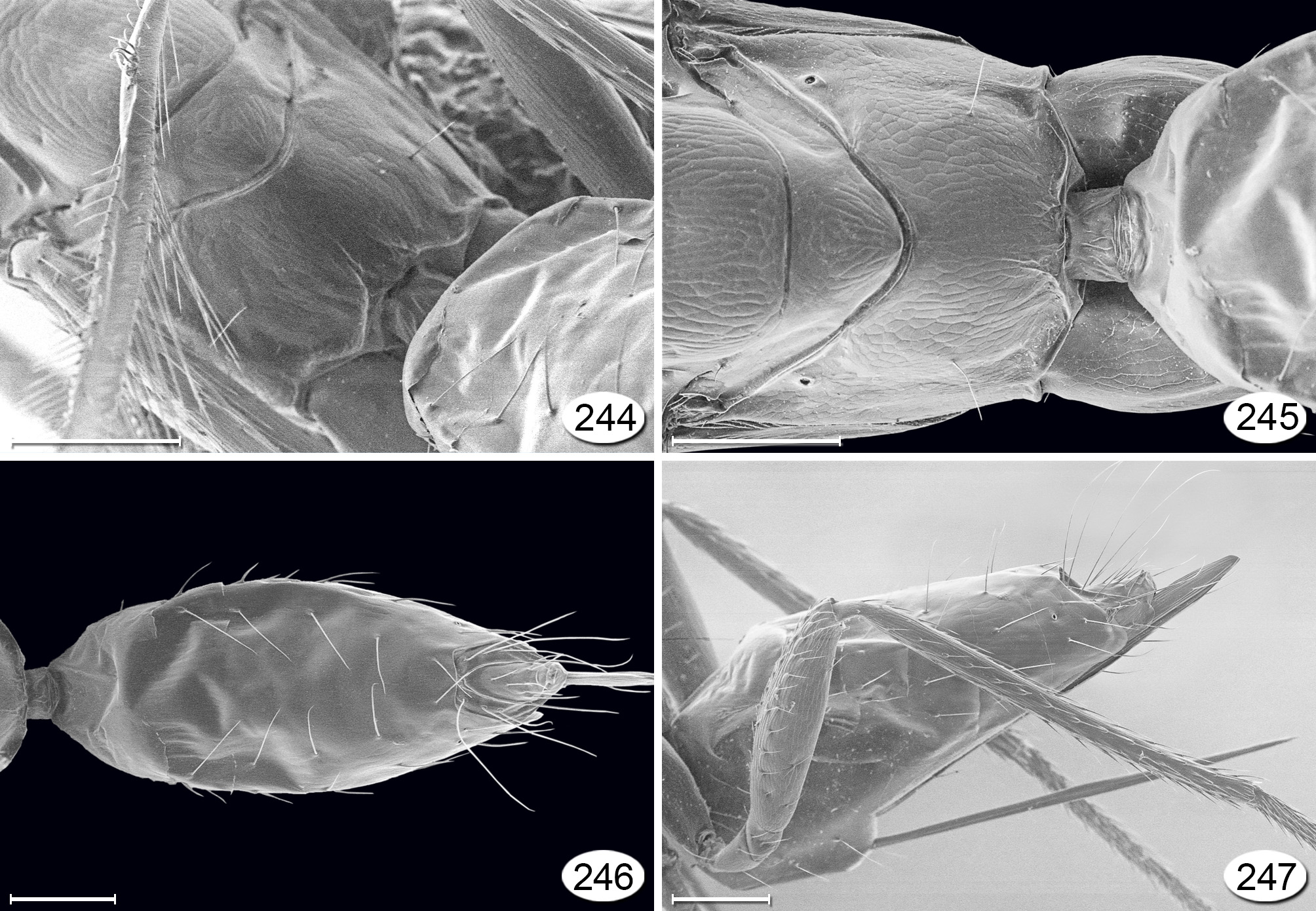
Diagnosis. Within Gonatocerini, the species of Progonatocerus are distinguished by the following combination of features: pronotum entire ( Figs 239, 241, 242 View FIGURES 238 – 243 ); scutellum with two short longitudinal subcuticular lines lateral 50 to the campaniform sensilla, visible in cleared slide mounts ( Fig. 227 View FIGURES 227, 228 ). Males have an unusually long flagellum ( Fig. 235 View FIGURES 232 – 237 ).
Description. FEMALE. Moderately sized specimens, 973–1306 Μm in length. Colour. Body yellow with various amounts of brown on gaster, and antenna except clava dark brown; clava usually white or cream-coloured ( Figs 221 View FIGURE 221 , 226 View FIGURES 222 – 226 , 234 View FIGURES 232 – 237 ).
Head. Head ( Figs 222 View FIGURES 222 – 226 , 228 View FIGURES 227, 228 ), about 1.52–1.73× as wide as long and about 0.83× as wide as high; in lateral view with anterior surface quite strongly convex ventral to torulus ( Fig. 240 View FIGURES 238 – 243 ). Face as wide as high ( Fig. 222 View FIGURES 222 – 226 ); subantennal sulci present, slightly converging, the distance between them at mouth margin less than their distance from preorbital sulci; preorbital sulcus closely following inner orbit almost to lower margin of eye then curving slightly inward to dorsolateral corner of mouth. Toruli almost touching transverse trabecula. Eye in lateral view slightly higher than long, dorsally extending to back of head ( Figs 239, 240 View FIGURES 238 – 243 ). Malar space about 0.3× eye height; malar sulcus almost straight, extending from posteroventral angle of eye to mouth margin about midway between mandibular condyles. Vertex in lateral view oblique, forming obtuse angle with face (separated by transverse trabecula), posteriorly acutely angled at junction with occiput ( Figs 240, 241 View FIGURES 238 – 243 ), with a sulcus extending behind lateral ocelli from eye to eye. Ocelli with LOL about 0.5× POL and OOL about 0.6× POL, with 2 setae between lateral ocelli. Occiput entire. Labrum with 3 setae. Mandible with 3 normal teeth ( Fig. 222 View FIGURES 222 – 226 ). Antenna. Scape about 4.3× as long as wide ( Figs 226 View FIGURES 222 – 226 , 234 View FIGURES 232 – 237 ) with radicle distinct, narrow 0.4–0.5× remainder of scape length; pedicel about 0.20–0.25× scape length, longer and wider than fl1; funicle 8-segmented, each segment at most with 2 mps; clava about 0.5× funicle length, apparently with 8 mps ( Figs 239, 242 View FIGURES 238 – 243 ). Mesosoma. About 1.7–1.8× as long as wide, 1.5–1.7× as long as high, and 0.9–1.0× as wide as high ( Figs 227, 228 View FIGURES 227, 228 , 242, 243 View FIGURES 238 – 243 ). Pronotum ( Figs 238, 239, 242 View FIGURES 238 – 243 ) entire, in dorsal view short and almost vertical but still slightly visible, merging smoothly with lateral panel. Pronotal spiracle about same size as propodeal spiracle ( Fig. 243 View FIGURES 238 – 243 ). Propleura normal. Prosternum rhomboidal, divided posteriorly by longitudinal sulcus extending at least half its length. Mesonotum in dorsal view with fairly wide but shallow, fairly straight and slightly diverging notauli. Transscutal articulation straight. Scutellum longer than wide, anteriorly with two short longitudinal subcuticular lines extending from scutoscutellar suture just medial to posterior apex of notauli and lateral to scutellar campaniform sensilla towards anterior margin of frenum ( Fig. 227 View FIGURES 227, 228 ). Axilla normal. Prepectus narrow, slightly wider medially and ventrally than dorsally. Mesopleuron spindleshaped and truncate at both ends with fine sulcus separating mesepisternum from mesepimeron. Metanotum with triangular dorsellum, its posterior margin quite strongly convex. Metapleuron triangular, separated from propodeum by complete but fine line. Propodeal spiracle small, separated by about its diameter from metanotum. Wings. Fore wing 3.7–4.0× as long as wide, with microtrichia fairly dense and uniform over entire surface to base of parastigma except absent narrowly anterior to retinaculum ( Figs 225 View FIGURES 222 – 226 , 233 View FIGURES 232 – 237 ). Venation almost 0.4× wing length.
Submarginal vein with the usual two basal setae (a dorsal macrochaeta and a ventral hypochaeta) and a hypochaeta apically, next to proximal macrochaeta of parastigma. Remaining venation (parastigma + stigmal vein) about 0.9× length of submarginal vein, with hypochaeta closer to distal than proximal macrochaeta and 2–5 setae between the macrochaetae. Stigmal vein with apex truncate. Hind wing normal. Venation 4.3× wing length. Metasoma. Petiole short, wider than long ( Figs 227 View FIGURES 227, 228 , 229 View FIGURES 229 – 231 , 245 View FIGURES 244 – 247 ). Gaster about 1.4–1.5× as long as high. Terga with white membrane at most narrowly visible between them. Cerci abutting each other dorsally. Ovipositor sheaths ( Figs 230, 231 View FIGURES 229 – 231 ) about as long as to shorter than gaster, equal to or longer than metatibia, and slightly exserted beyond gaster apex, with 1 subapical seta.
MALE. Colour. Body yellow with flagellum, trabeculae and gaster medially dark brown. Antenna. Scape (including radicle) short, about 2.8× as long as wide, with radicle about 0.25× scape length; pedicel about 0.46× scape length; flagellum unusually long, flagellomeres each with about 8 mps. Metasoma. Genitalia with aedeagal apodemes about as long as aedeagus and apodeme of genital sternum absent ( Fig. 237 View FIGURES 232 – 237 ).
Discussion. Progonatocerus is similar in some ways both to Gonatocerus and to Lymaenon . Progonatocerus has three feature resembling Lymaenon species—the stigmal vein apex is almost truncate ( Figs 221 View FIGURE 221 , 225 View FIGURES 222 – 226 , 233 View FIGURES 232 – 237 ), fl1 is not obliquely truncate dorsoapically, and fl2, fl3 and fl4 are not longer than the subsequent flagellomeres ( Figs 226 View FIGURES 222 – 226 , 234 View FIGURES 232 – 237 ). Progonatocerus also has three feature resembling Gonatocerus species—a rhomboidal dorsellum ( Figs 221 View FIGURE 221 , 227 View FIGURES 227, 228 , 242 View FIGURES 238 – 243 ), pronotal spiracle about the same size as propodeal spiracle ( Fig. 243 View FIGURES 238 – 243 ), and two setae in the ocellar triangle ( Figs 222, 223 View FIGURES 222 – 226 , 239 View FIGURES 238 – 243 ).
Etymology. From Latin “pro” meaning before (in the sense of earlier) and Gonatocerus . The name refers to its supposed earlier ancestry from which Gonatocerus and Lymaenon may have arisen. The gender is masculine.
Distribution. Progonatocerus species occur in the Oriental region. About 25 specimens representing perhaps three species were examined from Indonesia (Sumatra), Malaysia, Thailand, Vietnam. Two species are described here.
Hosts and habitat. Hosts are unknown. Specimens have been collected in forests.
Included species:
Progonatocerus albiclava Huber ; holotype ♀ in CNC (examined). TL: Malaysia, Sabah, Danum Valley. Progonatocerus brunneiclava Huber ; holotype ♀ in QSBG (examined). TL: Thailand, Nakhon Nayok Khao Yai
Nat. Park, entrance to Hnong Pak Chee Trail.


No known copyright restrictions apply. See Agosti, D., Egloff, W., 2009. Taxonomic information exchange and copyright: the Plazi approach. BMC Research Notes 2009, 2:53 for further explanation.