
Heptagonatocerus Huber
|
publication ID |
https://doi.org/ 10.11646/zootaxa.3967.1.1 |
|
publication LSID |
lsid:zoobank.org:pub:809A05D1-3BAD-4A32-8D56-C91A6B609D00 |
|
DOI |
https://doi.org/10.5281/zenodo.6112317 |
|
persistent identifier |
https://treatment.plazi.org/id/03F587E3-354F-FFE5-41CB-FD5ECFE3F726 |
|
treatment provided by |
Plazi |
|
scientific name |
Heptagonatocerus Huber |
| status |
gen. nov. |
Heptagonatocerus Huber , gen. n.
( Figs 121–145 View FIGURES 121 – 123 View FIGURES 124 – 127 View FIGURES 128 – 130 View FIGURES 131 – 135 View FIGURES 136 – 141 View FIGURES 142 – 145 )
Type species. Heptagonatocerus pulchellus Huber , by present designation.
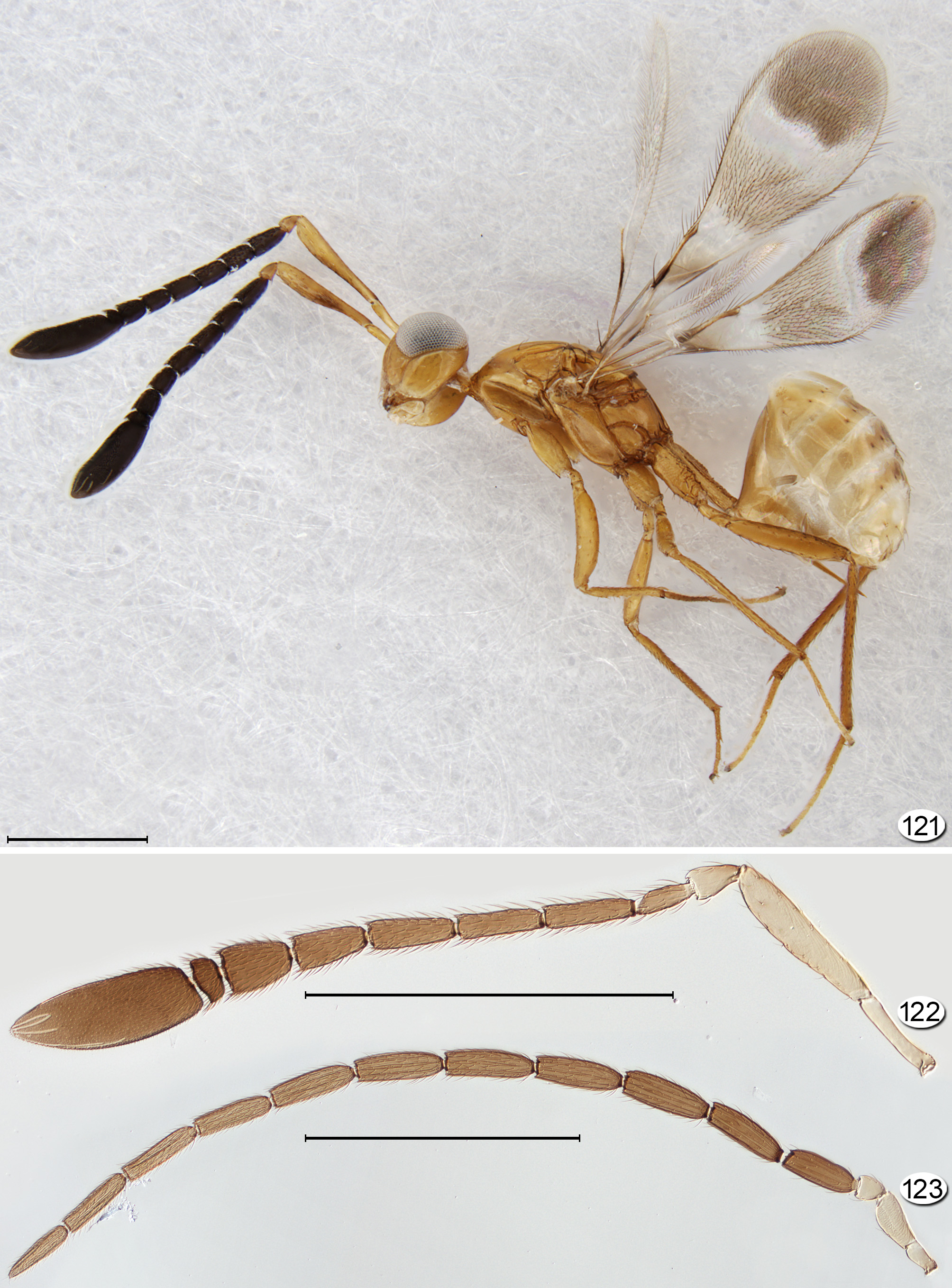
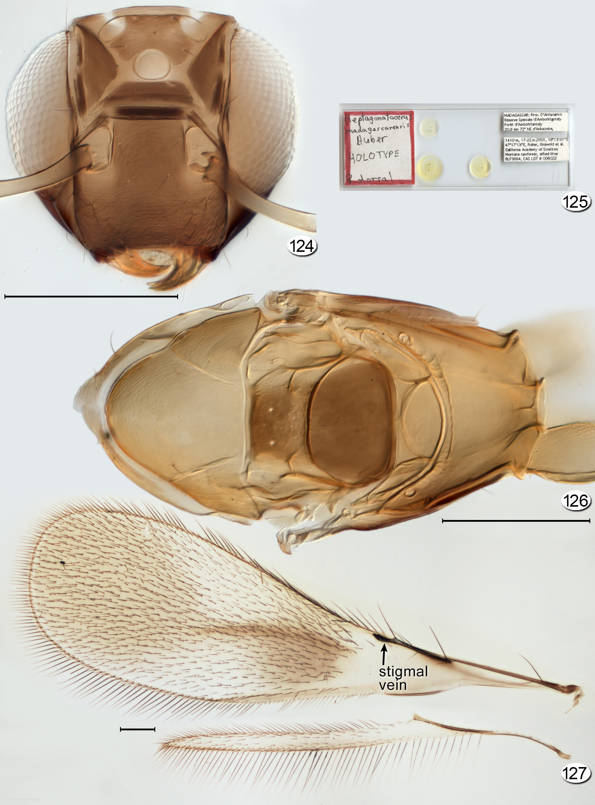
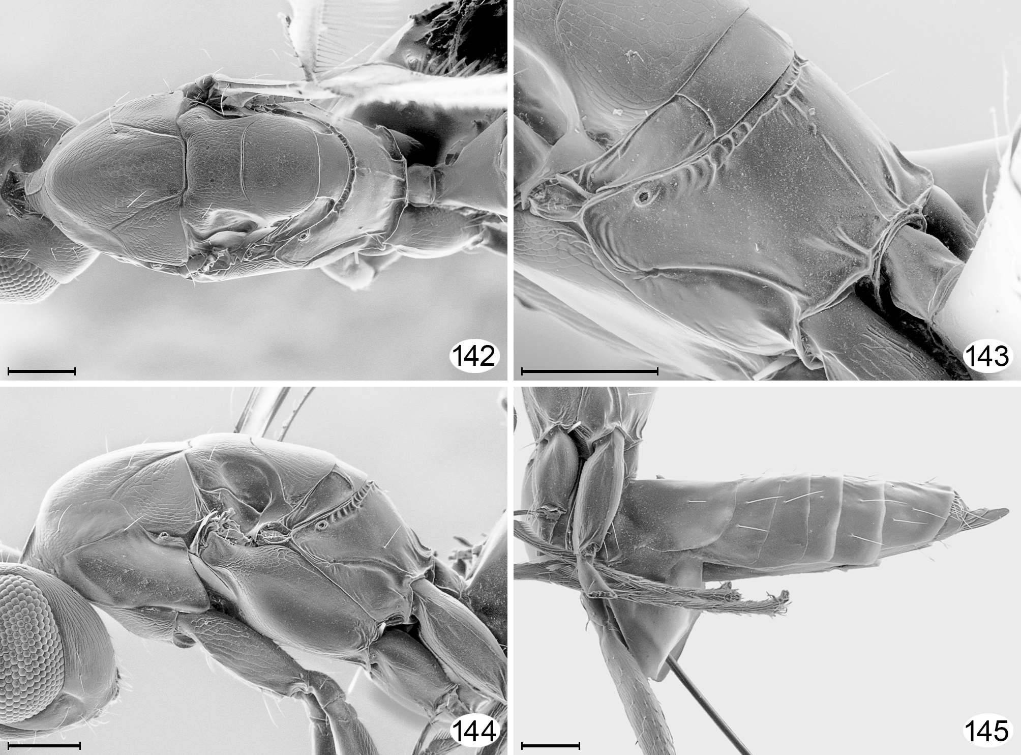
Diagnosis. Within Gonatocerini, the species of Heptagonatocerus are distinguished by the following combination of features: female antenna with 7-segmented funicle; fl7 wider than long and distinctly shorter than fl6 ( Figs 122 View FIGURES 121 – 123 , 131, 133 View FIGURES 131 – 135 , 140 View FIGURES 136 – 141 ); face without subantennal sulci ( Figs 124 View FIGURES 124 – 127 , 136 View FIGURES 136 – 141 ); propodeum with two short submedian carinae arising on side of petiole and usually also with a median longitudinal carina ( Figs 126 View FIGURES 124 – 127 , 142, 143 View FIGURES 142 – 145 ).
Description. FEMALE. Medium to large specimens, 768–2076 Μm in length. Colour. Body generally dark yellow to brown, usually with fore wing patterned with brown areas. Head. Head thick ( Fig. 137 View FIGURES 136 – 141 ), about 1.3–1.8× as wide as long and 1.1–1.3× as wide as high; in lateral view with evenly convex anterior surface ( Fig. 138 View FIGURES 136 – 141 ). Face about 0.8× as wide as high; subantennal sulci absent ( Figs 124 View FIGURES 124 – 127 , 136 View FIGURES 136 – 141 ); preorbital sulcus straight, appressed against eye to a little beyond lower level of torulus, then separating from eye and continuing to dorsolateral corner of mouth ( Figs 136, 138 View FIGURES 136 – 141 ). Toruli abutting transverse trabecula ( Fig. 124 View FIGURES 124 – 127 ). Eye in lateral view 0.86× as long as high to as long as high, well separated from back of head ( Fig. 138 View FIGURES 136 – 141 ). Malar space about 0.4–0.6× eye height; malar sulcus almost straight to distinctly curved and extending from ventral margin of eye to anterolateral corner of mouth ( Figs 121 View FIGURES 121 – 123 , 138 View FIGURES 136 – 141 ). Gena in lateral view narrow dorsally, wide ventrally and merging smoothly into occiput ( Figs 138, 139 View FIGURES 136 – 141 ). Vertex in lateral view oblique, forming right angle or obtuse angle ( Fig. 138 View FIGURES 136 – 141 ) with face (separated by transverse trabecula), posteriorly separated from occiput by transverse sulcus behind ocelli ( Fig. 137 View FIGURES 136 – 141 ). Ocelli with LOL about 0.5× POL and OOL about 0.55–0.75× POL, with 2 setae between lateral ocelli ( Figs 136, 137 View FIGURES 136 – 141 ). Occiput entire ( Fig. 139 View FIGURES 136 – 141 ). Labrum with 4 (possibly 5) setae. Mandible with 3 teeth. Antenna. Scape at least 6.0× as long as wide, with radicle distinct, narrow, about 0.36–0.47× scape length; pedicel about 0.19–0.26× scape length, about as short as but wider than fl1; funicle 7-segmented and fl7 wider than long and distinctly shorter than fl6 ( Figs 121, 122 View FIGURES 121 – 123 , 131, 133 View FIGURES 131 – 135 ), fl1 and fl7 without mps, fl2–fl8 each with 2 mps; clava about 0.37× funicle length, with 9 mps. Mesosoma. About 1.7–2.1× as long as wide, 1.8–1.9× as long as high, and 0.8–1.0× as wide as high. Pronotum in dorsal view short medially but visible, longitudinally divided medially, the lobes closely abutting (even slightly overlapping) ( Figs 126 View FIGURES 124 – 127 , 139 View FIGURES 136 – 141 , 142 View FIGURES 142 – 145 ), with dorsal area merging smoothly into lateral panel, the lateral panel more or less strongly concave dorsal to most of propleura and base of procoxa ( Fig. 121 View FIGURES 121 – 123 ). Pronotal spiracle small, about same size as propodeal spiracle ( Fig. 144 View FIGURES 142 – 145 ). Propleura normal. Prosternum rhomboidal, divided posteriorly by longitudinal sulcus extending at least half its length. Mesoscutum in dorsal view with fairly fine (clearly visible in micrographs), straight to anteriorly curved, diverging notauli ( Figs 126 View FIGURES 124 – 127 , 142, 144 View FIGURES 142 – 145 ). Transscutal articulation almost straight. Scutellum slightly longer than wide. Axilla normal. Prepectus fairly wide. Mesopleuron spindle-shaped and truncate at both ends, with wide, shallow femoral depression and apparently no line separating mesepimeron from mesepisternum ( Figs 121 View FIGURES 121 – 123 , 144 View FIGURES 142 – 145 ). Metanotum with dorsellum rectangular, its posterior margin distinctly convex ( Figs 126 View FIGURES 124 – 127 , 142, 143 View FIGURES 142 – 145 ). Metapleuron triangular, separated from propodeum by wide, curved sulcus becoming narrower anteriorly ( Figs 143, 144 View FIGURES 142 – 145 ). Propodeum in lateral view ( Figs 121 View FIGURES 121 – 123 , 143, 144 View FIGURES 142 – 145 ) sloping fairly strongly, in almost same plane as dorsellum; in dorsal view with three posterior carinae—a thin, short to long median carina, and a short, diverging submedian carina level with lateral margin of petiole about midway between median carina and junction with metapleuron—and anterior margin almost always with a distinct transverse sulcus abutting dorsellum (mostly between its lateral margins) usually containing several short longitudinal carinae ( Figs 126 View FIGURES 124 – 127 , 142–144 View FIGURES 142 – 145 ). Propodeal spiracle small, separated by less than its diameter from metanotum. Wings. Fore wing at most 3.9× as long as wide, and covered with microtrichia only to distinctly beyond apex of stigmal vein ( Figs 121 View FIGURES 121 – 123 , 127 View FIGURES 124 – 127 , 132, 135 View FIGURES 131 – 135 ). Venation about 0.4× wing length. Submarginal vein with the usual two basal setae (1 macrochaeta and 1 hypochaeta) and usually a hypochaeta apically, next to proximal macrochaeta of parastigma. Remaining venation (parastigma + stigma vein) distinctly shorter than submarginal vein, with 1 hypochaeta about midway between proximal and distal macrochaeta, and about 1 or 2 shorter setae between the macrochaetae. Stigmal vein with apex oblique. Hind wing normal. Venation about 0.4× wing length. Metasoma. Petiole wider than long ( Fig. 142 View FIGURES 142 – 145 ) to about 3.0× as long as wide ( Fig. 121 View FIGURES 121 – 123 ). Gaster about 1.1–5.0× as long as high. Terga without membrane visible between them. Ovipositor sheath as long as gaster, longer than metatibia and at most slightly exserted ( Fig. 145 View FIGURES 142 – 145 ), with 1 subapical seta.
MALE. Body length 845–1870. Colour. Body generally darker than in female.
Antenna. Scape 3.0–3.1× as long as wide, with radicle distinct, 0.14–0.34× as long as scape; pedicel small, 0.3–0.4× length of fl1; flagellomeres each with about 10 mps. Metasoma. Genitalia encapsulated in a sac-like phallobase (see genera other than Gonatocerus in Viggiani 1989) or at least with aedeagus not attached to apical sternum of gaster by apodemes ( Figs 129, 130 View FIGURES 128 – 130 ). [In this respect, Heptagonatocerus spp. differ from other genera of Gonatocerini.]
Etymology. From Greek “hepta” meaning seven, and “ Gonatocerus ” meaning elbowed antenna. The name refers to the 7-segmented funicle in the female antenna. The gender is masculine.
Distribution. Heptagonatocerus species occur in the Oriental and Afrotropical ( Madagascar, South Africa) regions.
Hosts and habitat. Hosts are unknown. Specimens have been collected mainly in forests.
Included species:
Heptagonatocerus madagascarensis Huber ; holotype ♀ in CAS. TL: Madagascar, Antanarivo, Ambohitantely
Forest Reserve.
Heptagonatocerus magnificus Huber ; holotype ♀ in QSBG. TL: Thailand, Chanthaburi, Khao Khitchakut Nat.
Park, entrance of youth camp.
Heptagonatocerus parvus Huber ; holotype ♀ in QSBG. TL: Thailand, Chaiyaphum Tat Tone Nat. Park, Phu Hang
Sing.
Heptagonatocerus pulchellus Huber ; holotype ♀ in QSBG. TL: “ Thailand, Nakhon Si Thammarat, Namtok Yong
Nat. Park, TV aerial, 966m.


No known copyright restrictions apply. See Agosti, D., Egloff, W., 2009. Taxonomic information exchange and copyright: the Plazi approach. BMC Research Notes 2009, 2:53 for further explanation.