
Heteromysis ( Olivemysis ) schoenbrunnensis, Wittmann & Abed-Navandi & Dubois & Chevaldonné, 2021
|
publication ID |
https://doi.org/10.11646/zootaxa.4980.3.3 |
|
publication LSID |
lsid:zoobank.org:pub:83C3E976-6AE1-4EA2-B7E8-301196255D80 |
|
DOI |
https://doi.org/10.5281/zenodo.5041498 |
|
persistent identifier |
https://treatment.plazi.org/id/C7AFB9EE-D0EC-4FF8-9D8B-511DCBBB3338 |
|
taxon LSID |
lsid:zoobank.org:act:C7AFB9EE-D0EC-4FF8-9D8B-511DCBBB3338 |
|
treatment provided by |
Plazi |
|
scientific name |
Heteromysis ( Olivemysis ) schoenbrunnensis |
| status |
sp. nov. |
Heteromysis ( Olivemysis) schoenbrunnensis sp. nov.
http://zoobank.org/ urn:lsid:zoobank.org:act:
Figs 8–13 View FIGURE 8 View FIGURE 9 View FIGURE 10 View FIGURE 11 View FIGURE 12 View FIGURE 13
Holotype. Adult male with 4.7 mm body length ( NHMW –27023), shrimp aquaria behind filtration sump of coral reef aquaria at Schönbrunn-Zoo, Vienna, Austria, May 2020, leg. Daniel Abed-Navandi.
Paratypes. 10 M ad. 3.2–5.3 mm, 1 F ad. 4.5 mm, 3 subad. ( USNM –1642206) in vials, same sample as for holotype; 2 F ad. 4.3–4.7 mm, 2 M ad. 3.9–4.7 mm ( MNINGA MYS 442 ) in vial, filtration sump of coral reef aquaria, remaining collection data as for holotype ; 3 F ad. 4.6–5.2 mm, 5 M ad. 3.8–5.1 mm, 1 F subad. ( USNM –1642205) in vials, plus 1 F ad. 4.9 mm ( NHMW –27022) and 1 M ad. 4.8 mm ( NHMW –27021) on slides, Feb. 2020, remaining collection data as before; 1 F ad. 6.0 mm ( NHMW –27024) in vial, collection data as for holotype ; 1 M ad. 4.9 mm ( NHMW –27025) in vial, collection data as for holotype ; 3 F ad. 5.5–6.0 mm, 3 M ad. 4.6–6.1 mm ( USNM –1642207) in vial, collection data as for holotype ; 3 F ad. 4.5–5.7 mm, 1 M ad. 4.5 mm ( NHMW –27026) in vial, Dec. 2020, remaining collection data as for holotype ; 3 F ad. 4.2–5.7 mm, 12 M ad. 3.2–5.6 mm, 1 F subad. ( USNM –1642208) in vial, filtration sump of coral reef aquaria, Dec. 2020, remaining collection data as for holotype .
Material sequenced. 18S and COI barcodes here provided for 1 M ad. from same sample as for holotype, deposited in GenBank under accession numbers MW591705 View Materials and MW596478 View Materials .
Type locality. Not defined because the locality of origin ought to be indicated according to Art. 76.1.1. of the nomenclatorial code ( ICZN, 1999). The species was first discovered from coral reef aquaria and service tanks at Schönbrunn-Zoo in Vienna ( Austria).
Derivatio nominis. The species name is a Latinized adjective with feminine location suffix, referring to the world’s oldest existing zoo, namely Schönbrunn-Zoo located in the historical imperial park of Schönbrunn.
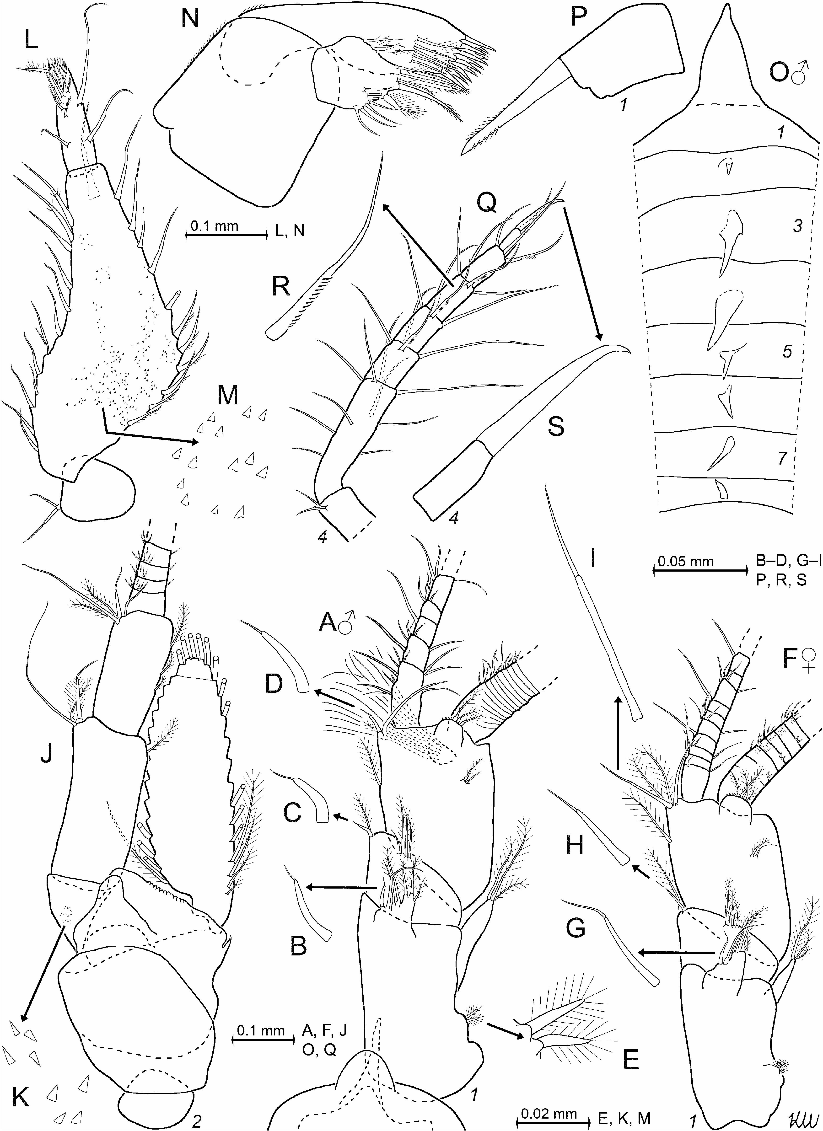
Diagnosis. Carapace normal, rostrum ( Fig. 9A, D View FIGURE 9 ) triangular with acute tip, 0.6–0.8 times length of terminal segment of antennular trunk (measured along dorsal median line). Antero-lateral edges of carapace well rounded. Eyes ( Fig. 9A–C View FIGURE 9 ) well developed, thick. Cornea occupies distal third of eye surface. Eyestalks with a prominent tooth on disto-mesial edge.
Antennulae ( Fig. 9A, G–K View FIGURE 9 ). The three segments of antennular trunk ( Fig. 9A, G View FIGURE 9 ) each with a setose dorsal apophysis. Apophysis of basal segment with blade-like spine (modified seta) apically bearing a minute spiniform process and 3–5 cilia ( Fig. 9J View FIGURE 9 ). Lateral lobe of basal segment, dorsal apophysis, and disto-mesial edge of the median segment, each with one whip seta ( Fig. 9I View FIGURE 9 ) plus barbed setae. Disto-mesial edge of terminal segment with blade-like, subapically flagellate, robust spine ( Fig. 9H View FIGURE 9 ). Appendix masculina small, weakly setose.
Antennae and mouthparts. Antennal scale ( Fig. 9L View FIGURE 9 ) reaches to 40–80% length of terminal segment of antennular trunk in both sexes. Scale length is 2.6–2.9 times maximum width. Scale with small apical segment. Mouthparts ( Figs 9M–N View FIGURE 9 , 10A–E View FIGURE 10 ) normal; labrum not produced into a spiniform process.
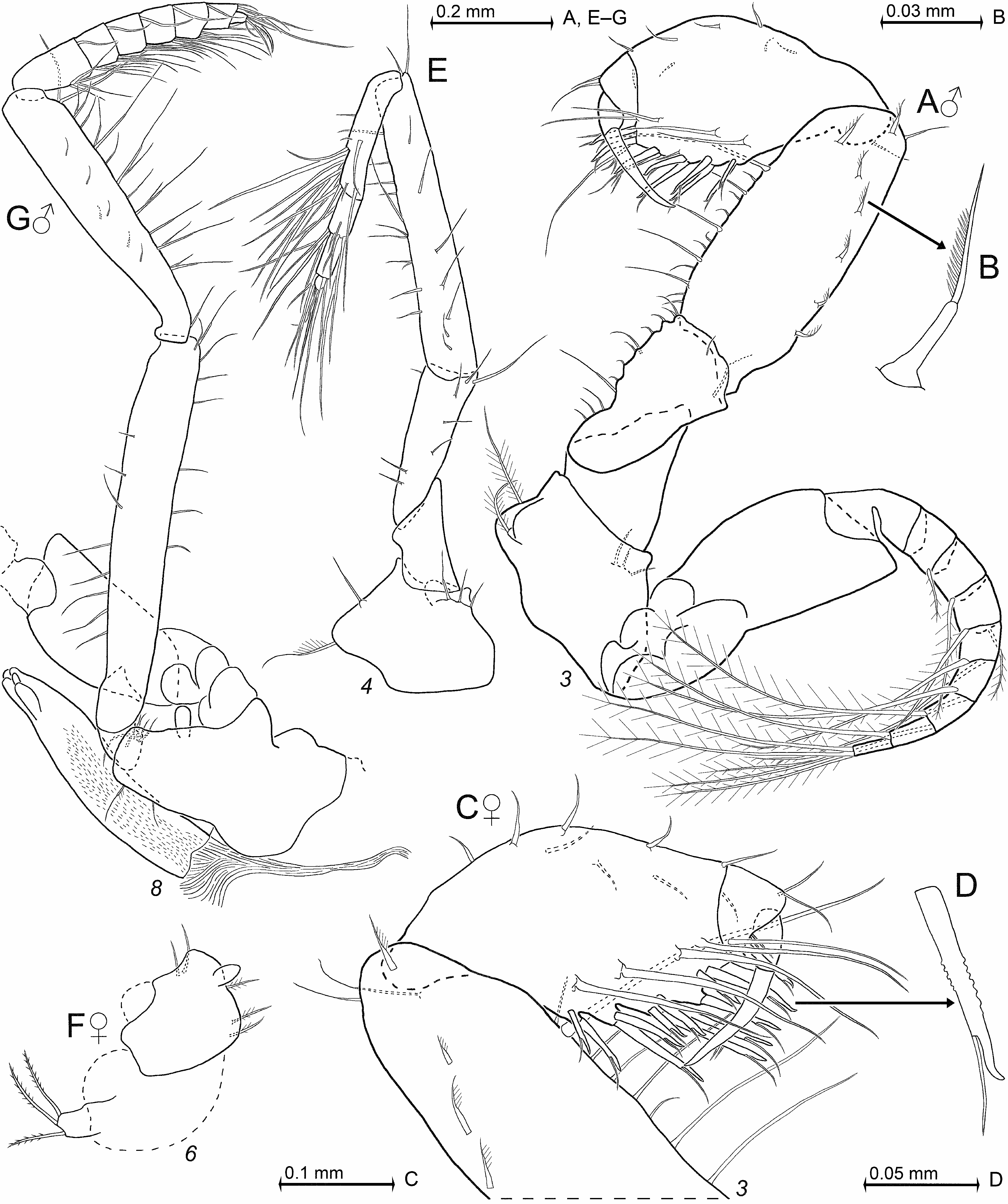
Thorax. Only the males with median processes from thoracic sternites 1–8, in part flanked by additional submedian processes ( Fig. 10F View FIGURE 10 ; anterior lobe of sternite 1 not counted as a process of that kind). Both sexes with flagellum of thoracic exopod 1 showing eight segments, exopods 2–8 with nine segments. Carpopropodus of thoracic endopods 1–8 with 2, 2, 2, 3–4, 6–7, 6–7, 6–7, and 6 segments, respectively. Claw 1 subapically, unilaterally ( Fig. 10H View FIGURE 10 ) or bilaterally ( Fig. 10G View FIGURE 10 ) serrated; claw 2 not detected (but potentially present in dense jungle of setae); claw 3 strong, smooth ( Fig. 10K View FIGURE 10 ); claw 4 needle-like, smooth ( Fig. 10L View FIGURE 10 ); claws 5–8 well-curved, slender, unilaterally serrated along medial to subapical portions ( Fig. 10M–P View FIGURE 10 ). Claw 3 (measured along its median line) 2.1–3.3 times dactylus length and 26–37% carpopropodus. Claws 1, 4–8 roughly half as long as claw 3 (note varying scales in Fig. 10 View FIGURE 10 ). Penes ( Fig. 11G View FIGURE 11 ) tube-like, 0.6–0.7 times length of ischium 8 and 0.8–1.0 times merus 8. Penes smooth all along, without setae, terminally blunt, ending in 4–5 lobes.
Gnathopods ( Fig. 11A–D View FIGURE 11 ). Shape of thoracic endopod 3 non-dimorphic except for the more rugose mesial margins of ischium and merus in males. Merus without disto-mesial ridge in both sexes. Carpus, propodus, and dactylus separated by distinct sutures. Carpopropodus 7–11% body length in both sexes. Carpopropodus strongly swollen, only 1.9–2.5 times longer than wide ( Fig. 11A, C View FIGURE 11 ). Carpus with 7–11 subapically flagellate spines along distal 50–70% of mesial margin. Propodus without paradactylary setae and without spines.
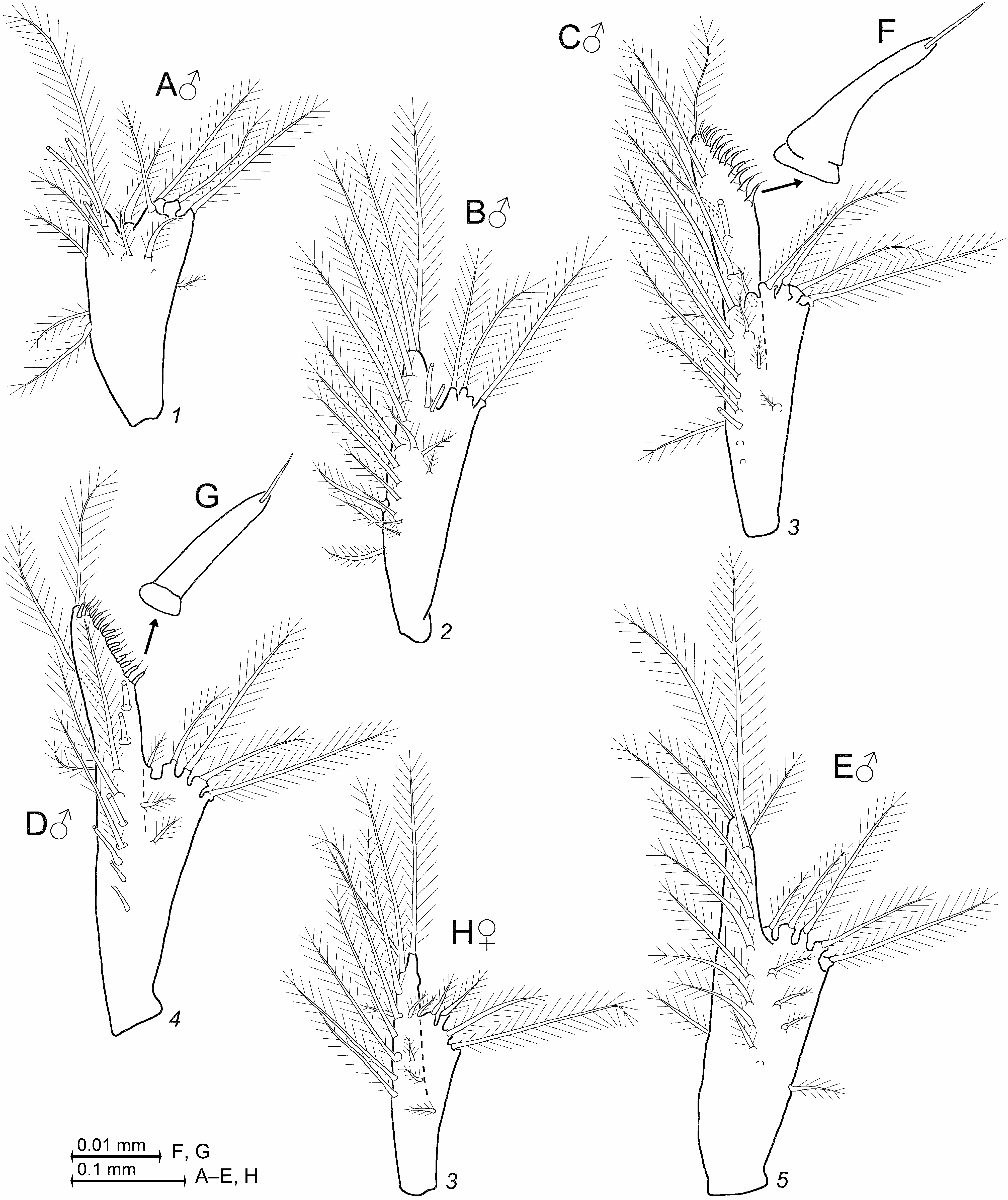
Pleopods ( Fig. 12 View FIGURE 12 ) rudimentary, unsegmented, with residual differentiation of endopod (pseudobranchial lobe). All female pleopods and male pleopods 1, 2, 5 without spines, with normal setae only. Male pleopod 3 ( Fig. 12C View FIGURE 12 ) with series of 8–11 small, flagellate spines along obliquely truncate, terminal margin; pleopod 4 ( Fig. 12D View FIGURE 12 ) with series of 13–16 even smaller flagellate spines in analogous position.
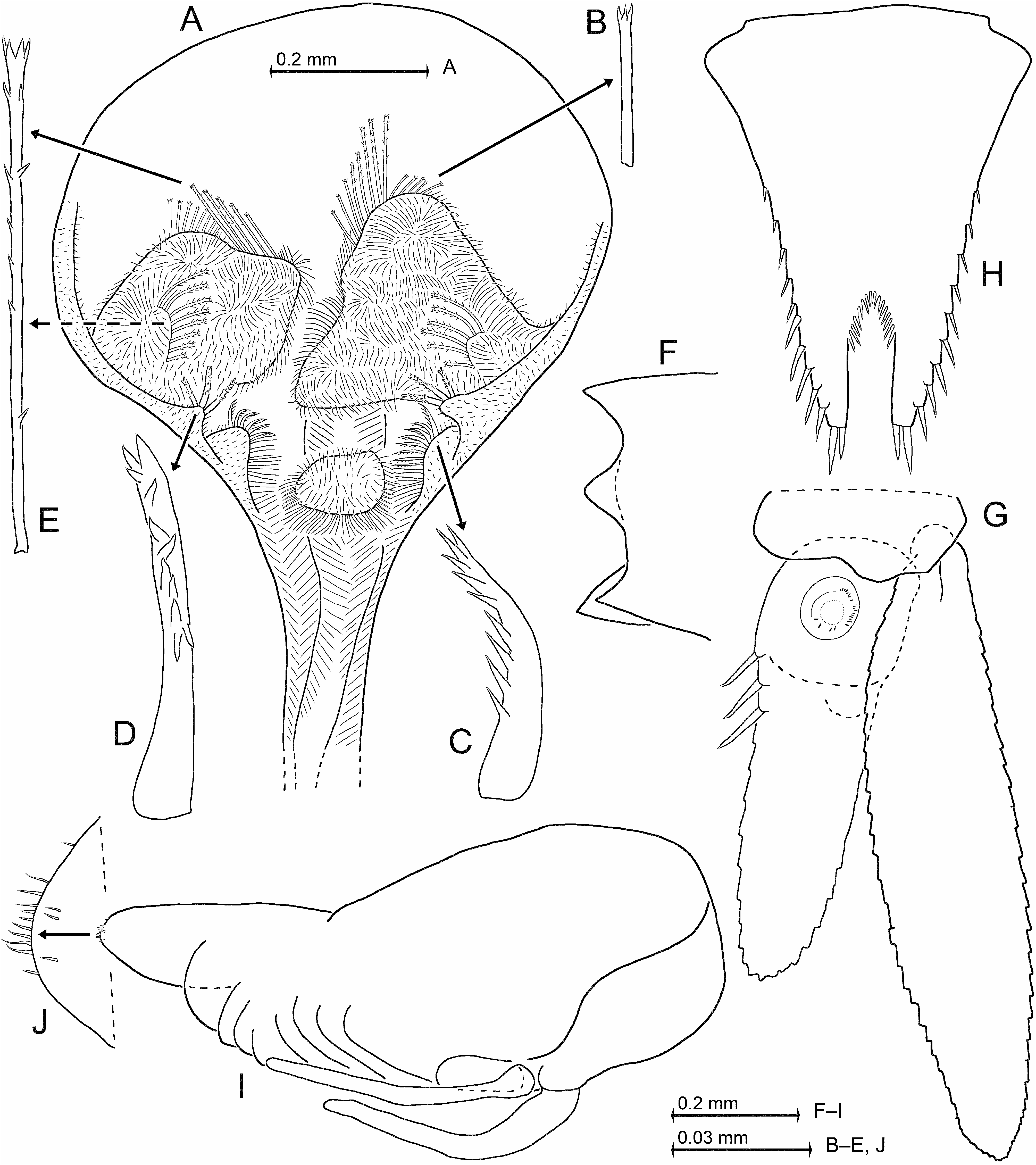
Tail fan. Uropods ( Fig. 13G View FIGURE 13 ) normal, entire; exopod extends by 18–27% its length beyond endopod; endopod with 3–4 spines near statocyst on mesial margin, distal spine-free portion is 2/3 length of endopod. Telson ( Fig. 13H View FIGURE 13 ) subtriangular, length 1.1–1.4 times its maximum width, 0.7–0.8 times exopod of uropod. Each lateral margin of telson with 9–10 spines on distal 50–70%, proximal portion smooth. Disto-lateral lobes each with two spines on narrowly truncate apex; latero-apical spines are 9–13% telson length; medio-apical spines are 0.6–0.8 times length of latero-apical spines. U-shaped apical cleft penetrates 30–33% telson length; proximal 25–40% of the cleft with 13–15 laminae, distally remaining portion with smooth parallel margins.
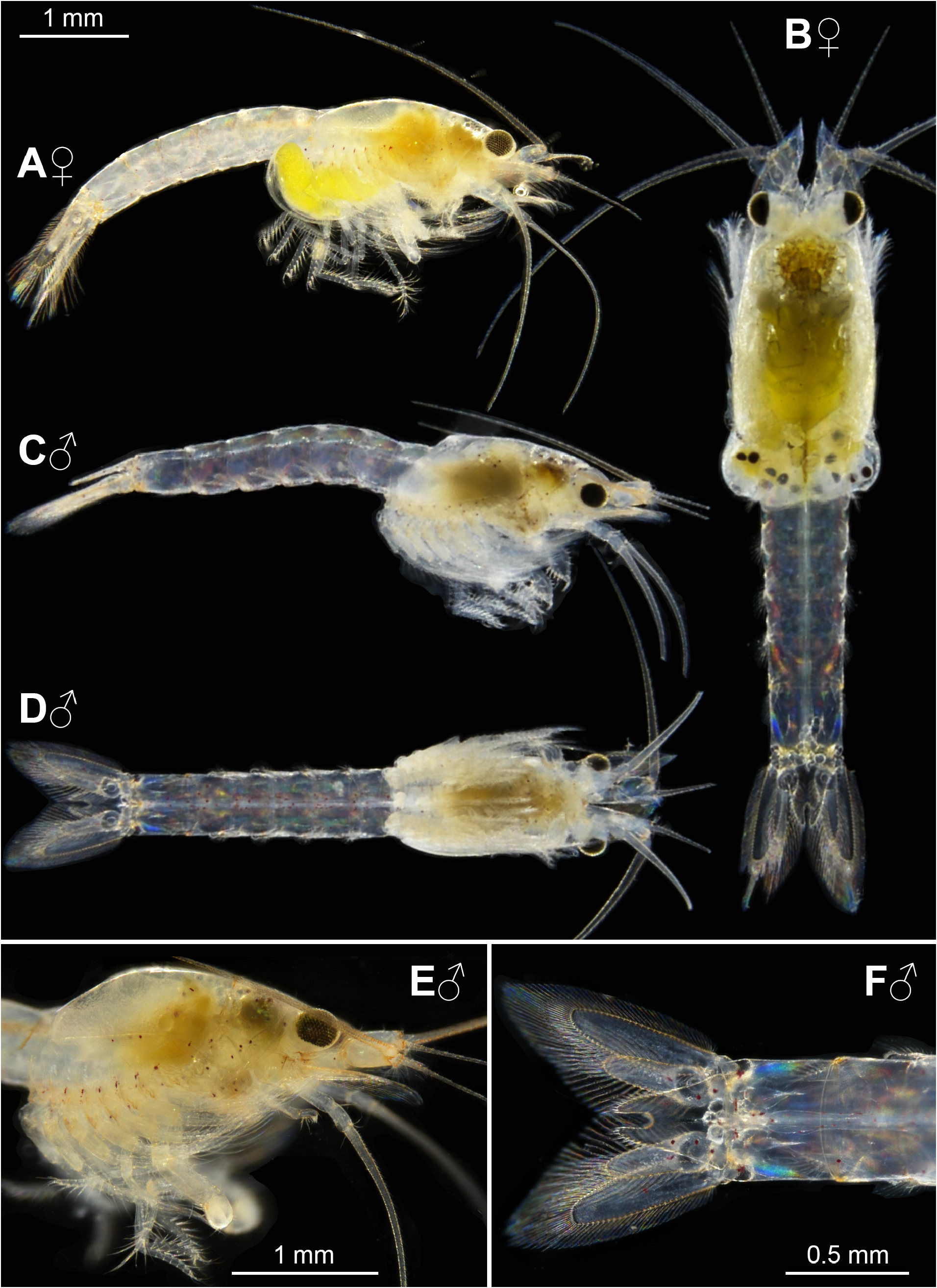
Description. All features of the diagnosis plus the above listed features shared by the three here described species. General appearance robust ( Fig. 8A–D View FIGURE 8 ). Size of adults is 4.2–6.0 mm (n = 17) in females and 3.2–6.1 mm (n = 36) in males. Cephalothorax comprises 30–42% body length, pleon 46–62%, carapace 27–38%, and rostrum 3–4%. Abdominal somites 1–5 measure 0.5–0.7, 0.5–0.8, 0.6–0.9, 0.7–0.9, and 0.7–0.9 times the length of somite 6, respectively.
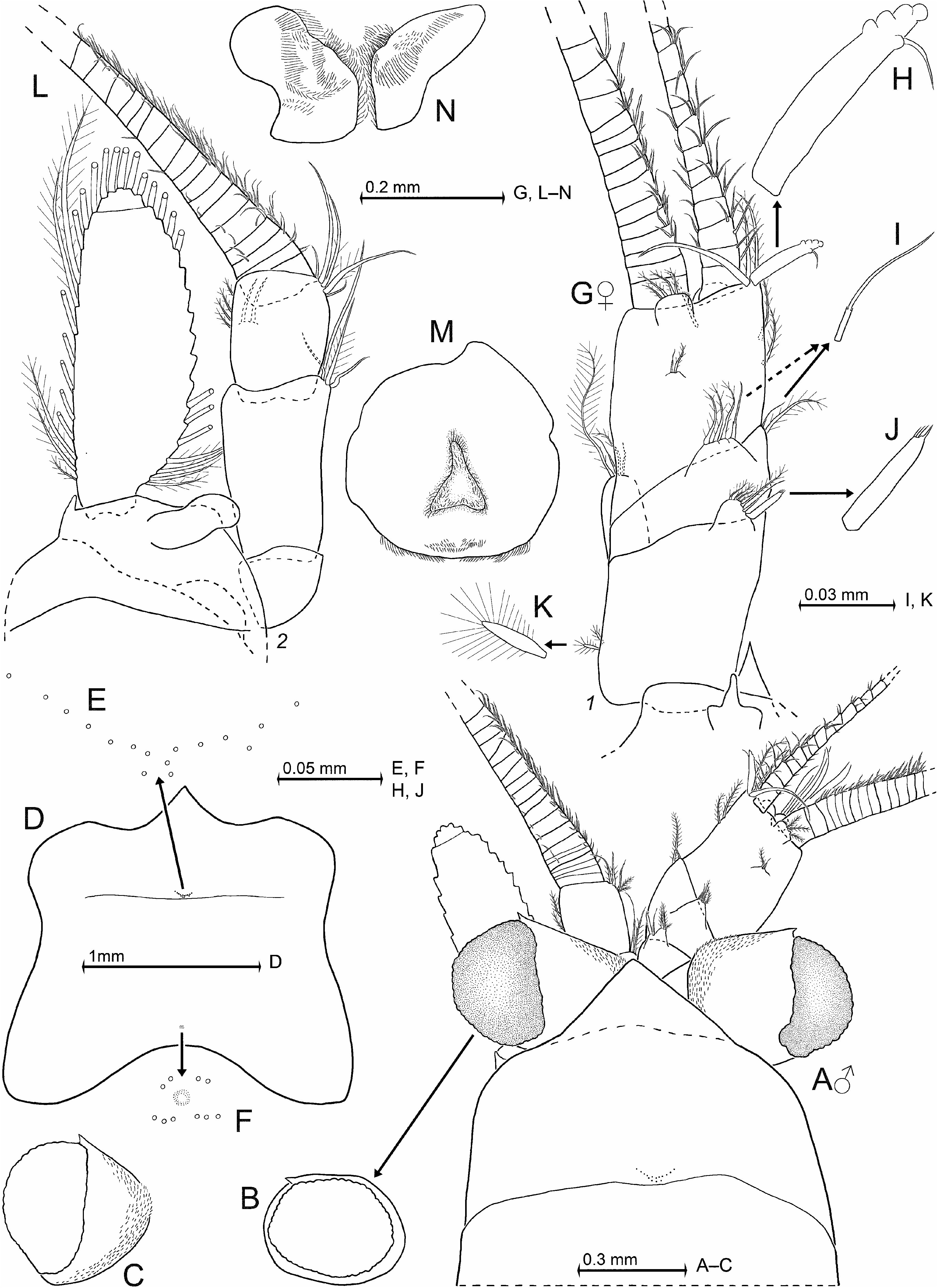
Carapace ( Fig. 9A, D–F View FIGURE 9 ). Rostrum covers basal portions of normal-oriented eyestalks. It reaches at most to the middle of artificially straight forward-oriented eyestalks (without cornea). Antero-lateral edges of carapace not visually projecting in situ whereas minimally projecting in artificially expanded carapace ( Fig. 9D View FIGURE 9 ). Posterior margin leaves 0.5–1.3 ultimate thoracic somite mid-dorsally exposed. As in many species of Mysidae , two characteristic groups of pores present medially on carapace. The anterior group ( Fig. 9E View FIGURE 9 ) is directly in front of the cervical sulcus and consists of 12–18 pores with about 1 µm diameter in a roughly V-shaped arrangement. The posterior pore group ( Fig. 9F View FIGURE 9 ) is more distant from the posterior margin of carapace; this group consisting of 10–12 such pores surrounding a larger but indistinct, rounded structure. Except for the here stated structures, outer surface of carapace smooth in both sexes.
Eyes ( Figs 8A–E View FIGURE 8 , 9A–C View FIGURE 9 ). Eyestalks and cornea dorsoventrally weakly compressed. Eyestalks with scales along mesial and proximal face. Cornea diameter 0.8–1.1 times the length of apical segment of antennular trunk. In dorsal view the cornea appears calotte-shaped, measuring 0.5–0.7 times eyestalk length (cornea not included; cornea appears enlarged in Fig. 9C View FIGURE 9 due to the pressure exerted by cover glass). In lateral view ( Fig. 9B View FIGURE 9 ), cornea oviform to oval with upper margin (= face) flattened.
Antennulae ( Fig. 9A, G–K View FIGURE 9 ). No sexual dimorphism except for the exclusive presence of the appendix masculina in males. Trunk extends 50–70% its length beyond eyes. Measured along dorsal midline, the basal segment is 42–47% trunk length, median 14–20%, and terminal 34–38%. Basal segment on basal half of its lateral face with 2–4 small setae bearing long barbs ( Fig. 9K View FIGURE 9 ). Its dorsal apophysis with 3–4 barbed setae, a whip seta, and a bladelike, apically modified spine (modified seta; Fig. 9J View FIGURE 9 ). Lateral lobe with one plumose, 2–3 shorter barbed setae, and a whip seta. Median segment with one plumose and one small, whip seta ( Fig. 9I View FIGURE 9 ) on disto-mesial edge. Dorsally it bears a large apophysis with 3–4 barbed plus a whip seta. Terminal segment 1.1–1.2 times longer than wide. Its mid-dorsal apophysis with 3–4 barbed setae, with small cilia lining the disto-mesial margin; no spiniform anterior projection. Disto-mesial edge of terminal segment with one flagellate, blade-like spine ( Fig. 9H View FIGURE 9 ) flanked by one large, smooth seta facing disto-laterally, plus two anteriorly directed plumose setae. Distal 20–30% of the flagellate spine with 3–4 tubercles on its anterior margin in addition to the distinct flagellum on posterior margin. In both sexes the lateral antennular flagellum is thicker than the mesial one by a factor of 1.4–1.8 when measured near basis of flagella. Male lobe weakly setose, inserts ventrally close to terminal margin of antennular trunk, broadly rounded, length is 17–21% width of terminal segment of trunk, its width 21–24%. Epi-antennular process (sub)-triangular with blunt tip, hypo-antennular process triangular with acute tip ( Fig. 9G View FIGURE 9 ).
Antennae ( Fig. 9A, L View FIGURE 9 ). Sympod ( Fig. 9L View FIGURE 9 ) dorsally with terminally rounded, tongue-like process. Lateral edge of sympod with short, anteriorly directed, subtriangular process; ventrally with crescent-shaped shield (dashed line in Fig. 9L View FIGURE 9 ) proximally behind antennal trunk; shield 3.0–3.5 times longer than its maximum width. Sympod caudally with bulbous lobe containing end sac of antennal gland. The three-segmented antennal peduncle with basal segment 16–22% peduncle length, second 42–48%, and third 31–35%. Antennal scale reaches clearly beyond antennal peduncle; scale with slightly convex, almost straight lateral margin and with strongly convex mesial margin; scale setose all around. Short, broad apical segment with 5–8% total scale length is separated by a transverse suture. Apical segment with five plumose setae.
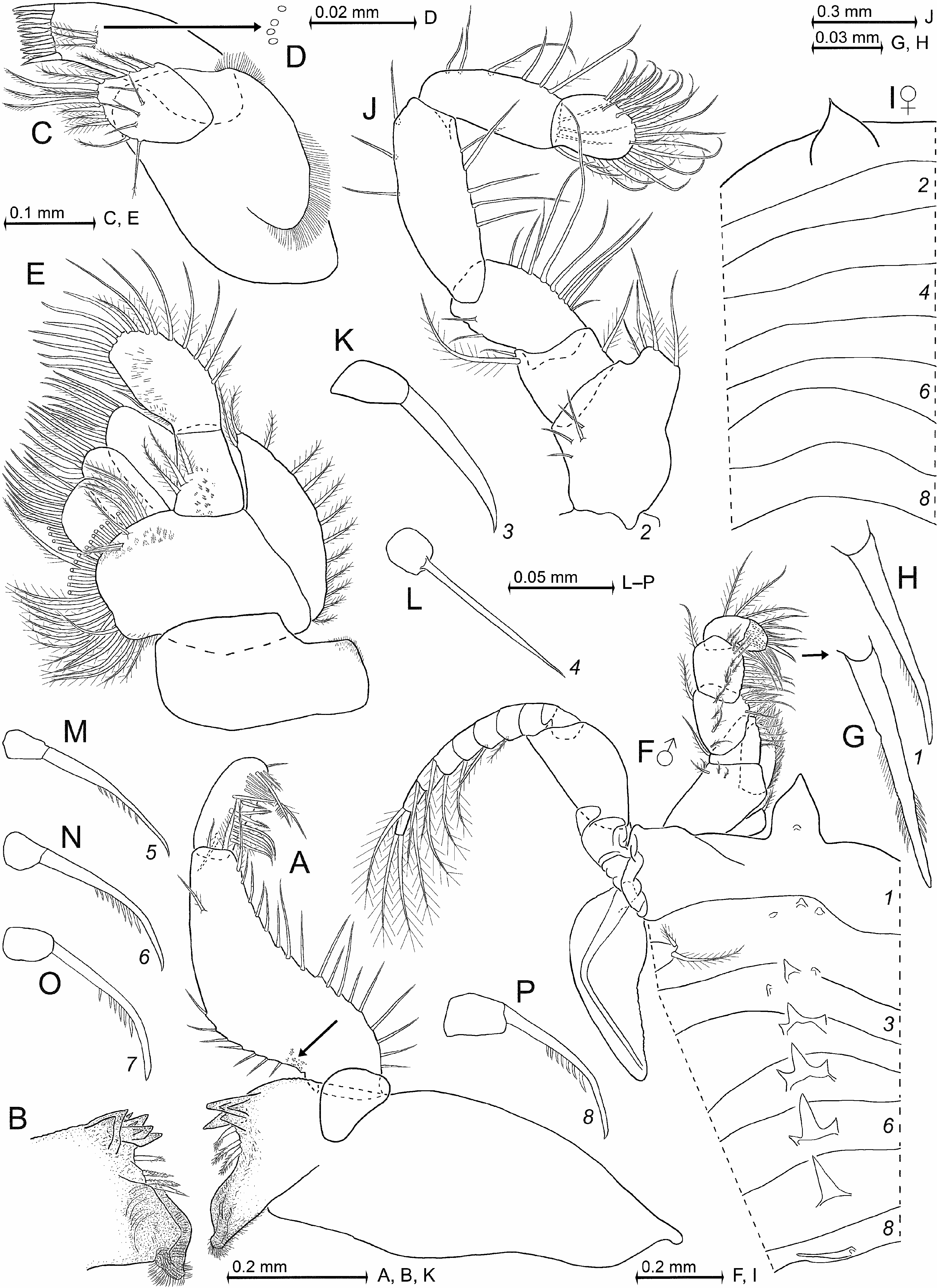
Mouthparts ( Figs 9M–N View FIGURE 9 , 10A–E View FIGURE 10 ). Mandibular palp three-segmented ( Fig. 10A View FIGURE 10 ). Its proximal segment without setae, 12–17% length of palp. Median segment 61–76% palp length. Its length 2.3–3.1 times maximum width. Its lateral margin all along with 15–17 whip setae. Mesial margin with one subapical seta, followed by a smooth median portion, and on proximal half by series of 6–8 setae increasing in length distally. Mid-caudal face of median segment with 0–2 setae (zero in Fig. 10A View FIGURE 10 ). Rostral face (below drawing plane in Fig. 10A View FIGURE 10 ) near mesio-basal edge with small field of minute triangular scales (arrow in Fig. 10A View FIGURE 10 ; size and shape of scales as in Fig. 2M View FIGURE 2 ). Terminal segment strongly setose, 19–25% palp length. Pars molaris with well-developed grinding surface in both mandibles. Pars incisiva with 4–5 teeth, digitus mobilis with 3–4 teeth, and pars centralis with 3–4 spiny teeth.
Maxillula ( Fig. 10C–D View FIGURE 10 ). Distal segment of maxillula ( Fig. 10C View FIGURE 10 ) terminally with 11–14 strong, mostly smooth spines, only the inner-most spine with two subapical denticles; this segment subterminally with three setae barbed on their distal 2/3; transverse series of three pores (diameter ca. 1.2 μm, Fig. 10D View FIGURE 10 ) closely adjoining the outer (= most ventral) seta. Endite of maxillula terminally with three distally spiny setae accompanied by three proximally thick barbed setae; mesial and lateral margins of the endite with numerous less thick setae.
Maxilla ( Fig. 10E View FIGURE 10 ) with 13–16 barbed setae all along lateral margin of exopod, the two apical setae larger than the remaining ones. Most basal portion of exopod with cilia (minute smooth setae) beneath and between the plumose setae. Basal segment of endopod with three basally thick, barbed setae. Terminal segment 1.6–1.9 times longer than wide.
Thoracic sternites ( Fig. 10F, I View FIGURE 10 ). Male sternites 2–7 with tooth-like median processes increasing in size caudally. Sternites 2–6 with additional, smaller, paired, submedian processes, fully tooth-like only at sternites 4–6 where they merge with the median process to form a strong trident. Sternite 8 with a single, elongate spine-like process in median position. Sternite 1 with large anterior lobe whose distal half is triangular with blunt apex in both sexes. Only in males this lobe bears a small, rounded ventral process, probably homologous with the median processes on sternites 2–8.
Thoracopods general ( Figs 10F–H, J–P View FIGURE 10 ; 11 View FIGURE 11 ). Length of flagella as well as of basal plates increases from exopod 1 to 6, and decreases from exopod 6 to 8. Basal plates ( Figs 10F View FIGURE 10 ; 11A, G View FIGURE 11 ) weakly expanded, length 1.5–1.8 times width in both sexes. Lateral margin of the plates ends in a narrowly rounded edge. The first thoracopods with large, leaf-like, smooth epipod ( Fig. 10F View FIGURE 10 ). Length of endopods increases in series of thoracopods 1, 2, 4, 3, 5, and remains subequal among endopods 5–8. Basis of endopods 4–8 ( Fig. 11E–G View FIGURE 11 ) with a small, lappet-like apophysis on rostral face below endopod; no such apophysis in endopods 1–3 ( Figs 10F, J View FIGURE 10 ; 11A View FIGURE 11 ). Ischium becomes increasingly slender from endopods 1 to 5, and length of ischium increases in series of endopods 1, 2, 3≈4, 5; both these measurements remain (sub)-equal among endopods 5–8. Ischium shorter than merus in endopods 1–4 ( Figs 10F, J View FIGURE 10 ; 11A, E View FIGURE 11 ), subequal in endopod 5, but longer than merus in endopods 6–8 ( Fig. 11G View FIGURE 11 ). Thoracic endopods 1–3 each with dactylus ( Figs 10F, J View FIGURE 10 ; 11A, C View FIGURE 11 ) larger than that of endopods 4–8 ( Fig. 11E, G View FIGURE 11 ). Combined praeischium plus ischium of endopod 2 ( Fig. 10J View FIGURE 10 ) are 0.7–0.9 times merus length, carpopropodus plus dactylus 1.0–1.3 times merus. Dactylus very large, with dense brush formed by great numbers of normal setae and 10–14 modified setae, the latter apically bent, bearing two symmetrical series of denticles (stiff barbs) on either side in subbasal to median portions. Carpopropodus of endopod 4 ( Fig. 11E View FIGURE 11 ) densely furnished with smooth simple setae and whip setae, no other types of setae. When stretched anteriorly, endopod 8 ( Fig. 11G View FIGURE 11 ) reaches to the maxilla or up to basis of antenna; when stretched posteriorly to basis or end of pleonite 5.
Gnathopods ( Fig. 11A–D View FIGURE 11 ). Thoracic endopod 3 forms a powerful subchela. Basis with distinct but much shorter endite compared to that ( Fig. 10J View FIGURE 10 ) of endopod 2. Ischium and merus strong, as normal in gnathopods. Ischium 1.7−2.0 times as long as wide; merus 2.4−2.7 times as long as wide and 1.5−1.8 times length of ischium. Series of 5−6 unilaterally barbed whip setae ( Fig. 11B View FIGURE 11 ) along (near) lateral face of merus, plus a single whip seta in subterminal position; series of normal barbs (cilia) along median to subapical portions of the setae; no modified barbs. Distal half of ischium with 4–6 short whip setae on mesial margin, each whip seta on the tip of a short triangular projection ( Fig. 11A View FIGURE 11 ) in males, projections even shorter in females. Proximal 50–60% of merus with 4–7 short whip setae on mesial margin, the whip setae alternating with longer smooth setae; no triangular projections. Carpopropodus length 0.9–1.1 times merus, 1.1–1.5 times ischium. Carpus ( Fig. 11A, C View FIGURE 11 ) with spines arranged in series of 3–5 pairs in proximal position and 1–3 stand-alone spines in more distal position. Within pairs, the rostral spine larger than the caudal one. Most of these spines irregularly ‘serrated’ (rugose; Fig. 11D View FIGURE 11 ) along their anterior and posterior margins. The most proximal spines may be weakly serrated or not so.
Marsupium ( Fig. 11F View FIGURE 11 ). The large oostegites 1, 2 near basis with 3–6 setae which are microserrated along distal half. Ventral and rostral portions of the outer face of only the second oostegite with total of 12–15 small whip setae whose length increases towards the tip of this oostegite. Thoracopod 6 with rudimentary oostegite represented by a small, rounded lobe with three microserrated setae apically ( Fig. 11F View FIGURE 11 ).
Pleopods ( Fig. 12 View FIGURE 12 ). Reduced to small setose, bilobate, or obscurely bilobate plates in both sexes, generally larger in males than in females. Length without setae or spines increases from first to fifth pleopods in females. This series discontinuous in males, with size increasing in series of pleopods 1, 2, 5, 3, 4. For potential presence and numbers of flagellate spines on pleopods, see ‘Diagnosis’ above. Pleopods 3, 4 knife-shaped in males only; each with a single seta at apex ( Fig. 12C–D View FIGURE 12 ); their spines with a subapical flagellum ( Fig. 12F–G View FIGURE 12 ). Compared with pleopod 4 ( Fig. 12G View FIGURE 12 ), the spines of pleopod 3 are larger ( Fig. 12F View FIGURE 12 ), more subtriangular, and on the average less crowded. All setae are plumose or barbed in both sexes (not counting flagellate spines).
Tail fan ( Figs 8F View FIGURE 8 , 13F–H View FIGURE 13 ). Scutellum paracaudale ( Fig. 13F View FIGURE 13 ) sinusoid, well rounded. Exopod of uropods ( Fig. 13G View FIGURE 13 ) with slightly convex lateral margin and clearly convex mesial margin. Its length 1.2–1.4 times endopod. Exopod extends 38–44% its length beyond telson, endopod 21–42% its length beyond telson (partly due to telson inserting more rostrally). Endopod basally with large statocyst, containing normally only one statolith with diameter of 71–92 µm (n = 8 statoliths from five specimens). Statoliths discoidal with shallow fundus and distinct tegmen. Statolith formula 2 + 3 + (5–9) + (4–6) = 16–19 (n = 7). One additional statocyst examined contained a small ( 61 µm) statolith penetrated by sensory hairs as usual, and a spherical mineral concretion with diameter 32 µm not containing any trace of sensory hairs; both objects mineralized with fluorite. Mysid statocysts with such aberrations are rarely found in nature ( Wittmann & Ariani 2019). Telson ( Fig. 13H View FIGURE 13 ) length 1.2–1.3 times sixth pleonite and 0.8–1.0 times endopod of uropod. Spines on lateral margins almost continuously increasing in size distally. Apical cleft much deeper than wide. Distal portions of cleft smooth; proximal portions lined by acute laminae which are shorter than the spines on distal third of lateral margins of telson.
Color ( Fig. 8 View FIGURE 8 ). The animals appeared reddish to transparent upon aquarium observation. Non-transparent specimens became more light-colored during transport to the lab for microphotography. Eggs and nauplioid larvae yellow, in part with a slight green tinge. Content of foregut, gut, and gonads visible through the (semi)-transparent body. Iridescence of parts of body and appendages visible in Fig. 8 View FIGURE 8 varied with the direction of incident light.
Larvae ( Figs 8B View FIGURE 8 , 13I–J View FIGURE 13 ). In the ethanol-fixed material, 15 out of 17 adult females examined had 2– 9 eggs or larvae in the brood pouch; two lacked a brood. Totals of nine eggs, 31 nauplioid larvae, and 28 postnauplioid larvae were available for measurements. Two females with 4.2–5.7 mm body length carried 2– 7 eggs with diameter 0.38–0.43 mm. Two females 4.7–4.8 mm carried 3–4 nauplioid larvae at substage N1 with body length 0.76–0.89 mm, four females 4.5–5.7 mm had 2–6 N2 with 0.83–1.04 mm, one female 6.0 mm had nine N3 with 0.89–1.07 mm. Two females with 4.9–6.0 mm carried 4–8 postnauplioid larvae at substage P2 with 1.13–1.52 mm, four females with 4.5-5.7 mm had 2–6 P3 with 1.40–1.80 mm. A crude estimate suggests that the larvae attain 1/3 parent length shortly before the moult that leads to the free-living juvenile stage. Six mounted nauplioid larvae ( Fig. 13I View FIGURE 13 ) with smooth cuticle all around except for tip of abdomen, which bears numerous spine-like setae ( Fig. 13J View FIGURE 13 ). No caudal furca. Remaining features in Fig. 13I View FIGURE 13 are typical of the state of development.
Foregut ( Fig. 13A–E View FIGURE 13 ). Lateralia anteriorly with dense series of slender, apically coronate spines arranged in a lateral group of short spines with smooth shaft ( Fig. 13B View FIGURE 13 ) and a mesial group of long spines armed with loose series of small denticles along the shaft ( Fig. 13E View FIGURE 13 ). The latter type of spines also on median lobes of the lateralia. Lateralia more caudally with separate group of 6–9 apically pronged, serrated spines ( Fig. 13C View FIGURE 13 ). Dorsolateral infoldings with three longer, apically pronged spines which are serrated ( Fig. 13D View FIGURE 13 ) along distal 50–60% of the shaft.
Gut contents of four specimens were mainly crustacean remains (copepods, Artemia ), minor amounts of unidentifiable material and mineral particles.
Aquarium observations. The mysids hovered close to surfaces in dark areas of the aquarium and the connected filtration sumps: they cruised mainly above the bottoms and walls with the ventral side of the body facing the substrate. The movement patterns were parallel to the respective surface, and of a stop-and-go type. The mysids swam to and fro, mostly along predominantly linear, constant courses. Loose aggregations were formed by less than five specimens; in these aggregations a size-specific segregation was apparent. Occasionally, larger animals showed a chasing-like behavior, during which the speed was quickly accelerated and a short direct encounter occurred.
No known copyright restrictions apply. See Agosti, D., Egloff, W., 2009. Taxonomic information exchange and copyright: the Plazi approach. BMC Research Notes 2009, 2:53 for further explanation.
|
Kingdom |
|
|
Phylum |
|
|
Class |
|
|
Order |
|
|
Family |
|
|
SubFamily |
Heteromysinae |
|
Tribe |
Heteromysini |
|
Genus |
|
|
SubGenus |
Olivemysis |