
Microstonyx major ( Gervais, 1848 )
|
publication ID |
https://doi.org/10.5281/zenodo.4665351 |
|
persistent identifier |
https://treatment.plazi.org/id/03F587AF-FF9B-FFC2-8A89-F9E4FBE4FADA |
|
treatment provided by |
Felipe |
|
scientific name |
Microstonyx major ( Gervais, 1848 ) |
| status |
|
Microstonyx major ( Gervais, 1848) ( Figs 1-5 View FIG View FIG View FIG View FIG View FIG ; Appendix: Tables 1-6)
Sus major Gervais, 1848 : pl. XII, fig. 2.
Microstonyx major – Kazancı et al. 1999: 507. MATERIAL EXAMINED. — Skull: AK3-131, skull with all cheek teeth; AK5-501, broken skull with partial face and all cheek teeth.
Maxilla: AK7-153, maxilla with all cheek teeth; AK11- 66, right maxilla with P2-M3; AK5-443, juvenile right maxilla with DP3-DP4 and M1; AK5-623, left maxilla with left P3-M3; AK7-100, right maxilla with DP4 and M1.
Mandible: AK2-112, left mandible with p2-m3; AK11-72, mandible with both rami and complete dentition; AK11-67, juvenile mandible with both rami, which bears i1 (right), i2 (left) and both side dp3, dp4, m1; AK3-126, left mandible with p3-m3; AK4-187, mandible with both rami, with both side p2-m3, incisor and canine are missing; AK4-251, left mandible with p3-m3; AK5-270, juvenile left mandible with dp2-dp4, m1-m2; AK5-442, left mandible with p2-m3; AKB-51, right mandible with p3-m2; AKK-120, female(x) mandible with both rami and all incisors (di2), canine, and p2-p4 (half left p4); AKK-121, left mandible with m1-m3.
Isolated teeth: AK12-5, right I1; AK2-488, left i3; AK4-186, right M3; AK5-624, left I1; AK6-85, a right broken m3; AK7-154, left m2; AK7-183, right m3; AKA-1, right M3; AKK-192, germ fragment of left M2; AKK-286, left m1; AKK-287, left m2; AKK-288, right m2; AKK-83, right I2.
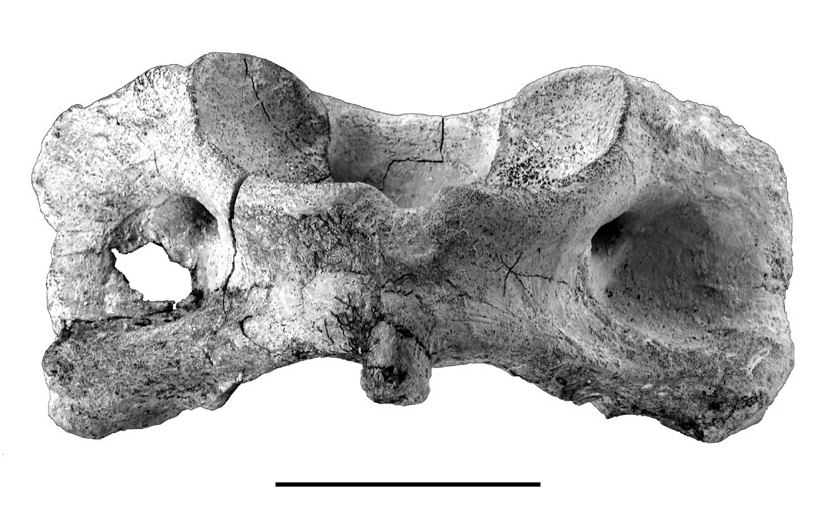
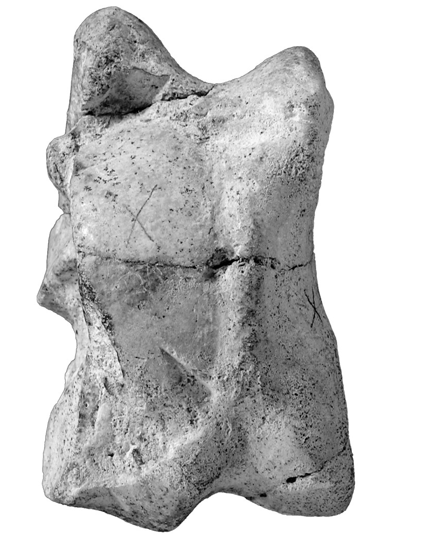
Postcranial: AK3, AK5-236, AK5-258, AK5-346, AK6-91, AK5-628, AK5b-838, atlases; AK6-96, vertebra centrum; AK4-109, left humerus; AK7-184, left distal humerus; AK11-52, right radius; AK3-302, left radius and ulna; AK5-188, left distal radius; AK5-570, AK6-258, left proximal radii; AK5-625, right proximal metacarpal III; AKB-54, left metacarpal III; AKK- 82, right metacarpal III; AK5-48, right metacarpal IV; AK2-489, left tibia; AK4-88, left astragalus; AK5-149, right metatarsal IV; AK5-199, metapodial.
AGE. — Late Miocene, radiometric age 7.1 ± 0.15 Ma ( Karadenizli et al. 2005).
LOCALITY. — Akkaşdağı, Çankırı-Çorum Basin, Turkey.
DESCRIPTION
According to the mandibular data, seven adult, one young adult and two young individuals compose the local population, which includes one mature male, and at least two mature females.
Skull
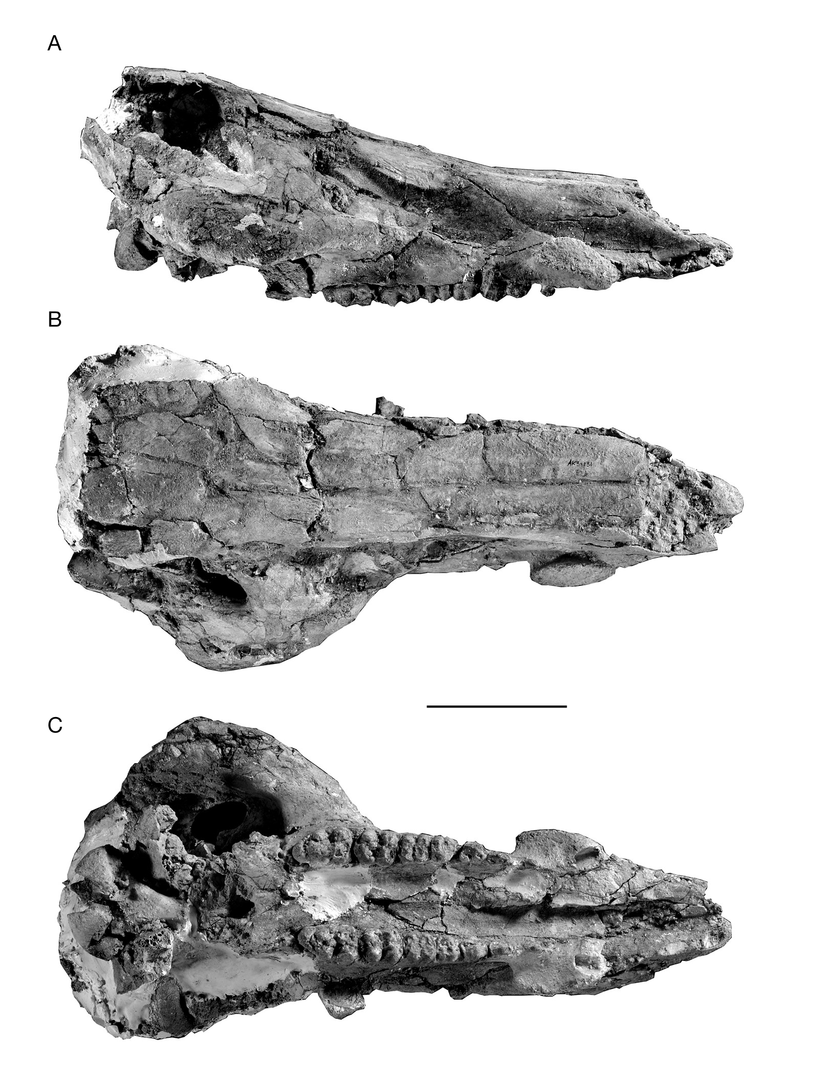
There are two skulls in the Akkaşdagwı suid collection, AK3-131 and AK5-501. The latter is only a middle part of a skull with the face partially preserved.Apart from showing clearly that the occiput is very high, it adds little to what is seen in the well preserved skull AK3-131 ( Fig. 1 View FIG ). The latter skull is almost complete but dorso-ventrally compressed, especially in the occipital region ( Fig. 1A View FIG ). Judging from the completely erupted M3 it belongs to an adult individual. In dorsal view ( Fig. 1B View FIG ), the caudal part of the skull is broken at the posterior part of the parietal, and much of the braincase is not preserved. The left zygomatic arch is missing, while the right side is well preserved. The nasals are long, of nearly constant width almost to the tip. In ventral view ( Fig. 1C View FIG ), the specimen is almost complete from the apices of the premaxillae to the occipital condyles, but the pterygoid process is heavily deformed by compression and difficult to investigate. The right zygomatic arch is robust and extends strongly towards lateral. There are no facial crests, and the anterior rim of the zygomatic arch originates at the anterior end of M3. The orbit is small and far behind M3. The rim of the orbit is incomplete but there is a distinctive, deep lachrymal notch (infraorbital fossa). The occipital condyles and sphenoid surfaces are of typical suid form. Although broken, the jugular processes seem robust and the tympanic bullae are oriented downwards, both suggesting a modern suid form. The choanae open posteriorly, far behind M3. The cheek dentition (P1-M3) is well preserved, but all the canines and incisors are missing. The small and shallow canine alveolus suggests that the canine was small. The alveolar crest is elongated and relatively slender.
Mandible
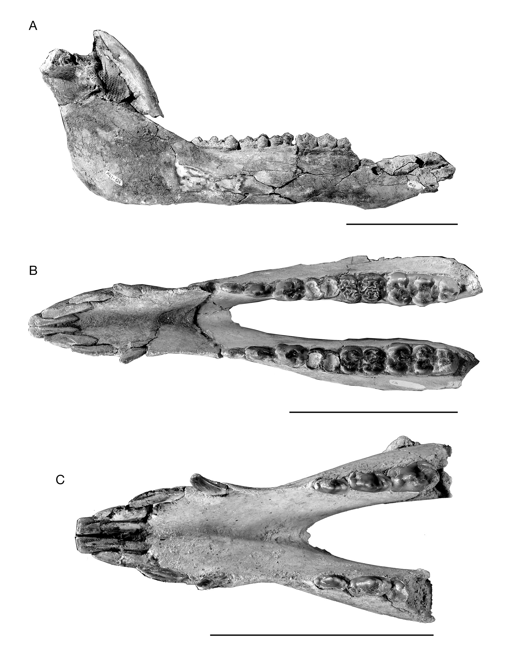
The mandible is the most common element in the Akkaşdagw ı suid collection. The best preserved specimen is labelled as AK3-1 2 6 ( Fig. 2A View FIG ), and is certainly associated with the skull AK3-131. The mandible is almost complete except for minor damage, but the canine is unfortunately missing. Two mandibles preserve canines, AK11-72 ( Fig. 2B View FIG ) and AKK- 120 ( Fig. 2C View FIG ). The description of the mandibular morphology of the Akkaşdagwı suid is mainly based on specimens AK3-126, AK11-72, and AKK-120.
The horizontal ramus is shallow and slim, while the ascending ramus is high, about three times the height of the horizontal ramus. The ascending ramus rises gently upwards and backwards, with a mandibular angle of about 120°.The ascent begins well behind m3, so that this tooth is completely visible in lateral view. The glenoid condyle of the mandible is broken at the surface, but what remains clearly shows its robustness. The pointed coronoid process is a little higher than the glenoid condyle, and the mandibular notch is quite shallow. The symphysis is elongated, ending before p2. There seem to be two kinds of canine, but the distinction is not very sharp. The canine in mandible AKK- 120 is short and narrow, almost symmetrical, with a very narrow posterior facet separated from the lateral facets by two crests. Since the tooth is narrow, the boundary of the lateral facets forms a sharp anterior crest. Enamel covers the whole tooth and ends above the alveolus. It is virtually identical with the canine that we described from Hezheng as a female individual ( Liu et al. 2004). The canine in mandible AK11-72 is slightly more robust, with an oval transverse section and no obvious crests separating the posterior facet from the lateral ones.
Dentition
The dental morphology of Microstonyx is greatly variable, and has been shown extensively in previous publications (e.g., Van der Made et al. 1992; Kostopoulos et al. 2001; Liu et al. 2004, and literature listed herein), and the Akkaşdagwı suid fits well within the known range. Notable characteristics of the Akkaşdagwı population include a somewhat complicated M3/m3 occlusal pattern, and the main lingual cusp of p4 being placed as far forward as the labial one.
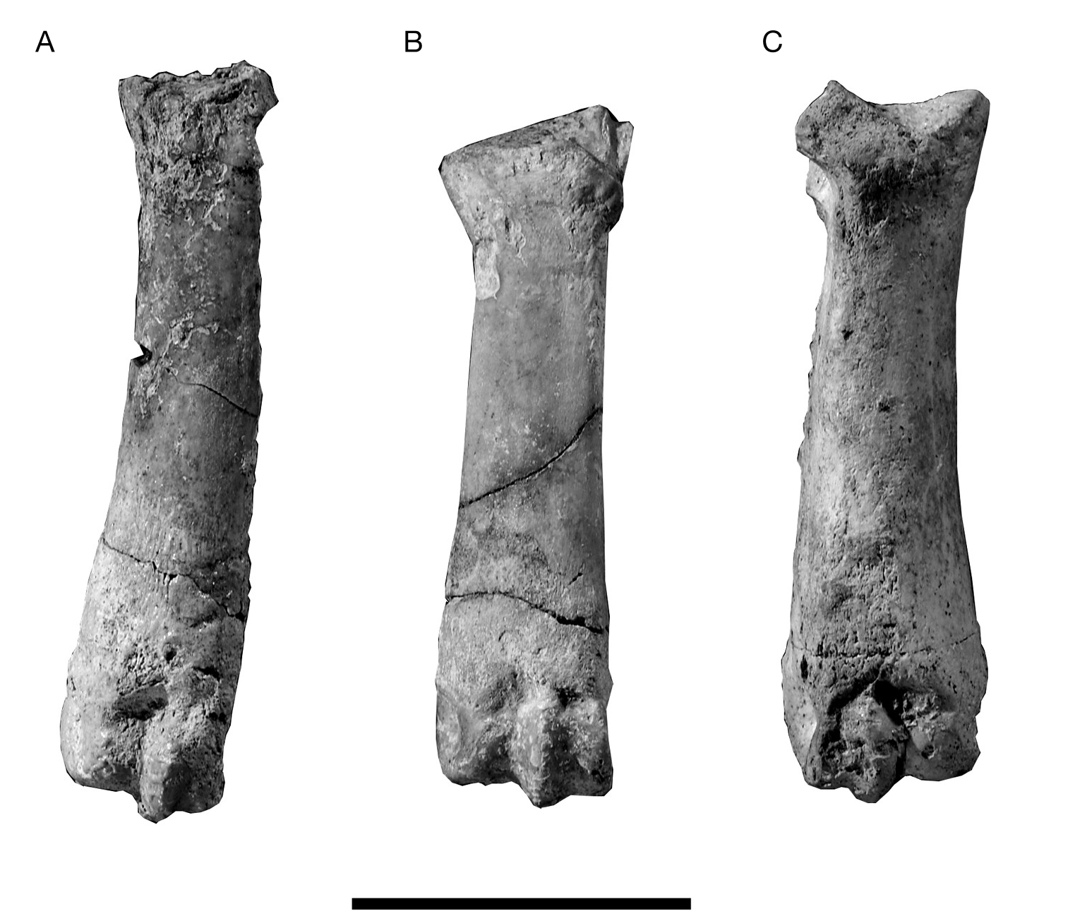
Postcranials
Late Miocene suid limb bones are rarely described and figured, making it difficult to supply a comparative study of the abundant postcranial material of the Akkaşdagwı suid. In order to facilitate future comparative work we here present figures and measurements of the material (Appendix: Tables 1; 2; Figs 3-5 View FIG View FIG View FIG ).
No known copyright restrictions apply. See Agosti, D., Egloff, W., 2009. Taxonomic information exchange and copyright: the Plazi approach. BMC Research Notes 2009, 2:53 for further explanation.
|
Kingdom |
|
|
Phylum |
|
|
Class |
|
|
Order |
|
|
Family |
|
|
Genus |
Microstonyx major ( Gervais, 1848 )
| Liu, Liping, Kostopoulos, Dimitris S. & Fortelius, Mikael 2005 |
Sus major
| Gervais 1848 |