
Xenorhyncocoris Miller, 1938
|
publication ID |
https://doi.org/10.5852/ejt.2021.746.1315 |
|
publication LSID |
lsid:zoobank.org:pub:FE4F837A-DB93-431E-AB0D-C2DCD0987B96 |
|
DOI |
https://doi.org/10.5281/zenodo.4700903 |
|
persistent identifier |
https://treatment.plazi.org/id/03F1CE55-FFA3-FF9E-FD9A-FC3FFA8A57D3 |
|
treatment provided by |
Plazi |
|
scientific name |
Xenorhyncocoris Miller, 1938 |
| status |
|
Xenorhyncocoris Miller, 1938 View in CoL
Figs 1–59 View Figs 1–4 View Figs 5–11 View Figs 12–17 View Figs 18–25 View Figs 26–36 View Figs 37–43 View Figs 44–45 View Figs 46–49 View Figs 50–56 View Figs 57–59
Xenorhyncocoris Miller, 1938: 135 View in CoL .
Type species by original designation: Xenorhyncocoris caraboides Miller, 1938 View in CoL .
Xenorhyncocoris View in CoL – Cook 1977: 64, 70. — Maldonado Capriles 1990: 78. — Putshkov & Bérenger 1999: 92.
Revised diagnosis
This genus can be recognized within Ectrichodiinae by the following combination of characters: macropterous (male, based on X. attractivus sp. nov.) or micropterous (female); head club-shaped, subapically widened, distinctly longer than pronotum; ventral surface of anteocular part flat; antenniferous tubercle surrounded by lump-form process laterally; antennae four-segmented, with basiflagellomere shorter than other segments; labial segment II longest and dorsoventrally flattened, segment III inflated, segment IV flattened laterally; apex of prosternum acute, distinctly surpassing fore coxae; metathoracic gland evaporatorium invisible in lateral view; femora not thickened; apexes of tibiae bulbous; fossula spongiosa present on fore and mid tibiae; abdominal tergite II with three longitudinal ridges. In macropterous male, anterior pronotal lobe distinctly shorter but more than half as long as posterior lobe; scutellum broad, with 1+1 widely separated apical prongs and 1+1 lateral prongs; fore wing nearly reaching apex of abdomen. In micropterous female, anterior pronotal lobe distinctly longer than posterior lobe, swollen; scutellum broad, suberect or erect, with 1+1 widely separated apical prongs; fore wing not reaching apex of scutellum.
Redescription
Macropterous male (based on X. attractivus sp. nov.)
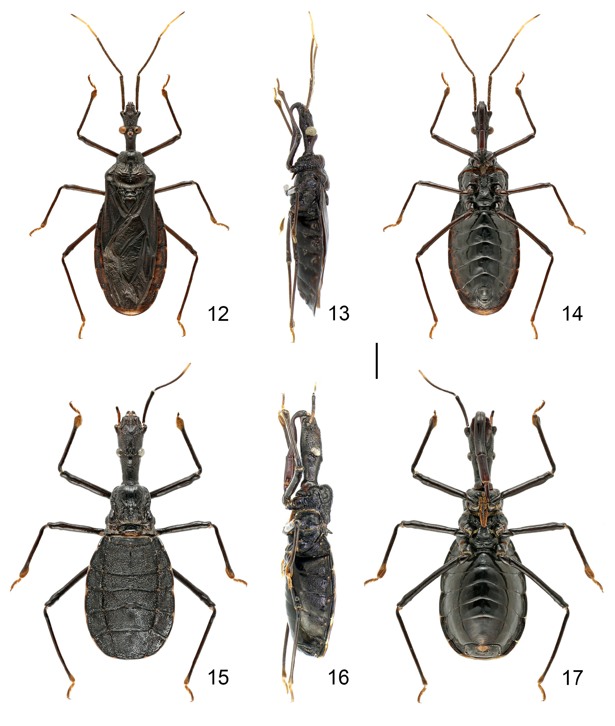
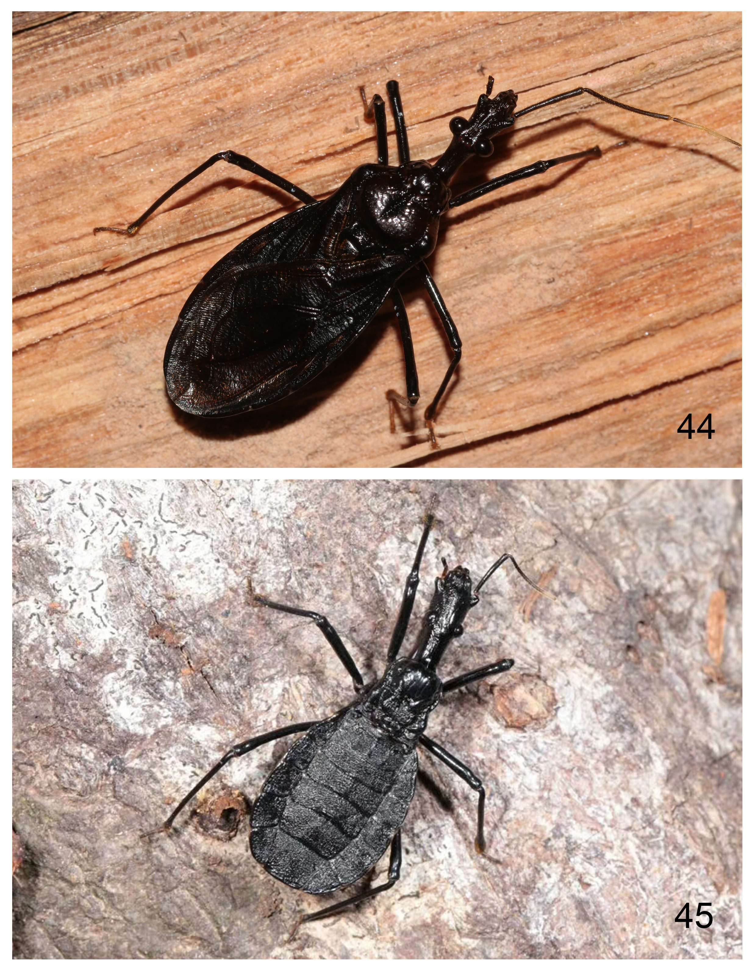
COLORATION. Body generally blackish brown ( Figs 12–14 View Figs 12–17 ); ocelli, basi- and disti- (except apical ⅓) flagellomeres, tarsi, anterolateral angles of each connexival segment and spiracles yellowish brown; connexivum faintly tinged with brown; apex of abdomen lighter.
STRUCTURE. Body oblong; body surface generally glabrous, moderately shining and wrinkled. Body surface with decumbent, tiny pubescence, difficult to observe; antennae densely covered with decumbent and erect, short, white setae; inner surfaces of fore trochanter and femur with several sparsely distributed, erect, long, white setae; inner surfaces of tibiae (except basal ⅓) with decumbent and suberect, short setae; apexes of tibiae and ventral surfaces of tarsi densely covered with short golden setae ( Figs 21–22 View Figs 18–25 ).
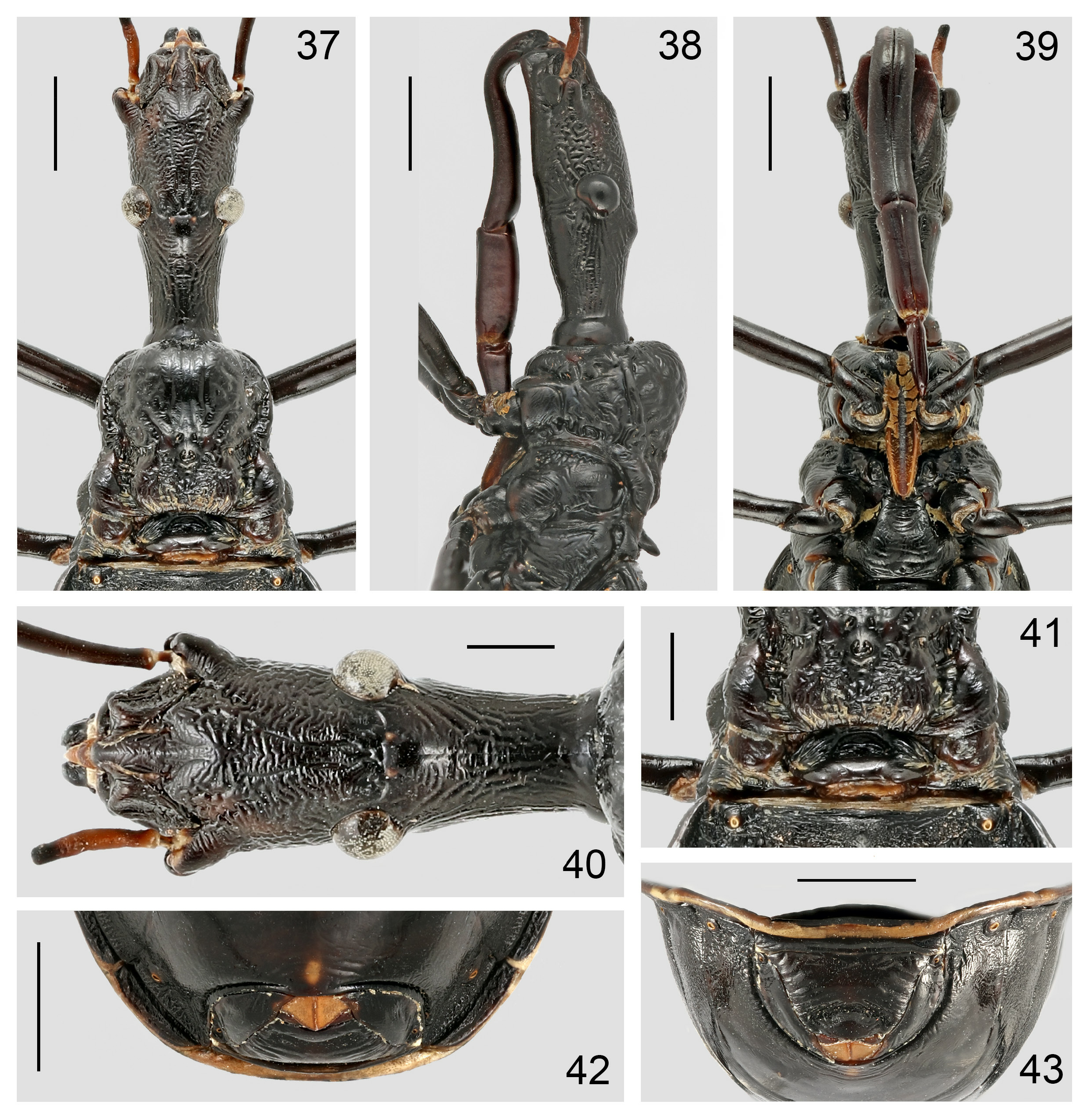
HEAD ( Figs 18–20 View Figs 18–25 ). Elongate, club-shaped, widened at antenniferous tubercles, 1.35 times as long as pronotum; anteocular part distinctly longer than postocular part, ventral surface flat, midpoint slightly concave; eyes large, strongly protruding laterally, ventral margin far remote from ventral surface of head; width across eyes slightly broader than width between antenniferous tubercles; ocelli situated on tubercle; anteclypeus slightly elevated. Antennal insertion situated before middle of anteocular part but relatively far from apex of head; antenniferous tubercle produced, surrounded by lump-form process laterally ( Fig. 19 View Figs 18–25 ); antennae ( Figs 12, 14 View Figs 12–17 ) four-segmented, with antennal scape slightly shorter than head, pedicel longest and slightly curved, basiflagellomere shortest and distiflagellomere slightly longer than basiflagellomere. Labium ( Figs 19–20 View Figs 18–25 ) robust; labial segment II longest and dorsoventrally flattened, apex reaching posterior margin of eye, strongly curved at base; segment III strongly inflated; segment IV strongly flattened laterally, knife-like.
PRONOTUM ( Figs 18–19 View Figs 18–25 ). Trapezoidal, wider than long; anterior pronotal lobe distinctly shorter but more than half as long as posterior lobe, slightly swollen, with medial longitudinal sulcus restricted in extreme base; posterior lobe broad, with deep, carinulate, medial longitudinal sulcus and a pair of deep, carinulate, lateral sulci; transverse sulcus distinct; lateral pronotal margins constricted; posterior margin slightly convex. Prosternum ( Fig. 20 View Figs 18–25 ) strongly developed, distinctly surpassing fore coxae, apically acute. Scutellum ( Fig. 18 View Figs 18–25 ) broad, with 1+1 widely separated apical prongs and 1+1 lateral prongs; midpoint of scutellum depressed. Anterior margin of mesopleuron with a row of distinct punctuations ( Fig. 19 View Figs 18–25 ). Mesosternum ( Fig. 20 View Figs 18–25 ) with a shallow, medial, longitudinal furrow. Metapleuron ( Fig. 19 View Figs 18–25 ) longer than high. Metasternum ( Fig. 20 View Figs 18–25 ) slightly swollen on both sides. Metathoracic gland callus present in lateral view; metathoracic gland evaporatorium small, not extend dorsally in lateral view.
LEGS. Slender ( Figs 12–14 View Figs 12–17 ). Femora not thickened, slightly sinuated subapically; tibiae slenderer than respective femora, straight; apex of fore tibia bulbous, laterally compressed forming a blunt, weak dorsal carina ( Fig. 21 View Figs 18–25 ); tarsomere III subequal to combined length of tarsomeres I and II; fore and mid tibiae with fossula spongiosa occupying about apical 0.15 of their ventral surface ( Fig. 22 View Figs 18–25 ).
WINGS. Well developed. Fore wing ( Fig. 23 View Figs 18–25 ) nearly reaching apex of abdomen; corium with majority parts of M and Cu separate; membrane with base of outer cell distinctly shorter than inner cell, distal part of R forming a close cell with M, distal part of M extending beyond apex of outer cell. Hind wing ( Fig. 24 View Figs 18–25 ) with distal parts of Sc, R and M reaching outer margin; hamus nearly reaching base of hind wing; only one secondary vein.
ABDOMEN. Ovoid, with lateral outline rounded ( Figs 12, 14 View Figs 12–17 ). Abdominal tergite II with three longitudinal ridges. Ventral laterotergites II to VI distinctly separate from respective sternites. Intersternal sutures of segments II to VI carinulate; midpoint of anterior margins of sternites IV to VI curved anteriorly; anterior margin of sternite VII strongly curved anteriorly; segment VII distinctly expanded posteriorly ( Fig. 25 View Figs 18–25 ); segment VIII invisible at resting state ( Fig. 25 View Figs 18–25 ). Spiracles round.
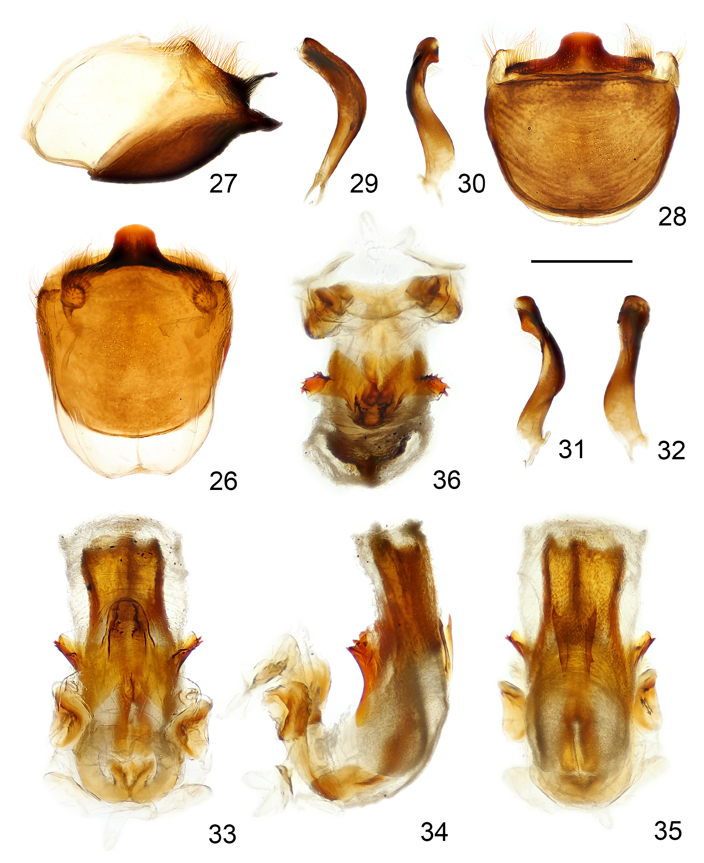
MALE GENITALIA. Pygophore ( Figs 26–28 View Figs 26–36 ) short, oblong; median process directed dorsoposteriad. Parameres ( Figs 29–32 View Figs 26–36 ) relatively stout, bent, with a subapical process.
Micropterous female
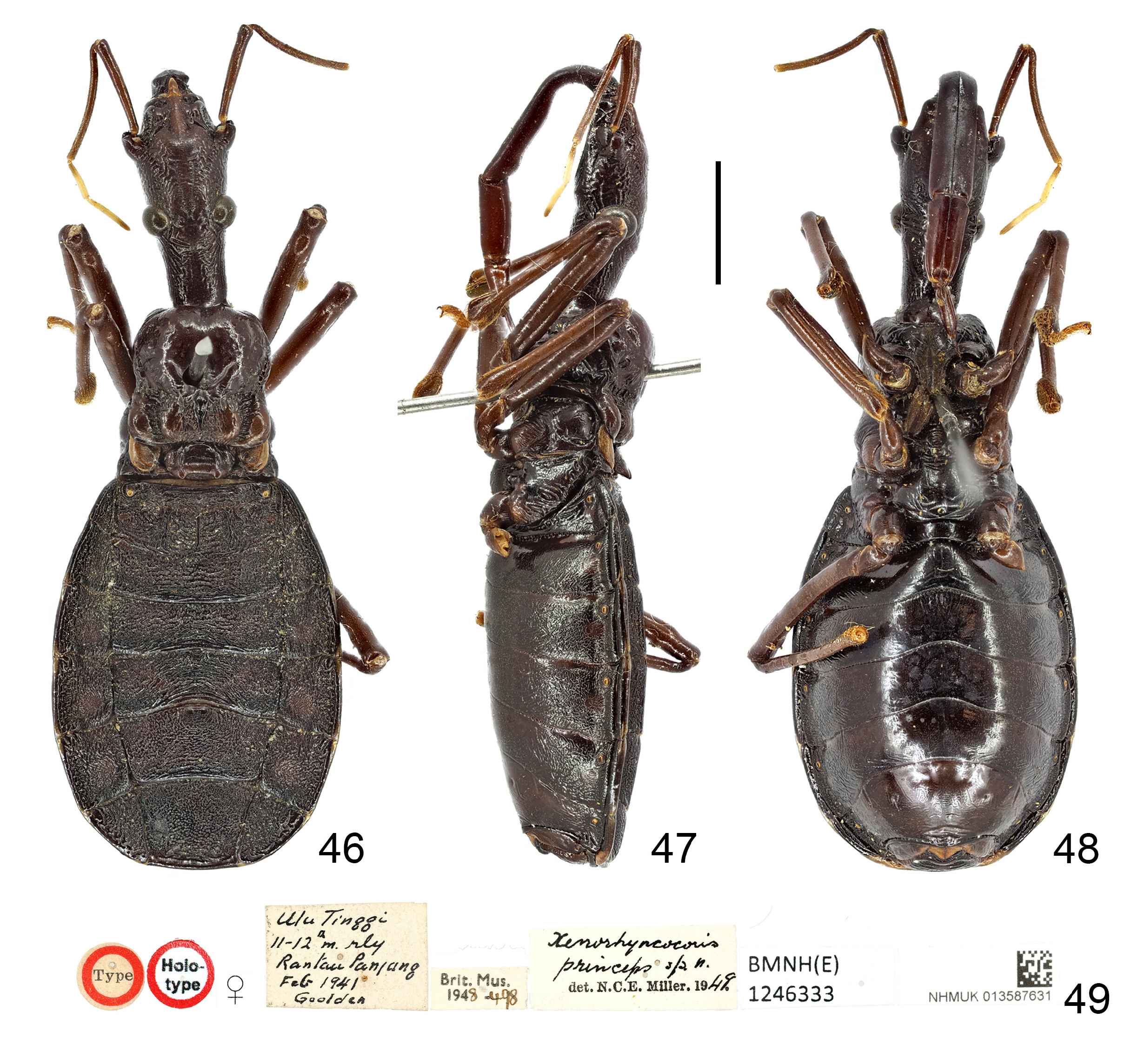
COLORATION. Body generally brown to blackish brown ( Figs 1–3 View Figs 1–4 , 15–17 View Figs 12–17 , 46–48 View Figs 46–49 , 57–59 View Figs 57–59 ); tarsi, anterolateral and posterolateral angles of each connexival segment, apical margin of abdomen, spiracles and valvula I yellowish brown.
STRUCTURE. Body shape and vestiture similar to those in male, but differs in the following characteristics: body robust; antennal scape and basal half of pedicel bare; lateral margins of prosternum with suberect, short, golden setae; head thickened, 1.8–2.2 times as long as pronotum; eyes small; width between antenniferous tubercles broader than width across eyes; ocelli strongly reduced; anteclypeus not elevated; antennal scape distinctly shorter than head; pronotum nearly square; anterior pronotal lobe much longer than and as wide as or slightly narrower than posterior lobe, swollen dorsally; medial longitudinal sulcus of pronotum reduced to deep, medial depression; lateral margins of anterior pronotal lobe marginated; posterior margin of pronotum concave; prosternum much longer, reaching or surpassing anterior margins of mid coxae; scutellum suberect or erect, with prongs weakly developed; fossula spongiosa larger, occupying about apical 0.2 of ventral surface of fore and mid tibiae; fore wing not reaching apex of scutellum; abdomen broader, with ventral surface slightly flattened in middle.
FEMALE GENITALIA. Platelike ( Figs 10–11 View Figs 5–11 , 42–43 View Figs 37–43 , 55–56 View Figs 50–56 , 59 View Figs 57–59 ); tergite VIII transverse, anterior and posterior margins nearly straight; tergite IX large, trapezoidal, with transverse depression subapically; posteromedian margin of valvifer I slightly sinuate; valvula I small, triangular, with posterior margin slightly concave; styloid visible in resting state.
Diversity and distribution
Four species, occurring in the Oriental Region ( Fig. 60 View Fig ).
Female-based key to species of Xenorhyncocoris Miller, 1938
1. Body length about 37 mm; head 2.2 times as long as pronotum; anteocular part 1.3 times as long as postocular part; labial segment II surpassing posterior margin of eye, curved dorsally, approaching ventral surface of head; apex of prosternum almost reaching posterior margins of mid coxal cavities ..................................................................................................... X. caraboides Miller, 1938 View in CoL
– Body length about 34 mm or less; head 1.8–1.9 times as long as pronotum; anteocular part 1.4–1.7 times as long as postocular part; labial segment II reaching posterior margin of eye, straight or curved dorsally; apex of prosternum reaching middle of mid coxae or less .................................... 2
2. Body blackish brown; labial segment II curved dorsally, approaching ventral surface of head; pronotum 1.15 times as broad as its length along midline; scutellum erect; fore wing reaching middle of scutellum ..................................................................................................... X. attractivus sp. nov.
– Body brown; labial segment II straight; pronotum as broad as its length along midline; scutellum suberect; fore wing surpassing middle of scutellum ......................................................................... 3
3. Anteocular part 1.4 times as long as postocular part; labial segment II 1.9 times as long as segment III, with ventral surface thickened at apical ⅔; prosternum reaching anterior margins of mid coxae ...................................................................................................................... X. princeps Miller, 1949 View in CoL
– Anteocular part 1.7 times as long as postocular part; labial segment II 1.6 times as long as segment III, with apex bulbous; prosternum reaching middle of mid coxae .................................................... ........................................................................................ X. schoenitzeri Putshkov & Bérenger, 1999 View in CoL


No known copyright restrictions apply. See Agosti, D., Egloff, W., 2009. Taxonomic information exchange and copyright: the Plazi approach. BMC Research Notes 2009, 2:53 for further explanation.
|
Kingdom |
|
|
Phylum |
|
|
Class |
|
|
Order |
|
|
SubOrder |
Heteroptera |
|
Family |
|
|
SubFamily |
Ectrichodiinae |
|
Tribe |
Ectrichodiini |
Xenorhyncocoris Miller, 1938
| Chen, Zhuo, Liu, Yingqi & Cai, Wanzhi 2021 |
Xenorhyncocoris
| Putshkov P. V. & Berenger J. - M. 1999: 92 |
| Maldonado Capriles J. 1990: 78 |
| Cook M. L. 1977: 64 |
Xenorhyncocoris Miller, 1938: 135
| Miller N. C. E. 1938: 135 |