
Meroloba suturalis (Snellen van Vollenhoven, 1858)
|
publication ID |
https://doi.org/10.5281/zenodo.5301164 |
|
publication LSID |
lsid:zoobank.org:pub:402537AD-557B-4F95-8EB1-EC67EAEE0DDB |
|
persistent identifier |
https://treatment.plazi.org/id/03ED87D8-6B7A-0935-FEC1-9952ADDEFD57 |
|
treatment provided by |
Marcus |
|
scientific name |
Meroloba suturalis (Snellen van Vollenhoven, 1858) |
| status |
|
Meroloba suturalis (Snellen van Vollenhoven, 1858)
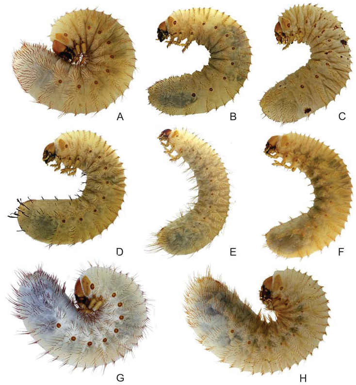
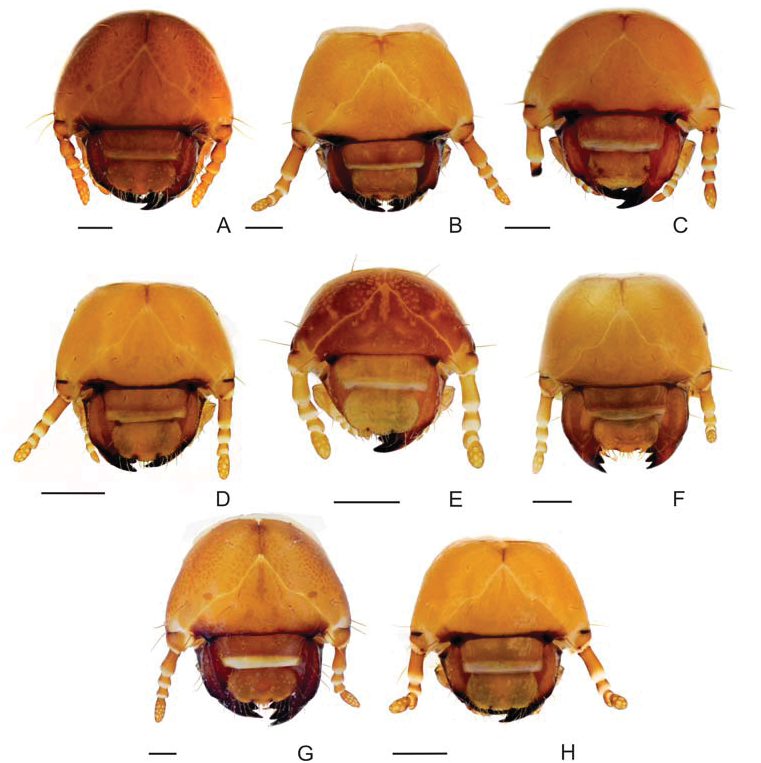
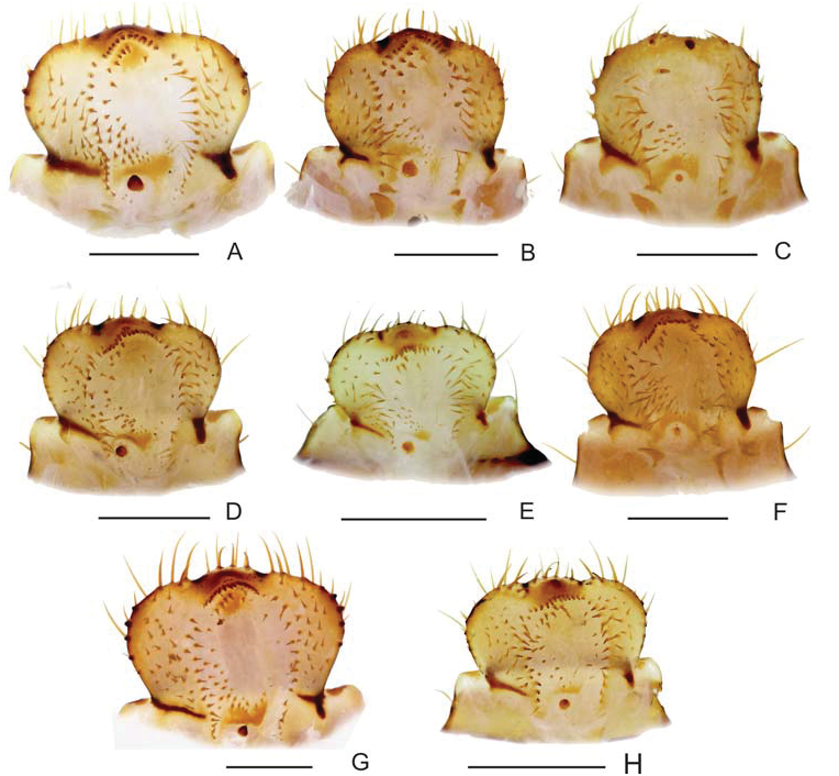
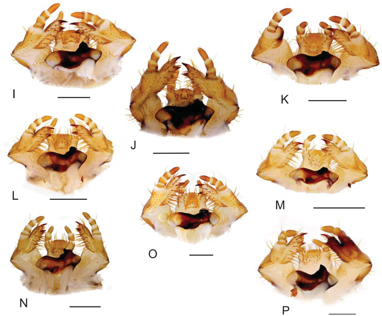
( Figs 2F View Fig ; 3F View Fig ; 4F View Fig ; 5F View Fig ; 7A–C,P View Fig ; 8J–L,S View Fig ; 9J,R View Fig ; 10F View Fig ; 11F View Fig ; 12L,O View Fig ; 13D,K View Fig )
Material examined. 7 third instar larvae reared from adults collected in: MALAYSIA: BORNEO ISLAND: SABAH SULTANATE: Kinabatangan River Reserve, Sukau (Kota Kinabatangan), 5°31ƍ13ƎN 118°17ƍ41ƎE, 50 m a.s.l., 20.–22. ii.2010, P. Šípek leg.
Description of third instar larva. Body ( Fig. 2F View Fig ). Length 43.0–51.0 mm, dorsoventral interval of body segments increasing slightly towards the penultimate body segment (thus abdominal segments appear to be slightly thicker; abdominal segments VII and VIII are the thickest). Body with numerous setae; setae of ventral and dorsal body parts of equal shape and size.
Head capsule ( Fig. 3F View Fig ). Maximum width 3.5–3.7 mm, smooth, yellowish brown. Frontal sutures deeply bisinuated. Epicranial insertions of antennal muscles indistinct. Cranial chaetotaxy summarized in Table 2. Anterior and exterior frontal setae with minute single seta.
Antennae ( Figs 3F View Fig ; 9J View Fig ). Relative length of antennomeres I–IV (an I–IV): an I> an IV> an II> an III), ultimate antennomere with three to seven dorsal and six to nine ventral sensory spots.
Epipharynx ( Fig. 4F View Fig ). Haptomerum: Zygum convex, more or less protruding, with angulate or arcuate row of 17–19 stout conical setae and several similar setae proximad to the row. Sensilla of zygum grouped in two groups distad to the row. Acroparia: Lateral lobes of epipharynx with three to ¿ve long setae, medial lobe with four and four to ¿ve setae on ventral and dorsal side, respectively. Acanthoparia with four to eight setae; the size of setae increasing towards apex of epipharynx. Chaetoparia:Asymmetric, right side composed of approximately 45–60 setae, left side with around 28–33 setae in three to four rows. Both parts of chaetoparia with medial, more or less regular row of prominent long setae. Pedium large, with isolated row of two to ¿ve slender setae on the left side. Dexiotorma straight, right pternotorma well developed. Laeotorma absent, left pternotorma well developed.
Haptolachus: Sense cone small, conical, with four pores. Plate-like sclerite large, horse- -shoe shaped.
Mandibles ( Figs 7A–C View Fig ; 8J–L View Fig ; 13K View Fig ). Stridulatory area well developed, distinctly paler than the other parts of ventral mandibular face, with approximately ten regular ridges, bordered with numerous irregular rugosities at both proximal and distal margin of ¿eld. Scrobis with one or two lateral setae. Longitudinal furrow shallow. Lateral outline of both mandibles without external tooth. Scissorial teeth of left mandible subequal, large; right mandible with two large and one small scissorial tooth.
Maxilla ( Figs 5F View Fig ; 12L; 12O View Fig ; 13D View Fig ). Dorsal surface of cardo and labacoparia with zero to four, 13–20 setae, respectively. Ventral surface of cardo and labacoparia with zero to three and 5–14 setae, respectively, labacoparia often with one or two stiff setae. Dorsal surface of stipes with approximately 14–19 slender hair-like setae, one anterolateral stout seta may be present. Maxillary stridulatory apparatus with row of ¿ve to eight spine-like stridulatory teeth and one blunt tubercle ( Fig. 12L View Fig ), in some specimens some stridularory teeth outside the row. Ventral surface of stipes with single proximal and two distal setae. Galear portion of mala in dorsal aspect beside large falcate uncus with six (seven) large stiff and three to ¿ve medium-sized hair-like setae, respectively. Lacinial part of mala with around 16–18 mostly extremely long and stiff setae. Lacinial apex with single triangular uncus with short, conical seta at the base and another long, stout, conical seta next to it. Vental surface of mala with two longitudinal rows of four to ¿ve setae; setae of the exterior row long and stiff, setae of the interior row shorter and stout.
Hypopharyngeal sclerome ( Figs 5F View Fig ; 12O View Fig ). Four tegumentary expansions (phoba-like processes) present on the left lateral lobe of hypopharynx, tegumentary expansions on right medial portion of scleroma present.
Ligula ( Figs 5F View Fig ; 12O View Fig ; 13D View Fig ). Dorsal surface with two lateral groups of around 10 hairlike setae, two paramedial setae at the (ventro-) apical margin and central group of setae and sensilla. This group is composed of two paramedial rows of three stout, conical setae, and transverse, basomedial row with seven to eight conical or campaneiform setae.
Thorax ( Figs 2F View Fig ; 7P View Fig ; 8S View Fig ; 9R View Fig ). Prothoracic sclerite with four to seven setae on the anteroventral margin and another two to four setae in the posterio-dorsal area. Each sublobe of prothorax dorsal with one or two rows of medium-long and long setae interspersed with few short setae. Thoracic spiracle ( Fig. 7P View Fig ) 0.93 × 0.50 mm (height × width). Bullar opening distinct, approximately 0.1 mm wide, arms of respiratory plate thus well separated. Respiratory plate with approximately 20–40 holes across diameter. Venter of thorax (and the ¿rst abdominal segment) with paramedian pair of stiff setae, similar setae found also on legs. Pretarsus ( Fig. 8S View Fig ) conical, with two apical setae, falcate tip or similar structures absent.
Abdomen ( Figs 2F View Fig ; 10F View Fig ; 11F View Fig ). Each dorsal sublobe of abdominal segments I–VIII with two to three rows of setae. Setae in anterior row(s) short or medium-sized, posterior row also with several long setae. Venter of abdominal segments II–IX with several long stiff setae. Dorsum of abdominal segments IX–X, with numerous short to long setae. Spiracles on abdominal segments I–VI elongate, similar to thoracic spiracle, abdominal spiracles on segments VII–VIII almost circular, arms of respiratory plate almost concealed.
Raster ( Figs 10F View Fig ; 11F View Fig ). Pali, short, spine-like, smaller than setae of tegilla. Palidium monostichous, with approximately 20 pali arranged in two almost parallel rows, closed at the proximal end. Teges almost entirely separated by rows of pali. Septula opened posteriorly, about six times longer than broad. Tegilla with numerous apically recurved, pointed setae. Setae of the dorsal and ventral anal lip same as those on the dorsal abdominal lobes.


No known copyright restrictions apply. See Agosti, D., Egloff, W., 2009. Taxonomic information exchange and copyright: the Plazi approach. BMC Research Notes 2009, 2:53 for further explanation.
|
Kingdom |
|
|
Phylum |
|
|
Class |
|
|
Order |
|
|
Family |
|
|
Genus |