
Davisella spondia, Reis & Navia, 2010
|
publication ID |
https://doi.org/10.5281/zenodo.199630 |
|
DOI |
https://doi.org/10.5281/zenodo.6201403 |
|
persistent identifier |
https://treatment.plazi.org/id/03EC87A7-FFCD-B903-FF40-D8A887E8C080 |
|
treatment provided by |
Plazi |
|
scientific name |
Davisella spondia |
| status |
n. sp. |
Davisella spondia s Reis & Navia n. sp.
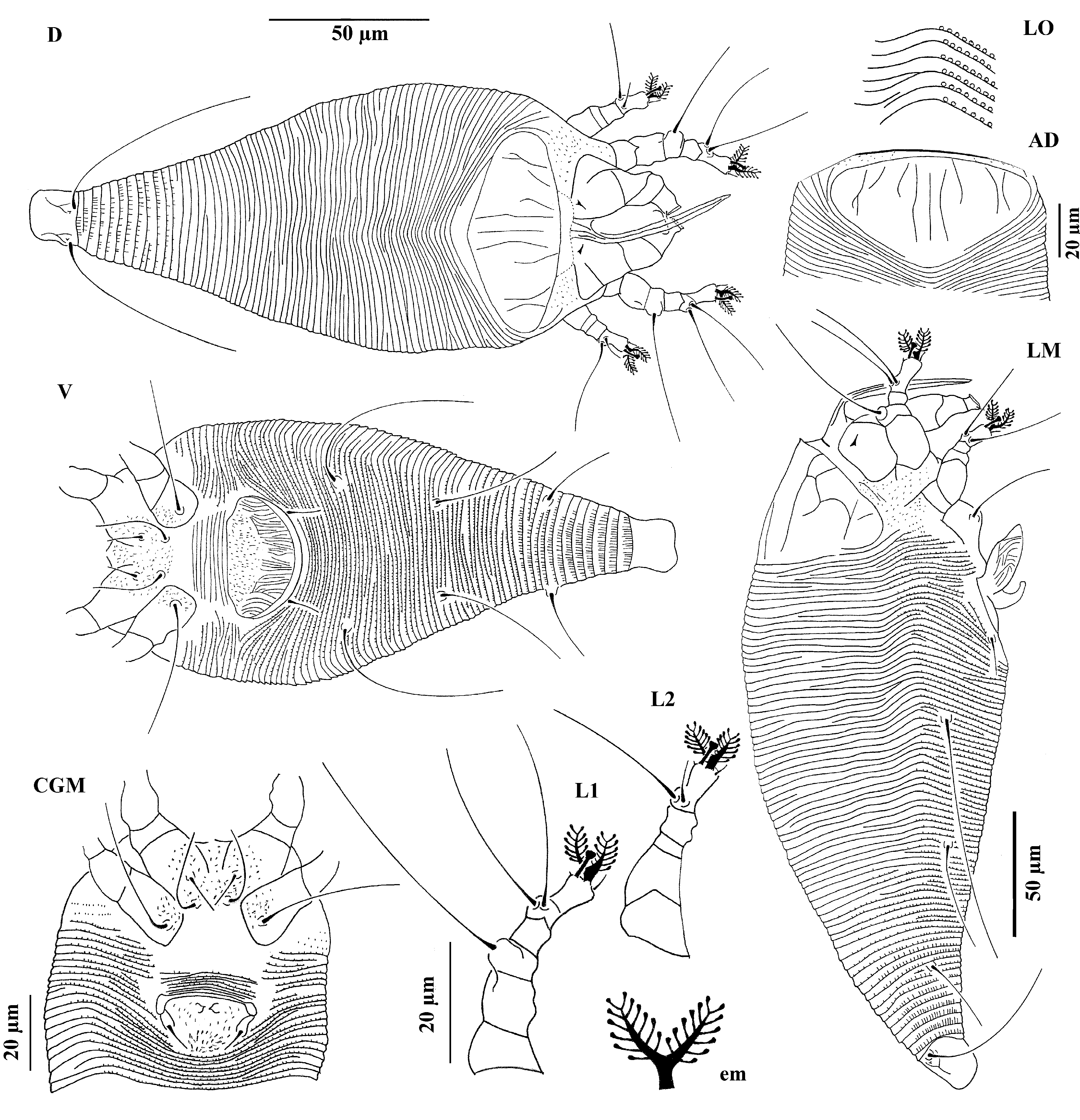
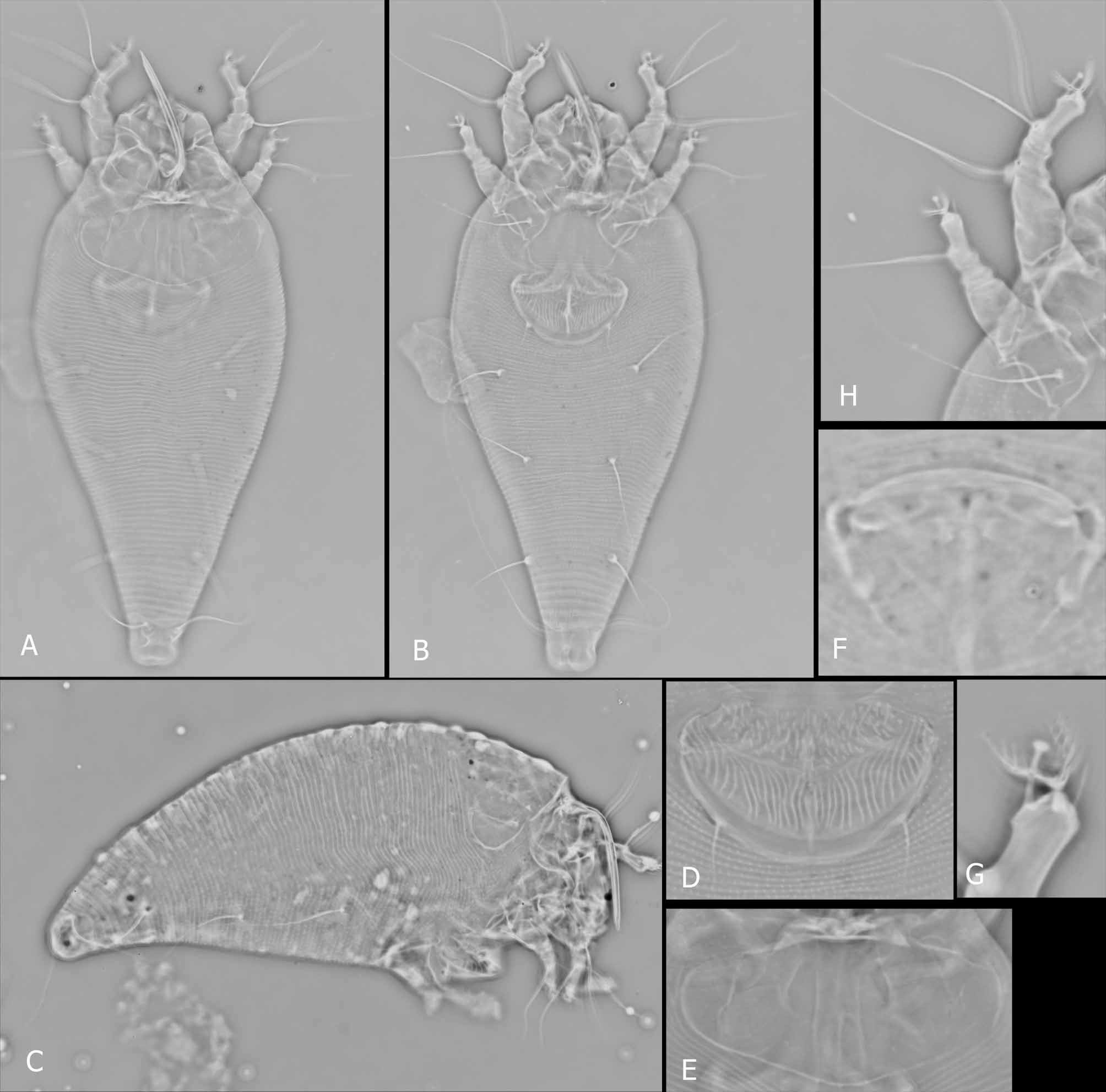
Diptilomiopidae , Diptilomiopinae ( Figs. 3 View FIGURE 3 & 4 View FIGURE 4 )
Diagnosis. Davisella spondias n. sp. is the seventh species to be assigned to this genus. It is distinctive in having the prodorsal shield ornamentation with free longitudinal lines (not joined forming cells or arches) and completely or partially circumscribed with a semi-elliptical line that surrounds almost the entire prodorsal shield. This species is near Davisella globosa ( Keifer, 1969) , Davisella haramotonis ( Keifer, 1974) , Davisella notosa ( Flechtmann, 1995) and Davisella paucisetosa ( Flechtmann, 1999) in the ornamentation of the coverflap with the basal region featuring granules (or dashes) and the distal region having radiating longitudinal lines. However, the new species has more distal longitudinal lines (35–38 lines) on the coverflap, while the other species have less than 30 longitudinal lines, except for D. haramotonis . The new species has a short, trapezoidal anterior lobe, while D. haramotonis has a broad, round anterior lobe over the gnathosoma base with the median line extending over almost the entire prodorsal shield length (only on the rear half in D. haramotonis ); the admedian lines are long as well, but not connected by curved cross lines as in D. haramotonis ; laterally on the prodorsal shield, only two longitudinal lines (no curved broken lines and short dashes as in D. haramotonis ). The new species is similar to D. haramotonis in having a 6-rayed empodium while that of Davisella breitlowi ( Davis, 1964) is 5–rayed, Davisella palmea ( Flechtmann, 1998) is 7–8 rayed and all the other species are 7 or 8–rayed.
FEMALE (n=10). Body fusiform, 160 (146–172), 77 (69–77) wide; colour in life, light-yellowish. Gnathosoma prominent, projecting well down, 34 (33–35); basal seta ( ep) 4 (3–4); apical seta ( d) 2 (2–3), simple. Prodorsal shield 29 (29–36), 61 (52–63) wide with sinuous, parallel median and admedian lines. Free longitudinal lines (extremities not joined forming cells or arches) and completely or partially circumscribed by a semi-elliptical line that surrounds almost entire prodorsal shield, except for the anterolateral sparsely granulated region. Scapular setae ( sc) and tubercles absent. Legs with all segments; femoral seta ( bv) and tibial seta ( l’) absent on both legs; genual seta ( l’’) absent on leg II. Leg I 35 (33–36); femur 13 (12–14); genu 5, genual seta ( l ”) 45 (42–47); tibia 6 (5–6), tibial seta ( l ’) absent; tarsus 11 (11–13), lateral seta ( ft ”) 34 (32– 34), dorsal seta ( ft ’) 34 (32–34), unguinal seta ( u’) 4 (4–5), solenidion (ω) 6 (5–6); empodium divided 9 (7–9), 6–rayed. Leg II 31 (29–32); femur 11 (10–11); tibia 4 (3–4); tarsus 9 (8–9), ft” 29 (26–29), ft ’ 10 (8–10), u’ 4 (4–5), ω 6 (5–6), empodium divided, 7 (7–8), each branch 6–rayed. Coxae I and II with numerous pointed granules. Coxal seta I ( 1b) 11 (11–14), 12 (11–13) apart; coxal seta II ( 1a) 23 (23–26), 10 (10–11) apart; coxal seta III ( 2a) 45 (40–47), 29 (29–32) apart; 8 (8–9) coxigenital annuli, without microtubercles. Genitalia 25 (21–25), 39 (39–40) wide, coverflap with proximal area granulated and distally with 38 (35–38) longitudinal radial lines; genital seta ( 3a) 10 (10–12). Opisthosoma with 73 (69–77) dorsal annuli, of which the posterior (about the last twelve) slightly microtuberculated, the first thirteen having a slight median ridge; 64 (63–69) ventral annuli, with pointed microtubercles on the posterior annulus margins, not present in the lateral region. Lateral seta ( c2) absent; ventral seta I ( d) 51 (51–68), on annulus 13 (13–14), 46 (39–48) apart, 32 (28–36) microtubercles apart; ventral seta II ( e) 61 (58–63), on annulus 34 (34–38), 29 (25–31) apart, 29 (23–29) microtubercles apart; ventral seta III ( f) 22 (22–24), on annulus 57 (55–60), 28 (24–30) apart, 28 (23–30) microtubercles apart. Caudal seta ( h2) 70 (65–70); accessory seta ( h1), minute.
MALE (n=5). Smaller than female, 142 (140–160), 69 (61–69) wide. Gnathosoma 28 (28–30); basal seta ( ep) 3 (2–3); apical seta ( d) 2 (2–3), simple. Prodorsal shield as in female, 31 (30–33), 59 (51–59) wide. Scapular setae ( sc) and tubercles absent. Legs as in female. Leg I 35 (32–38); femur 13 (11–13); genu 5 (4–5), genual seta ( l ”) 40 (36–41); tibia 5 (4–5); tarsus 9 (9–11), lateral seta ( ft ”) 29 (28–31), dorsal seta ( ft ’) 29 (28– 31), unguinal seta ( u’) 4 (4–5), solenidion (ω) 6 (5–6); empodium divided 7, 6–rayed. Leg II 27 (26–29); femur 10 (9–10); tibia 4 (3–4); genu 4 (3–4); tarsus 7 (7–8), ft ” 23 (23–26), ft ’ 7 (7–8), u’ 4 (4), ω 5 (5); empodium 7 (7), 6–rayed. Coxae as in female. Coxal seta I ( 1b) 10–11, 10–12 apart; coxal seta II ( 1a) 17–22, 8–11 apart; coxal seta III ( 2a) 35–36, 24–30 apart; 8–9 coxigenital annuli, microtubercles. Genitalia 16–19, 21–25 wide, posterior region granulated, eugenital setae as depicted; genital seta ( 3a) 8–10. Opisthosoma as in female, 58–65 dorsal annuli; 59–62 ventral annuli. Lateral seta ( c2) absent; ventral seta I ( d) 45–66, on annulus 10–13, 28–36 apart, 23–29 microtubercles apart; ventral seta II ( e) 48–58, on annulus 29–34, 19–23 apart, 17–21 microtubercles apart; ventral seta III ( f) 20–21, on annulus 50–55, 24–27 apart, 21–25 microtubercles apart. Caudal seta ( h2) 60–63; accessory seta ( h1), minute.
Type material. Female holotype, 41 female and 18 male paratypes, from Spondias mombin L. ( Anacardiaceae ), Recife, Pernambuco, Brazil. 08° 01’ 07” S, 34° 56’ 41” W, 19 August 2008, coll. A. C. Reis, on 8 microscope slides. Holotype and paratypes ( 36 specimens, 25 females and 11 males, on 5 microscope slides) deposited in the collection of the Laboratório de Acarologia, Departamento de Agronomia, Universidade Federal Rural de Pernambuco, Recife, PE, Brazil. Paratypes ( 23 specimens, 16 females and 7 males, on 3 microscope slides) also deposited in the collection of the Laboratório de Quarentena Vegetal, Embrapa Recursos Genéticos e Biotecnologia, Brasília, DF, Brazil.
Relation to host. Vagrant on the lower leaf surface; no visible damage.
Etymology. The specific designation “spondias ” refers to Spondias , the host plant genus.
No known copyright restrictions apply. See Agosti, D., Egloff, W., 2009. Taxonomic information exchange and copyright: the Plazi approach. BMC Research Notes 2009, 2:53 for further explanation.
|
Kingdom |
|
|
Phylum |
|
|
Class |
|
|
Order |
|
|
SubOrder |
Prostigmata |
|
SuperFamily |
Eriophyoidea |
|
Family |
|
|
Genus |