
Scaphisoma pandemum von Groll & Lopes-Andrade, 2021
|
publication ID |
https://doi.org/ 10.11646/zootaxa.4999.2.4 |
|
publication LSID |
lsid:zoobank.org:pub:C9A3640C-0297-4E3D-9210-AD3C5FC5F9B8 |
|
persistent identifier |
https://treatment.plazi.org/id/03E9EF6C-FFAC-D41F-4BDE-56C4FBAE2F55 |
|
treatment provided by |
Plazi |
|
scientific name |
Scaphisoma pandemum von Groll & Lopes-Andrade |
| status |
sp. nov. |
Scaphisoma pandemum von Groll & Lopes-Andrade View in CoL , sp. nov.
Figs 1–9 View FIGURE 1 View FIGURE 2 View FIGURE 3 View FIGURE 4 View FIGURE 5 View FIGURE 6 View FIGURE 7 View FIGURE 8 View FIGURE 9
Type locality. Mata da Biologia , Universidade Federal de Viçosa , Viçosa, state of Minas Gerais, Southeast Brazil .
Type material. HOLOTYPE: ♂ ( CELC) “ Brasil: MG, Viçosa ‘ Mata da Biologia’, 26.i.2017, S. Aloquio & W. Gomes leg. \ Scaphisoma pandemum von Groll & Lopes-Andrade HOLOTYPUS [red paper]” . PARATYPES: 20♂♂, 15♀♀, 8 with sex undetermined as follows: ♂, ♀, 1 ex. ( CELC), same data as holotype ; 2♂♂ ( CELC; one ♂ genitalia dissected and stored in glycerin) , 4♀♀ (2 CELC, 1 CAMB, 1 FMNH) “ BRASIL: MG, Viçosa ‘ Mata do Paraíso’, 11.ii.2015, S. Aloquio, A. Orsetti, C. Lopes-Andrade & M. Bento leg.” ; ♀, ♂, 1 ex. ( CELC; ♂ genitalia dissected and stored in glycerin) “ BRASIL: MG, Viçosa , Mata do Paraíso, 25.v.2016, A. Orsetti, C. Lopes-Andrade & Pecci-Maddalena leg. \ casca de tronco” ; ♂, ♀ ( CELC) “ BRASIL: MG, Viçosa , EPTEA Mata do Paraíso, 21.xi.2019, Leg. LabCol Fungo 10 \ Em Steccherinum undigerum ” ; 2♂♂, 4♀♀, 1 ex. ( CELC, 4♀♀ (2♀♀: genitalia dissected and stored in glycerin); 2♂♂: completely dissected and stored in glycerin, 1 ex.) “ BRASIL: MG, Viçosa , UFV, ‘ Mata da Biologia’, 26.vi.2019, Leg. E. von Groll & A. Orsetti \ Em Geesterania cf. carneola ( Steccherinaceae )” ; 5♂♂, ♀ ( CELC, 5♂♂ (1♂ genitalia dissected and stored in glycerin), ♀) “ BRASIL: MG, Viçosa , Mata da Biologia, 02.ix.2020 Leg. G.L.N. Martins & I.S.C. Pecci-Maddalena \ Em Geesterania cf. carneola ( Steccherinaceae )” ; 3♂♂, ♀ ( CELC) “ Mata da Biologia , 08.ix.2020, Leg. E. von Groll & A. Orsetti \ Fungo 05, Em Geesterania cf. carneola ” ; ♂ ( CELC; genitalia dissected and stored in glycerin), “ Mata da Biologia , 08.ix.2020, Leg. E. von Groll & A. Orsetti \ Fungo 12, Em Schizopora paradoxa ” ; 3♂♂ (1 CELC, 1 CAMB, 1 FMNH) , 2♀♀, 5exs ( CELC), “ BRASIL: MG, Viçosa , Mata da Biologia, 12.ix.2020, Leg. G.L.N. Martins & G.J. Figueiredo \ Em Geesterania cf. carneola ” ; ♂ ( CELC), “ BRASIL: MG, Viçosa , Recanto das Cigarras (Mata da Biol.), 20.xi.2019, Leg. LabCol, Fungo 25 \ Em Geesterania cf. carneola ( Steccherinaceae )”. All paratypes additionally labelled “ Scaphisoma pandemum von Groll & Lopes-Andrade PARATYPUS [yellow paper]” .
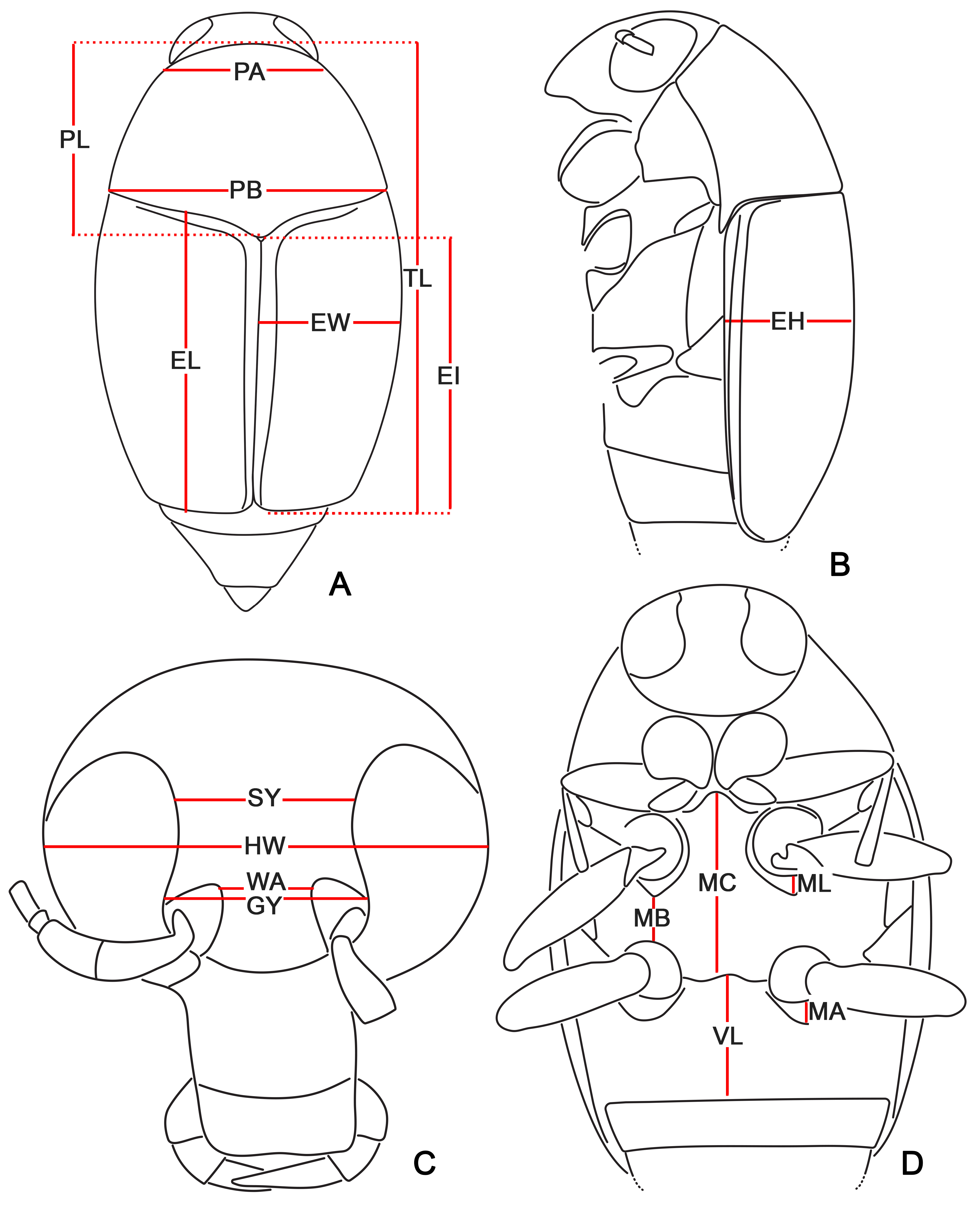
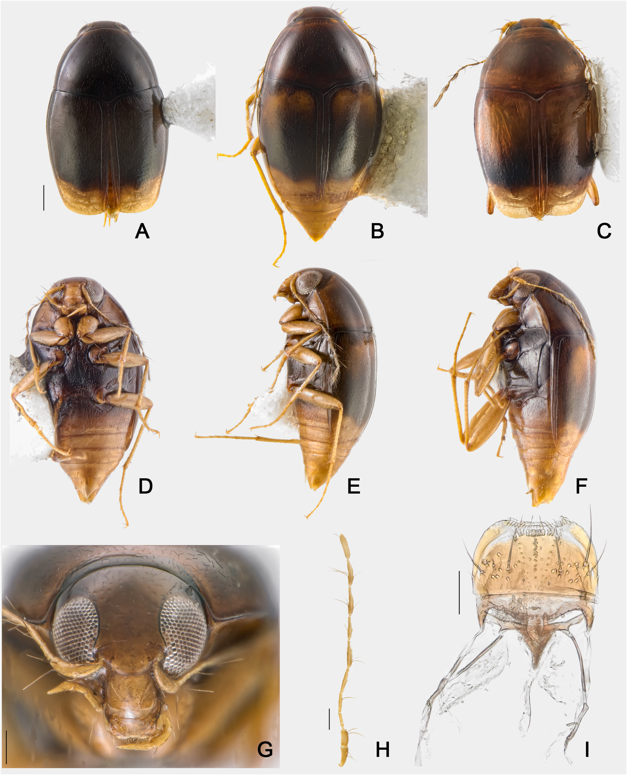
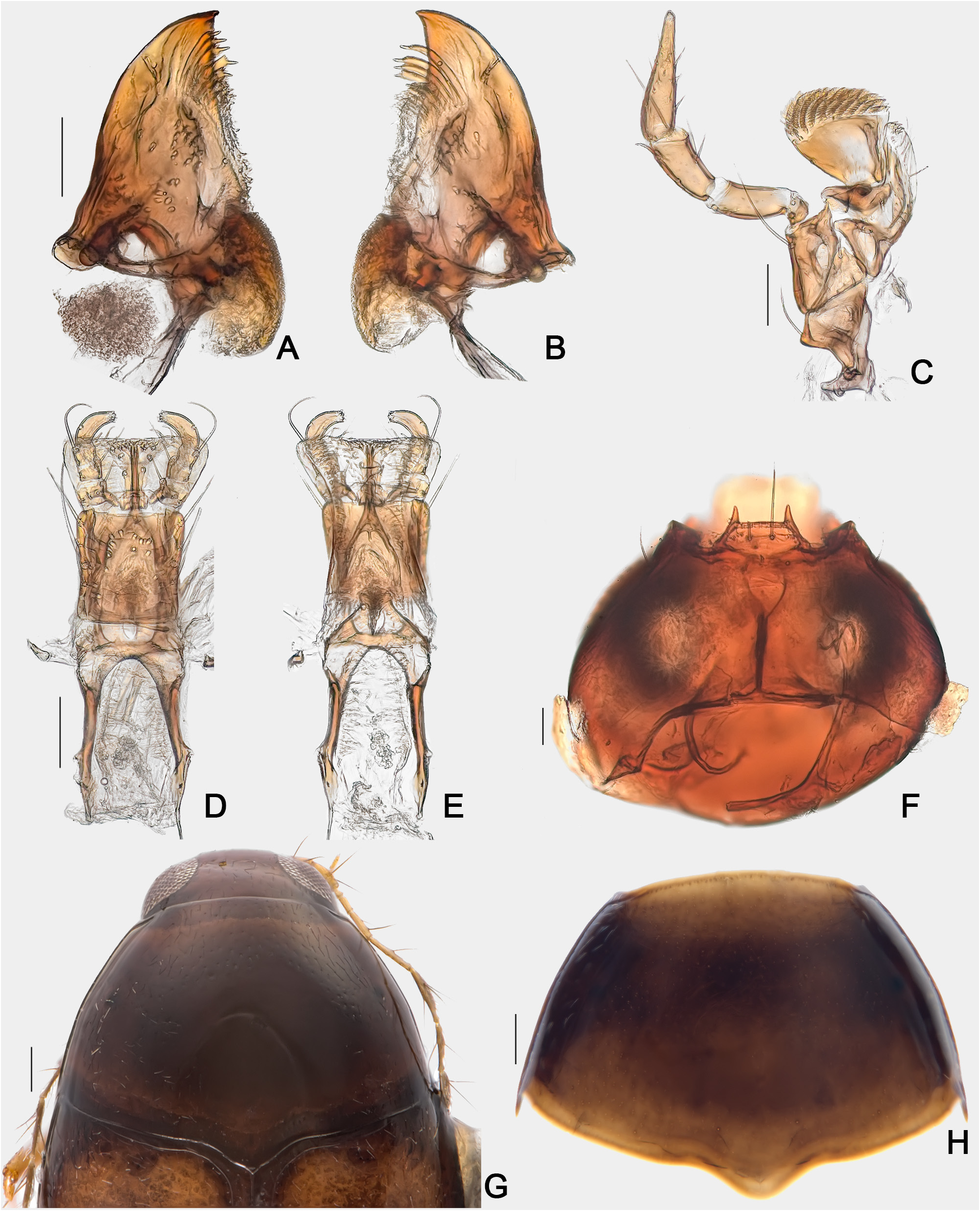
Diagnosis. TL: 1.38–1.56 mm in males and 1.50–1.64 mm in females. Elytral apex and sometimes the humeral areas yellow ( Figs 2A–C View FIGURE 2 ). First abdominal ventrite dark anteriorly and yellow at apex ( Fig. 2D View FIGURE 2 , 6F View FIGURE 6 ). Metaventrite densely pubescence on disc, but sparsely pubescent laterally ( Fig. 4D View FIGURE 4 ). Mesocoxal lines arcuate, impunctate ( Fig 4D View FIGURE 4 ); submetacoxal lines convex and punctate ( Fig. 6F View FIGURE 6 ). Aedeagus with internal sac simple ( Figs 7F, G View FIGURE 7 ), with conspicuous denticular structure ( Figs 7H View FIGURE 7 ).
Description. Coloration: brown to dark brown ( Figs 2A–F View FIGURE 2 ). Head castaneous ( Fig. 2G View FIGURE 2 ); antennae pale yellow to yellow ( Fig. 2H View FIGURE 2 ); mouth parts pale yellow ( Figs 2I View FIGURE 2 ; 3A–E View FIGURE 3 ). Thorax light brown to brown ventrally ( Fig. 2D View FIGURE 2 ); pronotum castaneous to brown ( Figs 3G,H View FIGURE 3 ); coxae and legs yellow ( Fig. 2D View FIGURE 2 ). Elytra mostly brown, with apical 1/4 yellow ( Figs 2A–C View FIGURE 2 ); humeral areas with or without yellow band extending to apex, more conspicuous in teneral specimens ( Fig. 2C View FIGURE 2 ). Abdomen mostly yellow; anterior portion of first segment dark ( Fig. 2D View FIGURE 2 ).
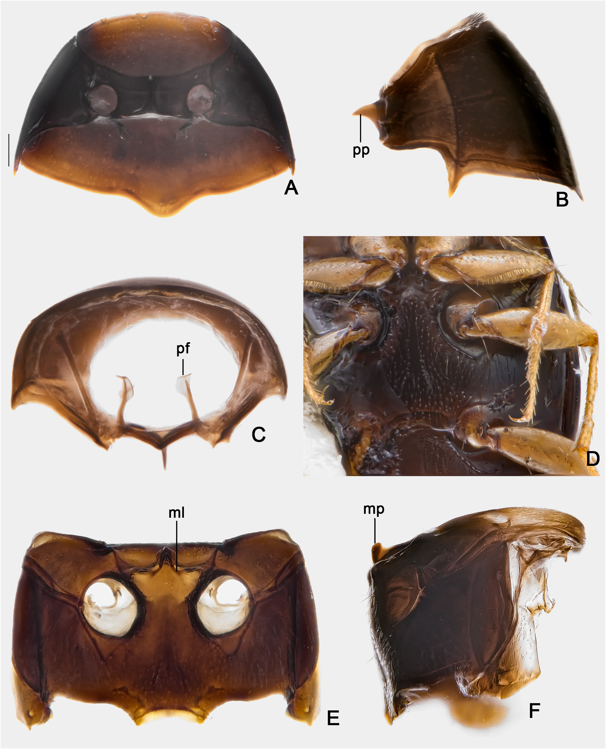
Head ( Fig. 2G View FIGURE 2 ). Subcylindrical, glossy and smooth; frons with fine and sparse punctation, pubescent. Labral setae present ( Fig. 2I View FIGURE 2 ). Mandibles unidentate ( Figs 3A,B View FIGURE 3 ), subapical serrations on left mandible very small, barely visible. Maxillae with aciculate palps (each apical palpomere narrower than the precedent), palpomere II with two apical setae ( Fig. 3C View FIGURE 3 ); galea wide, radulate ( Fig. 3C View FIGURE 3 ); lacinia with long and sparse lateral setae ( Fig. 3C View FIGURE 3 ). Hypopharynx setose ( Figs 3D,E View FIGURE 3 ). Labium with curved palps; palpomere I and II with one subapical seta each ( Fig. 3D View FIGURE 3 ). Mentum rectangular and setose ( Fig. 3D View FIGURE 3 ). Submentum with trapezoidal apex ( Fig. 3F View FIGURE 3 ). Gula not reaching submentum ( Fig. 3F View FIGURE 3 ), without pores ( Fig. 3F View FIGURE 3 ). Eyes notched anteriorly by antennal insertions ( Fig. 2G View FIGURE 2 ). Antennae filiform ( Fig. 2H View FIGURE 2 ), with measurements as follows (left antenna, from base to apex, in mm): 0.06, 0.07, 0.03, 0.05, 0.07, 0.08, 0.11, 0.09, 0.12, 0.11, 0.12.
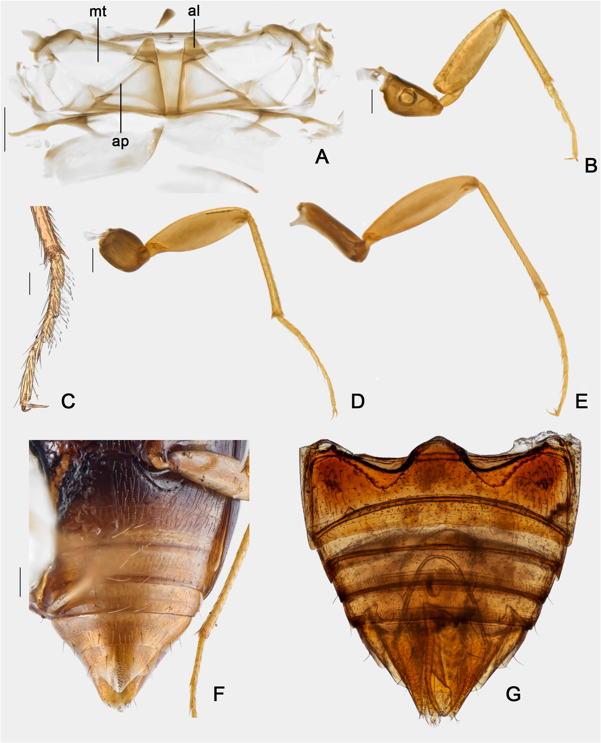
Prothorax. Pronotum trapezoidal, glossy; punctation dense, fine and somewhat deep; each puncture with one seta as long as interspace of two punctures ( Figs 3G,H View FIGURE 3 ); lateral margins rounded ( Figs 3G,H View FIGURE 3 ), with prominent carinae visible when seen from above, each carina with punctate lateral stria alongside. Posterior pronotal angle acuate, partially concealed bellow elytra ( Fig. 2F View FIGURE 2 ). Prosternum in front of procoxae ( Fig. 4A View FIGURE 4 ) shorter than procoxal cavity; prosternal process sickleform ( Fig. 4B View FIGURE 4 ). Hypomera smooth, glabrous ( Figs 2D View FIGURE 2 , 4A View FIGURE 4 ), completely visible in lateral view ( Figs 2E,F View FIGURE 2 ); apex not extended beyond posterior margin of pronotum. Profurca ( Fig. 4C View FIGURE 4 ) with stalk longer than apex; apex rounded.
Mesothorax. Mesoventral space (prepectus) absent. Procoxal rests subrounded ( Figs 4D,E View FIGURE 4 ). Mesoventral lines present, short, impunctate ( Figs 4D,E View FIGURE 4 ). Secondary lines absent. Mesoventral process paxillate, elongate-oval in lateral view ( Fig. 4F View FIGURE 4 ). Median lines short, connected to mesoventral process ( Fig. 4E View FIGURE 4 ). Mesanepisterna smooth, shinning; punctures, sparse; pubescence, fine ( Fig. 4E View FIGURE 4 ). Mesepimera shinning, smooth, about 1/2 the length of anapleural suture and 5 times as long as wide ( Fig. 4E View FIGURE 4 ). Distance between mesocoxae slightly shorter than width of each coxa ( Fig. 4E View FIGURE 4 ).
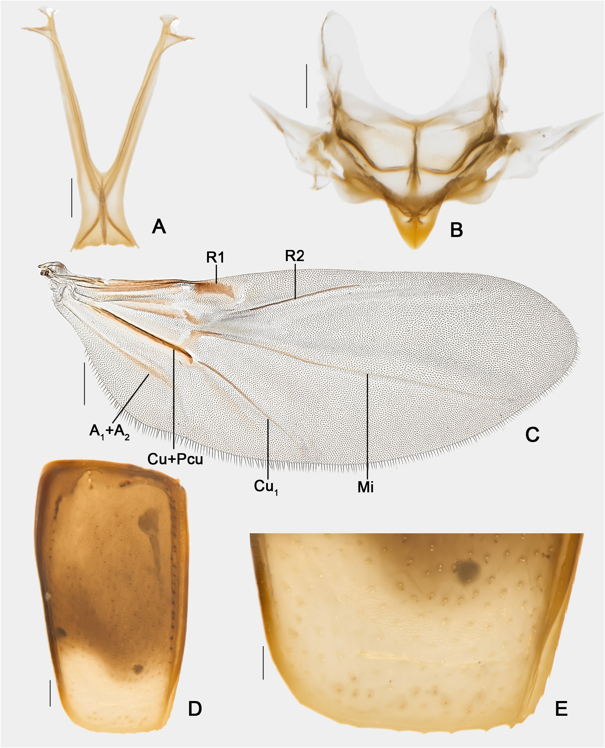
Metathorax. Metaventrite ( Fig. 4E View FIGURE 4 ) smooth, shinning; punctation dense and coarse on disc, fine and sparse laterally; pubescence dense and thick on disc, fine and sparse laterally. Metaventrite fused to mesoventrite. Mesocoxal lines arcuate, not punctate ( Fig. 4D,E View FIGURE 4 ). Primary setae setose ( Fig. 4D View FIGURE 4 ). Premetacoxal lines and discrimen absent. Metepimera expanding gradually posteriorly; metepimeral suture impunctate. Metanepisterna narrowed anteriorly ( Figs 2F View FIGURE 2 , 4E,F View FIGURE 4 ). Metendosternite with short stalk and well-developed arms ( Fig. 5A View FIGURE 5 ).
Pterothorax. Tip of scutellar shield exposed; tip width about one third the length of the pteronotum; scutellar suture trapezoidal ( Fig. 5B View FIGURE 5 ). Hind wings fully developed; R1 small, R2, M1 and Cu 1 thin, Cu+Pcu slightly thickened, A 1 +A 2 faint ( Fig. 5C View FIGURE 5 ). Elytra convex ( Figs 2A–C View FIGURE 2 , E-F, 5D); carinae of lateral margin visible in dorsal view; apical margins somewhat rounded; sutural striae somewhat expanding to disc and tapering to apex ( Figs 2A–C View FIGURE 2 ); basal striae reaching 2/3 of elytra, joined to sutural striae forming a curve ( Figs 2A–C View FIGURE 2 , 3G View FIGURE 3 ); adsutural area with single row of punctures, each puncture bearing a seta ( Figs 2A–C View FIGURE 2 ); lateral stria punctate; apical edge serrate ( Fig. 5E View FIGURE 5 ). Metanotum ( Fig. 6A View FIGURE 6 ) with alacristae trapezoidal; sides subrounded; metascutum, large; apodeme, oblique.
Legs ( Figs 6B–E View FIGURE 6 ). Profemora with ctenidium ( Fig. 4D View FIGURE 4 ). Mesofemora rounded in cross-section; subapical seta not sclerotized. Tibiae smooth, with spinose apex ( Figs 6B,D,E View FIGURE 6 ). Coxae, trochanters and femora with striate microsculpture.
Abdomen ( Figs 6F,G View FIGURE 6 ). Microsculpture transversely striate, densely pubescent; punctation fine, shallow; variable number of primary setae on disc of ventrites. Submetacoxal lines convex, punctate; submetacoxal areas finely microsculptured. Pygidium smooth dorsally.
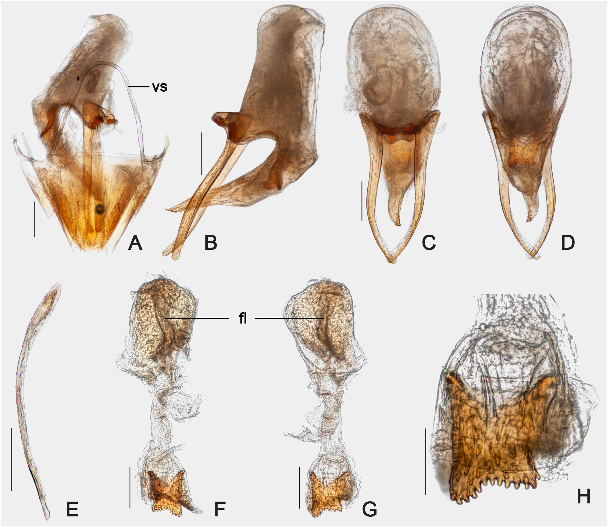
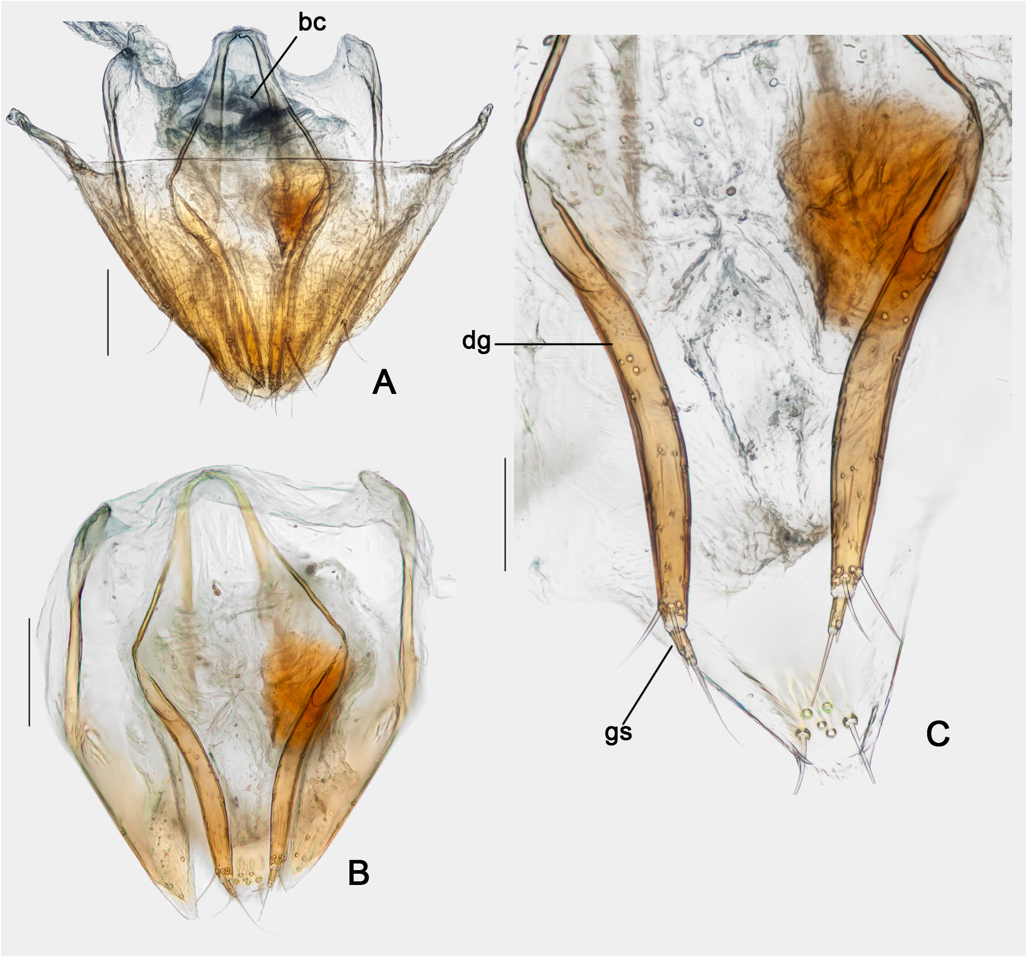
Males. Pro- and mesotarsomeres widened, with tenent setae ( Figs 6B,C View FIGURE 6 ). Abdominal ventrites 1–3 each with six thick primary setae, 4–5 each with two setae (variation may be due to loss of setae) ( Fig. 6F View FIGURE 6 ). Sternum IX long, narrow ( Fig. 7E View FIGURE 7 ). Tergite IX with broad ventral struts ( Fig. 7A View FIGURE 7 ). Aedeagus ( Figs 7 View FIGURE 7 B-D) symmetrical; basal bulb weakly sclerotized; apex well sclerotized, somewhat twisted, with subapical denticles ( Fig. 7B View FIGURE 7 ); parameres slightly arcuate and widened, overlapping apically; internal sac simple, with wide curved flagellum ( Figs 7F,G View FIGURE 7 ) and denticular structure bearing a row of teeth ( Figs 7H View FIGURE 7 ).
Females. Each abdominal ventrite with one pair of primary setae on disc, usually thinner than those of males. Ovipositor simple ( Figs 8A–C View FIGURE 8 ): distal gonocoxites elongated, with slender gonostyli; bursa copulatrix small, membranous ( Fig. 8A,B View FIGURE 8 ); spermatheca not detected.
Measurements. Males (n=6, unless otherwise specified; in mm): TL 1.38–1.56 (mean = 1.50, standard deviation ± 0.06), PL 0.56–0.65 (0.61 ± 0.03), PA 0.47–0.52 (0.50 ± 0.02), PB 0.84–0.92 (0.86 ± 0.03), EL 0.94–1.05 (1.00 ± 0.04), EI 0.76–0.93 (0.90 ± 0.06), EW 0.43–0.52 (0.47 ± 0.03), EH 0.35–0.42 (0.37 ± 0.03), HW 0.43–0.49 (0.46 ± 0.02), SY 0.17–0.18 (0.18 ± 0.01), GY 0.21–0.24 (0.22 ± 0.01), WA 0.10–0.12 (0.11 ± 0.01), MC (n=2) 0.15–0.47 (0.31 ± 0.23), MB (n=5) 0.13–0.15 (0.14 ± 0.01), VL (n= 5) 0.24–0.32 (0.28 ± 0.04), ML 0.05–0.09 (0.06 ± 0.02), MA 0.05–0.07 (0.06 ± 0.01). Females (n= 6, unless otherwise specified; in mm): TL 1.50–1.64 (mean=1.57, standard deviation ± 0.05), PL 0.62–0.66 (0.64 ± 0.01), PA 0.51–0.54 (0.52 ± 0.01), PB 0.86–0.95 (0.90 ± 0.03), EL 0.98–1.05 (1.02 ± 0.02), EI 0.91–0.97 (0.93 ± 0.02), EW 0.43–0.52 (0.49 ± 0.03), EH 0.36–0.43 (0.39 ± 0.03), HW 0.46–0.50 (0.48 ± 0.02), SY 0.18–0.21 (0.20 ± 0.01), GY 0.22–0.25 (0.23 ± 0.01), WA 0.10–0.15 (0.12 ± 0.02), MC (n= 4) 0.15–0.52 (0.40 ± 0.17), MB (n= 5) 0.15–0.19 (0.16 ± 0.02), VL 0.26–0.32 (0.30 ± 0.03), ML 0.05–0.07 (0.05 ±0.01), MA 0.05–0.07 (0.05 ± 0.01).
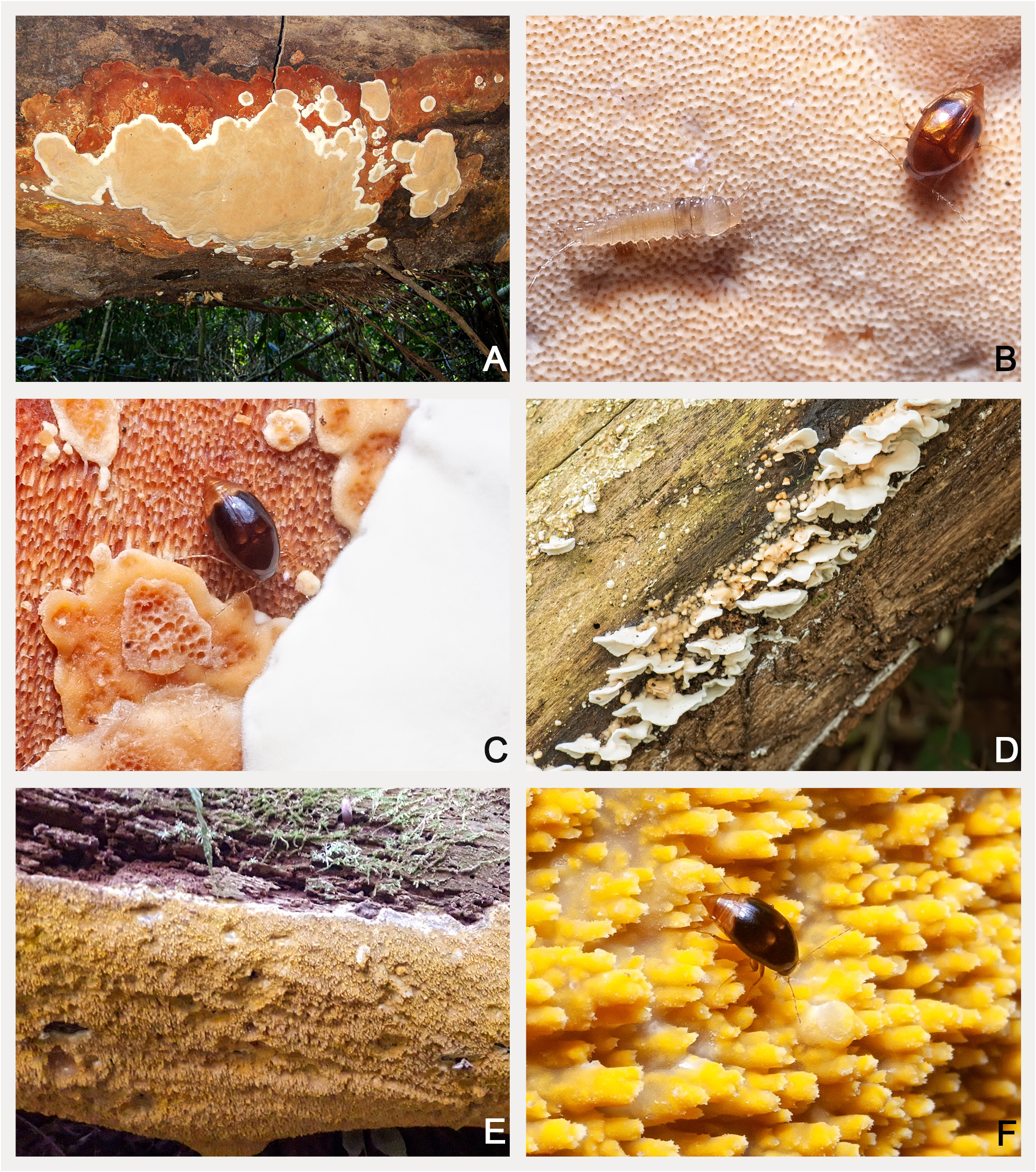
Host fungi. Adult specimens were collected six times from Geesterania cf. carneola (Bres.) Westphalen & Rajchenberg ( Steccherinaceae ; Figs 9A–C View FIGURE 9 ). Adults were also collected once in Steccherinum undigerum (Berk. & M.A. Curtis) ( Steccherinaceae ; Fig. 9D View FIGURE 9 ) and in Schizopora paradoxa (Schrad.) Donk ( Schizoporaceae ; Figs 9E–F View FIGURE 9 ).
Etymology. From the Latin “pandemus”, derived from the Ancient Greek πάνδημος and meaning “affecting all the people”, in the neuter nominative singular, in reference to the fact that the present article was written during the pandemic of covid-19, who drastically affected our ongoing works on the Atlantic Forest scaphidiines.
Remarks. Scaphisoma pandemum has been frequently collected in two sites: “Mata da Biologia” and “Mata do Paraíso”, both in Viçosa, Minas Gerais. The basidiomes identified as Geesterania cf. carneola, mentioned in the type material, correspond to a single fungus species, which was the most common host species for Scaphisoma pandemum and in which all color morphs were found. In order to make sure that the color morphs belong to a single species, two males from “Mata do Paraíso” and four males from “Mata da Biologia” were dissected. The dissected specimens from “Mata do Paraíso” were of different colors: one with the yellow humeral band and the other without it; they were found in different fungus species as well. Three males from “Mata da Biologia” were devoid of yellow humeral band, two of them collected in Geesterania cf. carneola and the other in Schizopora paradoxa . The fourth male had the humeral yellow band and was collected in Geesterania cf. carneola. We considered the observed color variation of the body as intraspecific variation of a single species.
Scaphisoma pandemum is similar to Scaphisoma lunatum (Matthews, 1888) from Nicaragua and Costa Rica. Both have similar coloration, but different mesocoxal lines: S. lunatum has parallel mesocoxal lines ( Fierros-López, 2006), while S. pandemum has arcuate lines. Scaphisoma pandemum is also comparatively smaller (1.38–1.64 mm; while S. lunatum is 1.75 mm). Their aedeagi are similar, but Fierros-López (2006) did not mention whether there is a denticular structure in the internal sac, present in S. pandemum ; parameres in S. pandemum are thinner and straighter than in S. lunatum . Scaphisoma pandemum is also similar to S. rubripes , but they have different body coloration: the body of S. rubripes is mostly black, and its antennae, labrum, tarsi and abdominal apex are red ( Pic, 1920a); while S. pandemum is brown to dark brown, its labrum, antennae and tarsi are pale yellow, and its abdomen and elytral apex are yellow. No specimen of S. pandemum examined by us had red body parts. Their body lengths are also different: S. pandemum is 1.38–1.64 mm long, while S. rubripes is 2 mm long.
| FMNH |
Field Museum of Natural History |
No known copyright restrictions apply. See Agosti, D., Egloff, W., 2009. Taxonomic information exchange and copyright: the Plazi approach. BMC Research Notes 2009, 2:53 for further explanation.
|
Kingdom |
|
|
Phylum |
|
|
Class |
|
|
Order |
|
|
Family |
|
|
Genus |