
VORTICELLIDAE Ehrenberg, 1938
|
publication ID |
https://doi.org/ 10.1080/00222930500239785 |
|
persistent identifier |
https://treatment.plazi.org/id/03DD1129-F014-FFDF-2F0E-23C45FCBF90A |
|
treatment provided by |
Felipe |
|
scientific name |
VORTICELLIDAE Ehrenberg, 1938 |
| status |
|
Family VORTICELLIDAE Ehrenberg, 1938 View in CoL View at ENA
Genus Pseudovorticella Foissner and Schiffmann, 1974 View in CoL Pseudovorticella cylindrica ( Dons, 1915) View in CoL nov. comb.
( Figures 1–18 View Figures 1–8 View Figures 9–18 ; Table I) Synonym Vorticella cylindrica Dons, 1915 View in CoL . Since no ciliature information was previously available for this organism, here we supply an improved diagnosis and detailed data on the infraciliature as well as on the morphology of living cells, based on the Qingdao population.
Revised diagnosis Inverted bell-shaped Pseudovorticella , length in vivo 40–50 mm and length: width ratio about
1:1; macronucleus J-formed; one contractile vacuole apically located; reticulate silverline system with 33–40 transverse striations between oral area and aboral ciliary wreath, and
14–19 striations between aboral ciliary wreath and scopula. The inner row of peniculus 3 conspicuously shorter than other two. Marine.
Description
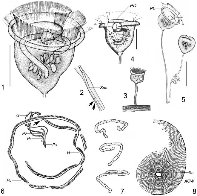
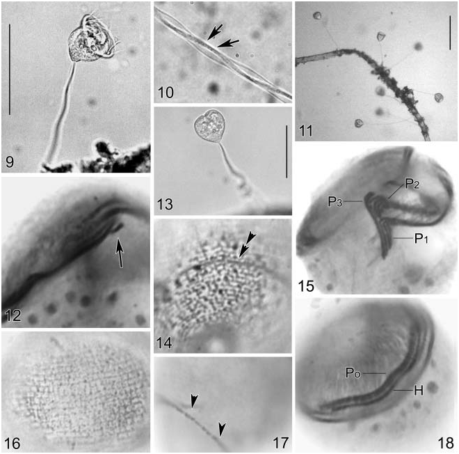
Cell constant in size and shape, about 40–50× 45–50 mm in vivo, usually with a slight constriction in the aboral region. Maximum width of cell at peristomial area with thin and relatively rigid peristomial lip (PL; Figures 1, 5 View Figures 1–8 , 9 View Figures 9–18 ). Peristomial disc (PD) flat and only slightly elevated when cell fully extended ( Figures 1 View Figures 1–8 , 9 View Figures 9–18 ). Pellicle generally smooth, while a finely reticulate pattern of striations can be observed only under high magnification (1000×) ( Figure 1 View Figures 1–8 ).
Cytoplasm colourless or greyish, usually containing several refractive particles, 4–7 mm in length ( Figures 1, 5 View Figures 1–8 , 13 View Figures 9–18 ). Single contractile vacuole large, apically located, contracting every 2–3 min. Macronucleus J-formed, variable in shape and generally situated transversely ( Figure 7 View Figures 1–8 ). Micronucleus not observed.
Stalk usually two to four times zooid length, relatively thick and about 5 mm in diameter ( Figure 11 View Figures 9–18 ). The spasmoneme (Spa) about 2 mm in diameter, with a string of conspicuous, dark grey thecoplasmic granules (ca 0.3 mm in diameter) ( Figures 2 View Figures 1–8 , 10 View Figures 9–18 , arrows).
Individuals often closely grouped together thus forming pseudocolonies of up to 30 zooids. Telotroch (swarmer) was not observed.
Infraciliature as shown in Figures 6 View Figures 1–8 , 12, 15, 17, 18 View Figures 9–18 . Haplokinety (H) and polykinety (Po) describing about 1.5 turns around peristomial disc before entering vestibulum, where they make a further turn. Similar to that of other congeners, polykinety forms three peniculi in lower half of vestibulum, each consisting of three rows. The posterior ends of peniculus 1 (P1) and P3 terminate at the same level whereas P2 situates between and terminates conspicuously above P1 and P3 ( Figures 6 View Figures 1–8 , 15 View Figures 9–18 ). The inner row of P3 is characteristically displaced relative to the other two: very short, about one-third length of the outer two rows which converge with P1. The haplokinety passing around the vestibulum on the opposite wall to the peniculi. Germinal kinety (G) consisting of zig-zag structure of kinetosomes and extending to the upper one-third of the vestibulum, where it passes immediately beyond the haplokinety ( Figure 6 View Figures 1–8 ). The epistomial membrane is short, located near the opening of the infundibulum ( Figures 6 View Figures 1–8 , 12 View Figures 9–18 , arrow). Aboral ciliary wreath encircles cell in posterior region and seems to be composed of a Z-rowed structure ( Figure 14 View Figures 9–18 , double arrowhead; Figure 17 View Figures 9–18 , arrowheads).
Silverline system reticulate ( Figure 16 View Figures 9–18 ). Transverse lines in the anterior region of the cell are more widely spaced than those in the posterior region ( Figures 8 View Figures 1–8 , 14, 16 View Figures 9–18 ). The number of transverse silverlines from peristome to aboral ciliary wreath (5aboral trochal band), 33– 40; from aboral ciliary wreath to scopula, 14–19 ( Table I).
Comparison
Pseudovorticella cylindrica View in CoL was originally described by Dons (1915) ( Figure 3 View Figures 1–8 ) under the name of Vorticella cylindrica View in CoL . Its infraciliature remained unknown though it was redescribed in the last decade by Song (1991a) ( Figure 4 View Figures 1–8 ). The Qingdao population resembles the previous descriptions in all aspects, e.g. body shape, size, number of contractile vacuoles, as well as marine habitat, hence we are confident that the identification is correct.
A summary of comparisons between P. cylindrica View in CoL and similar Pseudovorticella View in CoL and Vorticella spp. is given in Table II.
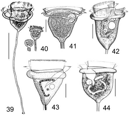
Morphologically, Pseudovorticella patellina (Müller, 1776) Song and Warren, 2000 ( Figure 39 View Figures 39–44 ) should be closely related to P. cylindrica . Nevertheless, the former differs from P. cylindrica in its larger size (55–110 versus 40–50 mm), the number of transverse silverlines between the aboral ciliary wreath and the anterior end (19–22 versus 33–40), number of contractile vacuoles (two versus one), and the extreme outwards extension of the peristomial lip. In addition, the structure of peniculus 3 is completely different (see Song and Warren 2000).
Pseudovorticella punctata (Dons, 1918) Warren, 1987 View in CoL ( Figure 43 View Figures 39–44 ) is most similar to P. cylindrica View in CoL , especially in terms of body size and the habitat. However, P. punctata View in CoL can be clearly distinguished from the latter by the appearance of the peristomial lip (considerably thick versus thin), the lower position of the contractile vacuole (below the vestibulum versus apically located), and having significantly fewer transverse lines (about 30 versus 52) ( Warren 1987).
Considering the general morphology, further comparisons should be also made with some Vorticella species. Vorticella campanula Ehrenberg, 1831 View in CoL ( Figures 40, 41 View Figures 39–44 ) corresponds very well in vivo with Pseudovorticella cylindrica View in CoL . In the past 80 years, P. cylindrica View in CoL was repeatedly considered to be a marine variety of V. campanula View in CoL ( Noland and Finley 1931; Warren 1987). In 1992, a thorough redescription of V. campanula View in CoL using modern techniques was given by Foissner et al. (1992). Thus, we have the opportunity to compare these two forms. Vorticella campanula View in CoL can be separated from P. cylindrica View in CoL by size (50–160 versus 40–50 mm), the habitat (freshwater versus marine), Vorticella View in CoL - type silverline system (versus Pseudovorticella View in CoL pattern), and dissimilarities considering the infraciliature ( Foissner et al. 1992).
Vorticella nebulifera Müller, 1786 also bears some resemblance to Pseudovorticella cylindrica , particularly with respect to the smooth pellicle and cell size. However, the two taxa can be clearly separated by the position of the contractile vacuole (lower ventral versus apically dorsal located) ( Figure 42 View Figures 39–44 ), different pattern of silverline system, and the body shape (slender in V. nebulifera ) ( Song 1991a).
With reference to body shape and the habitat, another similar form, Vorticella marina Greeff, 1870 whose silverline system and infraciliature remain unclear, should also be compared with P. cylindrica . The former differs from the latter, even at the in vivo level, in the lower position of contractile vacuole ( Figure 44 View Figures 39–44 ) and appearance of pellicle (distinctly striated versus finely striated) ( Song 1991a).
No known copyright restrictions apply. See Agosti, D., Egloff, W., 2009. Taxonomic information exchange and copyright: the Plazi approach. BMC Research Notes 2009, 2:53 for further explanation.
|
Kingdom |
|
|
Phylum |
|
|
Class |
|
|
Order |
|
|
Family |
VORTICELLIDAE Ehrenberg, 1938
| Sun, Ping, Song, Weibo, Ji, Daode & Hu, Xiaozhong 2005 |
Pseudovorticella cylindrica ( Dons, 1915 )
| Sun & Song & Ji & Hu 2005 |
Pseudovorticella cylindrica
| Sun & Song & Ji & Hu 2005 |
Pseudovorticella cylindrica
| Sun & Song & Ji & Hu 2005 |
cylindrica
| Sun & Song & Ji & Hu 2005 |
P. cylindrica
| Sun & Song & Ji & Hu 2005 |
P. cylindrica
| Sun & Song & Ji & Hu 2005 |
Pseudovorticella cylindrica
| Sun & Song & Ji & Hu 2005 |
P. cylindrica
| Sun & Song & Ji & Hu 2005 |
P. cylindrica
| Sun & Song & Ji & Hu 2005 |
patellina
| Song and Warren 2000 |
punctata
| Warren 1987 |
Pseudovorticella punctata (Dons, 1918)
| Warren 1987 |
P. punctata
| Warren 1987 |
Pseudovorticella
| Foissner and Schiffmann 1974 |
Pseudovorticella
| Foissner and Schiffmann 1974 |
Pseudovorticella
| Foissner and Schiffmann 1974 |
Pseudovorticella
| Foissner and Schiffmann 1974 |
Pseudovorticella
| Foissner and Schiffmann 1974 |
Pseudovorticella
| Foissner and Schiffmann 1974 |
Vorticella cylindrica
| Dons 1915 |
Vorticella cylindrica
| Dons 1915 |
Vorticella marina
| Greeff 1870 |
Vorticella campanula
| Ehrenberg 1831 |
Vorticella campanula
| Ehrenberg 1831 |
V. campanula
| Ehrenberg 1831 |
V. campanula
| Ehrenberg 1831 |
Vorticella campanula
| Ehrenberg 1831 |
Vorticella nebulifera
| Muller 1786 |