
Mnesilochus luwuense, Hennemann, 2021
|
publication ID |
https://doi.org/ 10.11646/zootaxa.5073.1.1 |
|
publication LSID |
lsid:zoobank.org:pub:AA3269D1-CA2F-4528-BC9D-3A4C75D05BD9 |
|
persistent identifier |
https://treatment.plazi.org/id/03DB87EE-FFDA-9D67-FF40-5FDCFB78F2C9 |
|
treatment provided by |
Plazi |
|
scientific name |
Mnesilochus luwuense |
| status |
sp. nov. |
Mnesilochus luwuense n. sp.
( Fig. 18 View FIGURE 18 )
Lonchodes haematomus, Günther, 1938: 78 .
HT, ♀: Sarasin, Luwu, Central-Celebes; Lonchodes hosei Kby. K. Günther det. [ NHMB, No. VI.D.133] .
Etymology: Named after the Luwu Regency, the type-locality and only known record of this new species.
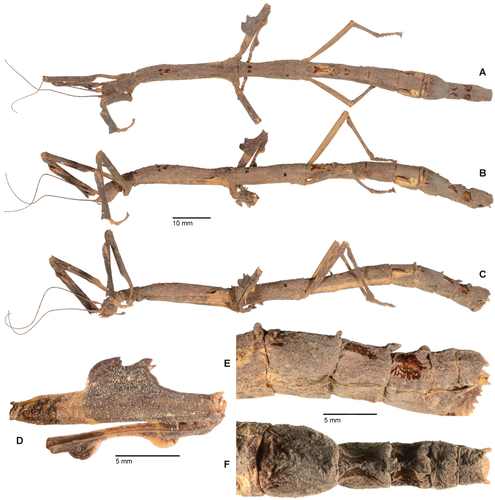
Differential diagnosis: Similar in general shape and appearance to the Philippine M. haedulus Stål, 1877 (as described and illustrated by Hennemann & Conle, 2007: 73, figs. 107–113) and the Palawanese M. palawanicus (Carl, 1913) but readily separated from these and from all other known members of the genus by the morphology of the mesofemora. While these species have the dorsomedian lobe roughly triangular in shape with the posterior margin dentate, this lobe is distinctively bifid with a median excavation in M. luwuense n. sp. ( Fig. 18D View FIGURE 18 ).
Description: ♀ ( Fig. 18 View FIGURE 18 ). The unique holotype has provisionally been preserved in spirits, what might have caused discolouration. Moreover, ♀♀ of this genus are known to show considerable morphological and chromatic variability, why the description here presented on basis of the holotype can in several aspects only be exemplary for this species.
Of medium size (body length 102.0 mm) and moderately stocky shape for the genus with a large, bifid dorsal lobe of the mesofemora. Entire body surface unevenly granulose, tuberculose, verruculose and rugulose; abdominal terga II and V– VIII with posteromedian protrusions. General colour of the holotype greyish mid brown with the posterolateral portions of the mesothorax, lateral portions of abdominal sterna II – IV buffy. Mesonotum with four small blackish dorsal markings in anterior half and a narrow black lateral marking some distance before the posterior margin. Metanotum with two black spots sub-anteriorly and a black spot at each posterolateral angle. Median portion of abdominal tergum II ochre with two elongate and washed blackish median markings and two smaller dark brown markings posterolaterally. Two similar small blackish markings in anterior portion of tergum III. Tergum V with lateral surfaces mostly pale ochre and with a blackish streak in anterior half. Terga VII and VIII each with a dark brown to black central marking. Cerci and epiproct straw. A blackish sub-apical marking also present on interior surface of protibiae. Mesofemora irregularly dark brown basally and with a buff transverse sub-basal band, the mesotibiae buff in the median portion and dark brown apically. Metafemora ochre on their interior surface. Antennae greyish mid brown ventrally, ochre dorsally with most antennomeres dark apically; scapus buff. Eyes dark grey .
Head: Elongate, sub-cylindrical, notably longer than wide and very slightly narrowed posteriorly. Between the eyes with a raised transverse area which has both outer ends protruded into an obtuse, slightly anteriad directed spine. Vertex flattened and irregularly set with granules, nodes and tubercles of variable sizes, also a few tubercles present on genae. Posterior margin with a transverse row of four obtusely rounded but enlarged tubercles. Eyes almost circular in outline, weakly protruding and their diameter contained 2.2x in length of genae.Antennae projecting somewhat over posterior margin of mesothorax. Scapus strongly compressed dorsoventrally, narrowed basally, expanded medially and roughly oval in outline. Pedicellus slightly oval in cross-section and less than half the length of scapus.
Thorax: Pronotum roughly of same dimensions as head, rectangular and 1.7x longer than wide; the transverse median sulcus distinctly impressed but narrow, straight and expanding over entire width of segment. Mesothorax 4.2x longer than pronotum and very weakly widened post-anteriorly. Mesonotum with a fine longitudinal median line and set with a few scattered, enlarged and somewhat wart-like tubercles; a transverse row of four enlarged tubercles just before posterior margin. Metanotum over 3x longer than wide and about 0.6x length of mesonotum, the posterior half weakly expanded and the median line like on mesonotum. Mesopleurae with a medio-longitudinal row of flat, elongate tubercles. Mesosternum densely granulose.
Abdomen: Median segment a little more than half the length of metanotum, 1.8x longer than wide with the lateral margins weakly concave and a small transverse swelling posteromedially. Abdomen excluding median segment slightly shorter than pro-, meso- and metathorax combined with segments V–VI notably swollen. Segment II about equal in length to median segment, II–IV uniform in length and V–VII gradually increasing in length; II parallel-sided and 2x longer than wide, III–V gradually widening with V only 1.3x longer than wide. Anterior portion of VI widest part of abdomen and tergum strongly narrowing towards posterior with a trapezoidal outline; lateral margins somewhat deflexed. VII parallel-sided, as broad as II and but shorter and hardly longer than wide. Tergum II with a transverse posterior lobe that bears five rounded tubercles, V and VI each with a similar but more obtuse transverse posteromedian protuberance. Terga VII and VIII each bear a slightly bilobed posteromedian excrescence, which posteriorly extends over the anterior portion of the following tergum ( Fig. 18E View FIGURE 18 ). Preopercular organ formed by a pair of low and obtuse, verruculose swellings near posterior margin of sternum VII. Tergum IX transverse and slightly narrowed towards the posterior. Anal segment narrower than all preceding segments, weakly keeled medio-longitudinally, almost 2x higher than long and with a widely rounded posteromedian excavation ( Fig. 18F View FIGURE 18 ); the posterolateral angles somewhat protruded and the lateral margins slightly deflexed. Epiproct fairly large, shieldshaped and broadly rounded with an obtuse medio-longitudinal bulge; slightly projecting beyond apex of abdomen ( Fig. 18F View FIGURE 18 ). Cerci small, compressed laterally and gently spiral in their length axis. Subgenital plate bulgy and with a prominent, irregularly dentate median keel in posterior portion; very slightly reaching beyond apex of abdomen ( Fig. 18E View FIGURE 18 ).
Legs: In general shape and anatomy rather typical for the genus. Posteroventral carina of profemora with a big triangular sub-apical tooth followed by a small tooth; the anterodorsal carinae strongly lamellate and weakly undulate. Dorsal carina of protibiae strongly rather unevenly deflexed and lamellate with the apical portion forming an obtuse, roughly triangular lobe; not deflexed in the very basal portion although. Two outer ventral carinae slightly lamellate and unevenly undulate. Mesofemora with a very large lobe that is roundly indented medially and thus is of a general bi-lobed outline; the raised apical portion with an additional tooth ( Fig. 18D View FIGURE 18 ). Posteroventral carina protruded into a moderately sized but fairly acute distal directed tooth at the apex. Two outer ventral carinae each with two triangular sub-apical teeth, the apical one being much smaller than the preceding. Mesotibiae with two almost semi-circular dorsal lobes, one sub-basally and one sub-apically ( Fig. 18D View FIGURE 18 ). Metafemora slender and strongly laterally compressed as usual for the genus; the two outer ventral carinae each with three sub-apical teeth, that decrease in size towards apex of femur. Probasitarsus with a moderately sized roughly triangular dorsal lobe; meso- and metabasitarsus simple and just slightly longer than following tarsomere.
Comments: Males and eggs unknown. Günther (1938: 78) erroneously assigned this specimen to Hermagoras hosei ( Kirby, 1896) , a species endemic to Borneo, and also in error referred to this species as a synonym of Staelonchodes haematomus ( Westwood, 1859) which however is a distinct species that also is an endemic of Borneo. Moreover, Günther (1938: 78) assumed this ♀ was the opposite sex of the ♂ paratype of Hermagoras celebensis ( Hennemann, 1998) n. stat. but examination of the concerned specimen clearly shows it is a distinct species, which for instance is at first glance seen by the morphology of the mesofemora. This latter character is known to be very constant in species of Hermagoras Stål, 1875 and Mnesilochus Stål, 1877 , thus being a useful character for species distinction within these two genera. Careful re-examination of this ♀ has shown it to represent an as yet undescribed species and the lack of a medio-longitudinal keel on the mesosternum places it in the genus Mnesilochus .
Distribution: South Sulawesi, Sulawesi Selatan, Luwu Regency [NHMB].
Genus Myronides Stål, 1875
( Figs. 19–23 View FIGURE 19 View FIGURE 20 View FIGURE 21 View FIGURE 22 View FIGURE 23 )
Type-species: Lonchodes pfeifferae Westwood, 1859: 44 , pl. 5: 6, by subsequent designation of Rehn, 1904: 38.
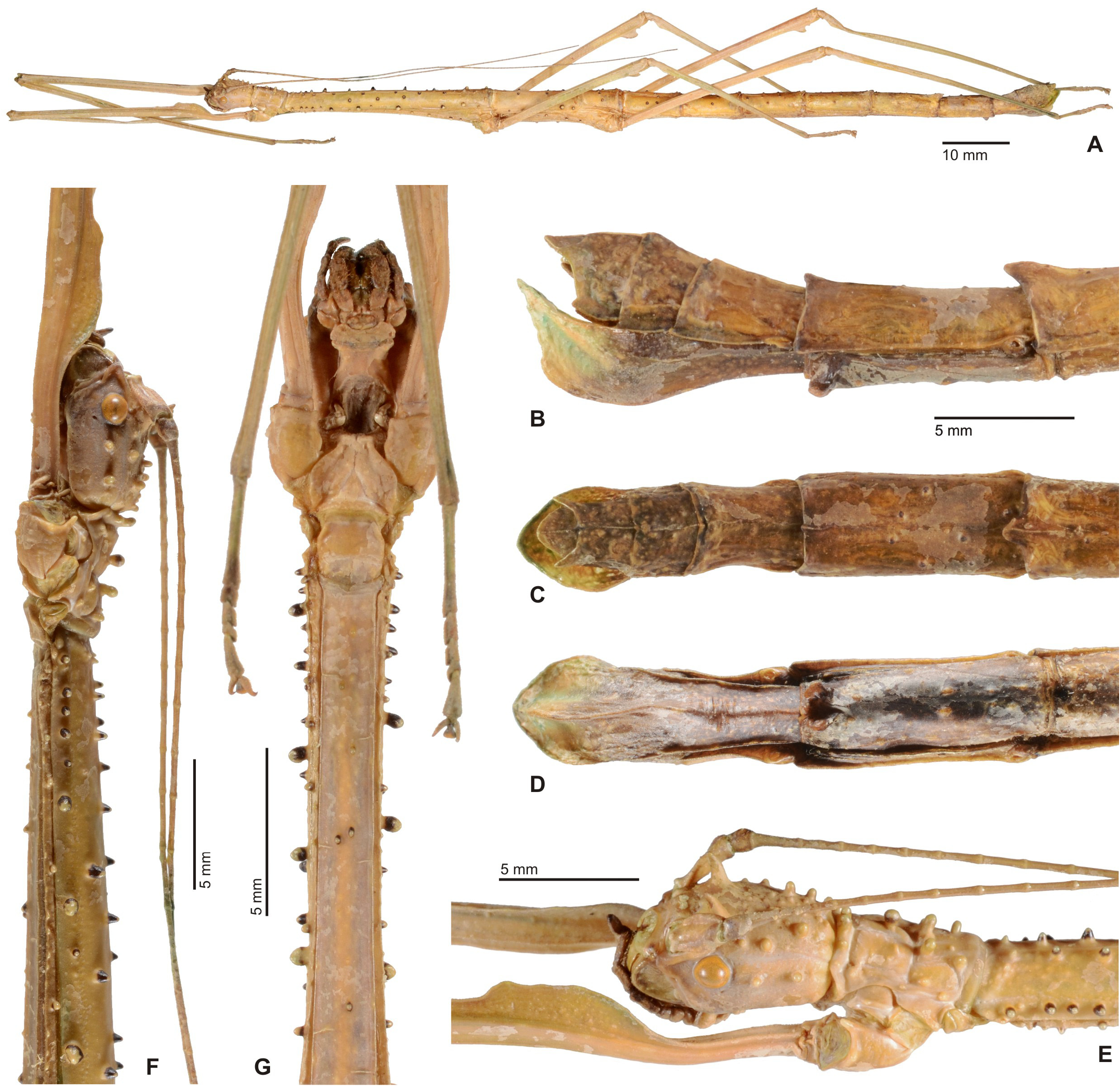
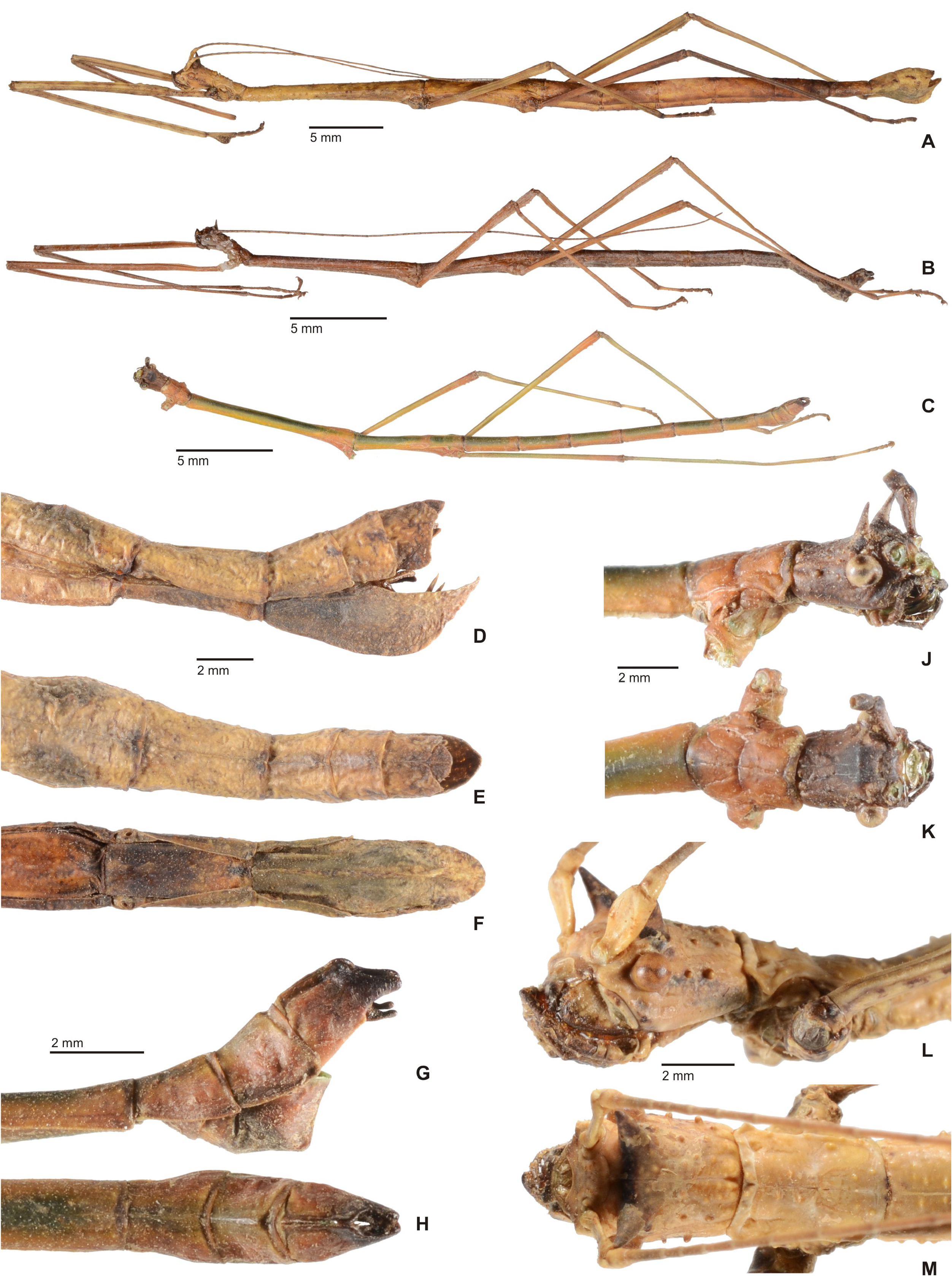
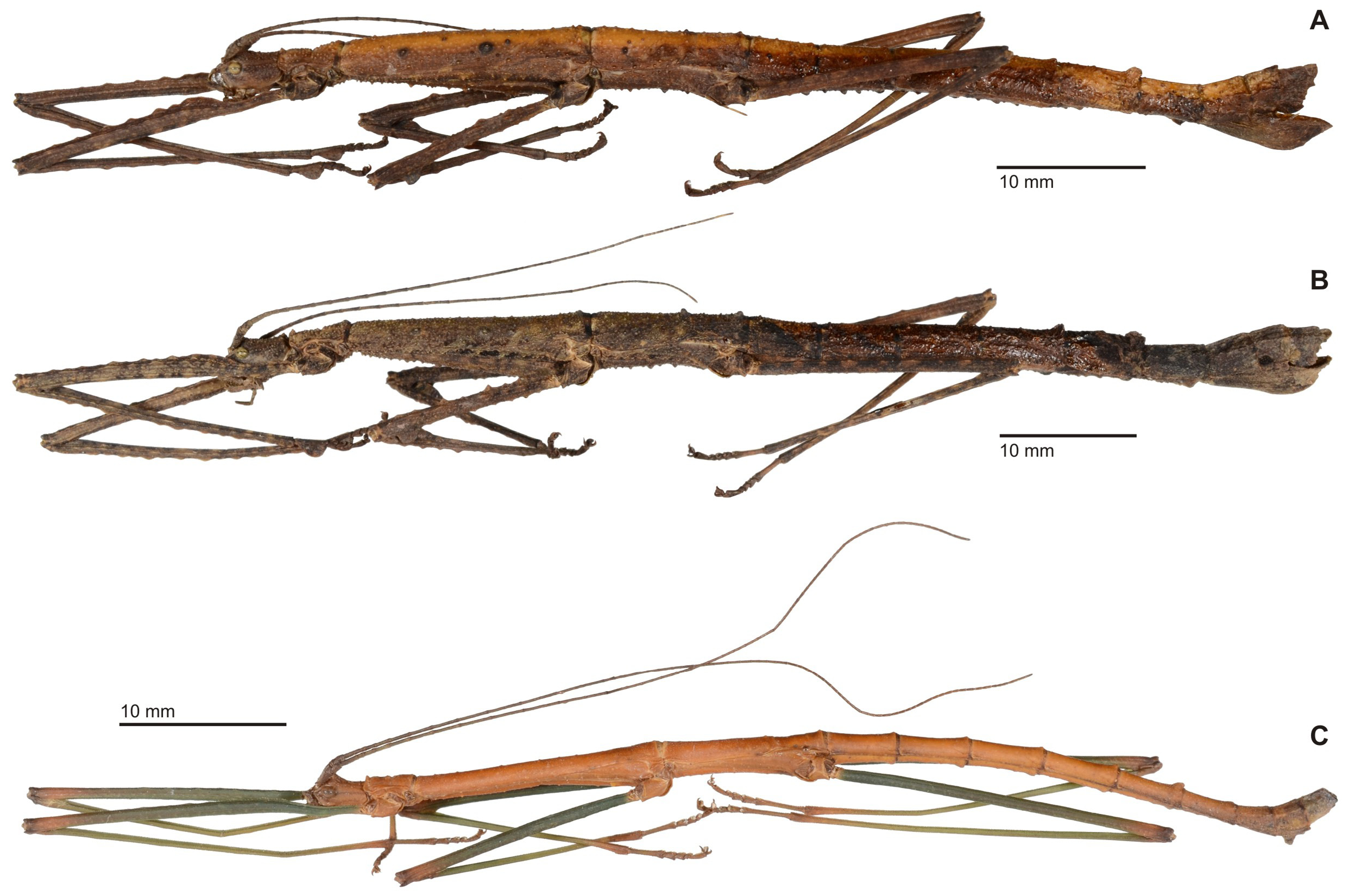
Description: ♀, ♂. Moderately sized (body length ♂♂ 60.0–85.0 mm, ♀♀ 88.0– 125.5 mm) and slender to fairly stocky Lonchodinae . Distinct sexual dimorphism with ♂♂ much smaller, more slender and less sculptured than ♀♀. Colouration various shades of brown, ochre, grey and green; variable intraspecifically in ♀♀, which often have the femora with a conspicuous pale transverse band in apical half of femora. In ♀♀ dorsal body surface with a ± defined longitudinal median carina or weakly tectinate longitudinally. Body surface of ♀♀ densely granulose and/or tuberculose, in ♂♂ smooth with only a few scattered granules on thorax. Head slightly longer than wide with genae ± parallel-sided; vertex flattened and between eyes with a pair of ± distinct spines. Antennae elongate, consisting of about 60 antennomeres and reaching at least to median segment (♀♀) or abdominal segment V–VI (♂♂). Prothorax about equal in length to head, rectangular and with a distinct median sulcus. Mesothorax 5.5–7.0x longer than prothorax; very slender in ♂♂. Meso- and metasternum ± distinctly tectinate longitudinally (very weak in ♂♂); in ♀♀ keel often granulose. Abdomen equal in length or a little longer than head and thorax combined. Median segment of moderate length, less than ½ the length of metanotum. Segments II–VI in ♀♀ very indistinctly increasing in length, all longer than wide and ± parallel-sided. At least terga V–VI with a posteromedian tubercle or swelling. Sternum VII with a distinct Preopercular organ formed by a pair of swellings, spines or lobes close to posterior margin. Terminalia of ♀♀: Terga VIII–X much shorter than previous and decreasing in length. Anal segment tectinate with anterolateral angles rounded and ± deflexed; posterior margin deeply excavated. Epiproct fairly large, rounded, scale-shaped and slightly projecting over apex of anal segment. Cerci very small and hidden under anal segment. Subgenital plate strongly keeled longitudinally, convex and bulgy. Terminalia of ♂♂: Segments II–VI considerably longer than VII and roughly uniform in width and length; VII–X much shorter than previous. Anal segment tectiform and split over entire length to form two hemi-terga that have the anterior margins roundly excavated in apical portion and are apically protruded into a ± long, slender and straight digitiform process. Cerci small, cylindrical and hidden under anal segment. Poculum small, convex, cup-shaped with posterior margin rounded and hardly reaching to posterior of tergum IX. Legs long and slender, profemora longer than mesothorax, mesofemora longer than mesothorax and hind legs slightly (♀♀) to distinctly (♂♂) projecting beyond apex of abdomen. In both sexes entirely unarmed, except for minute sub-apical lobe and/or spine on two outer ventral carinae of meso- and metafemora. In ♀♀ anterodorsal carina of profemora sometimes weakly undulate, occasionally deflexed and slight-ly lobate sub-basally just behind basal constriction of femur. Tarsi slender and simple in both sexes; probasitarsus in ♀♀ not lobed.
Egg: Small (overall length 2.5–3.5 mm), capsule ovoid to barrel-shaped,> 1.5x longer than wide, notably higher than wide and oval cross-section; polar area simple, without mound. Capsule surface coriaceous, granulose or minutely rugulose. Micopylar plate positioned centrally on dorsal egg surface, fairly small, elongately drop-, or pear-shaped with anterior portion progressively narrowing; ± half the length of capsule. Micropylar cup distinct, median line short. Operculum oval and inserted at in capsule at a positive angle of ± 10°. Capitulum irregularly shaped and on a short stalk. Colour of capsule shiny and often with a variable number of dark markings.
Differential diagnosis: Morphology suggests close relation to the Sulawesian endemic Neomyronides n. gen., Chondrostethus Kirby, 1896 (type-species: C. woodfordi Kirby, 1896 ) from the Solomon islands and New Britain as well as the Philippine Mithrenes Stål, 1877 (type-species: M. asperulus Stål, 1877 ).
From Neomyronides n. gen. it differs by the averaging larger size and more slender shape with relatively longer and more slender legs and longer tarsi as well as having a distinct medioventral keel on the mesosternum. Females are readily distinguished by the simple front and mid legs, slender probasitarsus and lacking tubercles in the posterior portion of the subgenital plate. Also, ♀♀ never have the struma or lobe-like posteromedian swelling on abdominal tergum VI occasionally seen in Myronides . Males differ by the more slender hemi-terga of the anal segment, which have a narrowed postero-apical protrusion, as well as the larger cerci. Eggs at once differ from those of Neomyronides n. gen. by the flat operculum ( Figs. 20D–E View FIGURE 20 ).
From Chondrostethus both sexes differ by the shorter median segment of both sexes (more than half the length of metanotum in Chondrostethus ), slender probasitarsus of ♀♀ (with a dorsal lobe in Chondrostethus ) and lacking the conspicuous and typical node-like remnants of alae seen in ♂♂ of Chondrostethus . Eggs of Myronides are on average much paler in colour, have the capsule considerably less distinctly sculptured and a relatively shorter and broader micropylar plate.
Mithrenes was redescribed by Hennemann & Conle (2007: 7) and shares with Myronides the longitudinal median keel of the meso- and metasternum. From this genus Myronides however differs by the longer median segment, which is about half as long as the metanotum (at best 2/5 the length of metanotum in Mithrenes ), more globose head and more distinctly laterally compressed meso- and metafemora of both sexes. Males have the posteroventral projection of the hemi-terga of the anal segment straight and ± horizontal, whereas they are finger-like and angled downward by at least 60° in Mithrenes . The eggs of Myronides differ significantly from those of Mithrenes by being simply ovoid or barrel-shaped.
The monotypical New Guinean Echinothorax Günther, 1932 is only known from a unique ♀ specimen, which seems to differ from Myronides only by the dorsally spinose head and thorax and paired spines on the abdominal terga.
Comments: A re-description of Myronides is presented here to properly define the genus and to provide a basis for removing species that are currently attributed but not congeneric. As previously defined (see Otte & Brock, 2005: 207) Myronides is apparently polyphyletic. All Indochinese species previously attributed to Myronides belong elsewhere and are here transferred to other genera. Myronides ashmeadi (Rehn, 1904) from Trong, Thailand, is a member of the genus Lopaphus Westwood, 1859 (subfamily Necrosciinae ), which is seen in the median segment being considerably longer than the metanotum und genital morphology of the ♂ (n. comb.). The same is the case for Myronides baucis ( Westwood, 1859) from Nepal, whose ♀ is a fairly typical representative of the Necrosciinae genus Lopaphus ; e.g. the mesosternum is entirely smooth and there is no Preopercular organ on abdominal sternum VII (n. comb.). Myronides magnificus Brunner v. Wattenwyl, 1907 from Vietnam is of somewhat questionable generic affiliation and a more detailed treatment of this and a few apparently closely related species is necessary for any broader discussion and confirmed generic affiliation. Several characters, such as the unsplit anal segment and presence of a vomer (small and notably reduced although) in ♂♂, trapezoidal cross-section of the profemora, which have the medioventral carina midways on the ventral surface of the femur and lack of a stalked capitulum in the eggs place it in the subfamily Necrosciinae and in close relation the Lopaphus . Hence, it appears best to provisionally place M. magnificus and the very similar and closely related Lonchodes elegans (Brunner v. Wattenwyl, 1907) in the genus Lopaphus (n. comb.). Myronides curvithorax Brunner v. Wattenwyl, 1907 from Sikkim in NE-India is a member of the subfamily Lonchodinae but is for now best placed in Phraortes Stål, 1875 (n. comb.). Finally, Myronides kaupii Stål, 1875 is here transferred to the genus Leprocaulinus Uvarov, 1940 (see above, n. comb.).
Distribution: Wallacea (Seram, Ambon, Kelang, Ambelau, Buru, Peleng).
Species included:
1. Myronides batesii ( Kirby, 1896: 452) [ Lonchodes ]. HT, ♀: Holotype; Bourou, 63.8; Lonchodes batesii Kirby ; batesii; BMNH #844660 [NHMUK]. n. comb.
Comments: This species is here transferred from the Indochinese genus Menexenus Stål, 1875 .
Distribution: Maluku Islands, Buru Island [NHMUK]
2. Myronides cristulatus Brunner v. Wattenwyl, 1907: 255. LT, ♀ (by present designation): Coll. Br. v. W., Ins. Buru, H. Kühne; det. Br. v. W., Myronides cristulatus ; 24.643 [NHMW, No. 481]; PLT, ♀: Coll. Br. v. W., Ins. Buru, H. Kühne; det. Br. v. W., Myronides cristulatus [NHMW, No. 481].
= Lonchodes reductus Brunner v. Wattenwyl, 1907: 261. HT, ♂: Coll. Br. v. W., Ins. Buru, H. Kühne; det. Br. v. W., Lonchodes reductus ; 24.646 [NHMW, No. 497]. n. syn.
Distribution: Maluku Islands, Buru Island [NHMW, coll. FH]
3. Myronides glaucus n. sp.
Distribution: Banggai Islands, Peleng Island [ZSMC, coll. FH].
4. Myronides pfeifferae ( Westwood, 1859: 44, pl. 5: 6 (♀). HT, ♀: Holotype; Ceram, 55.8; Lonchodes pfeifferae Westw. Ceram ; Myronides ; BMNH #844673 [NHMUK].
= Lonchodes steira Westwood, 1859: 40 , pl. 23: 5 (♂). HT, ♂: Bacteria steira ♂ Westw. ; Amboina [OXUM, No. 560]. (Synonymised by Brunner v. Wattenwyl, 1907: 253)
Distribution: South Maluku Islands, Prov. Maluku Utara: Seram [NHMUK, coll. FH]; Ambon [OXUM]; Kelang [coll. FH]; Ambelau [coll. FH].
5. Myronides pussulatus n. sp.
Distribution: Sula Islands Regency, Sanana, Waifara [ZSMC].
6. Myronides tomohonense n. sp.
Distribution: NE-Sulawesi, Prov. Sulawesi Utara, Minahasa, Tomohon & Lokon [NHMB].


| NHMB |
Natural History Museum Bucharest |
| VI |
Mykotektet, National Veterinary Institute |
No known copyright restrictions apply. See Agosti, D., Egloff, W., 2009. Taxonomic information exchange and copyright: the Plazi approach. BMC Research Notes 2009, 2:53 for further explanation.
|
Kingdom |
|
|
Phylum |
|
|
Class |
|
|
Order |
|
|
Family |
|
|
Genus |
Mnesilochus luwuense
| Hennemann, Frank H. 2021 |
Lonchodes haematomus, Günther, 1938: 78
| Gunther, K. 1938: 78 |
Lonchodes steira
| Westwood, J. O. 1859: 40 |