
Petrobunus hebei, Zhang & Zhang & Sharma, 2018
|
publication ID |
https://doi.org/10.11646/zootaxa.4524.1.3 |
|
publication LSID |
lsid:zoobank.org:pub:FD930399-D849-4ED1-83FE-4FD0FA9D1466 |
|
DOI |
https://doi.org/10.5281/zenodo.5958285 |
|
persistent identifier |
https://treatment.plazi.org/id/03D887AC-3D12-FF8E-78EE-F976F953FF34 |
|
treatment provided by |
Plazi |
|
scientific name |
Petrobunus hebei |
| status |
sp. nov. |
Petrobunus hebei View in CoL sp. nov.
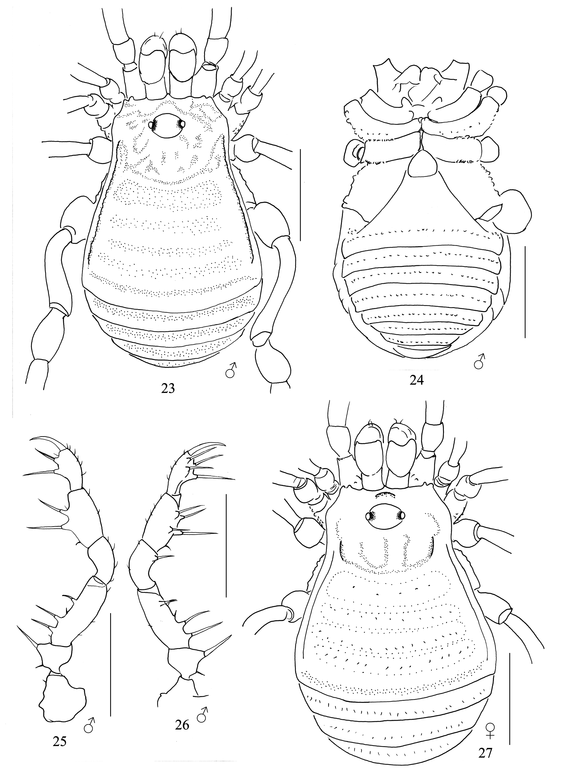
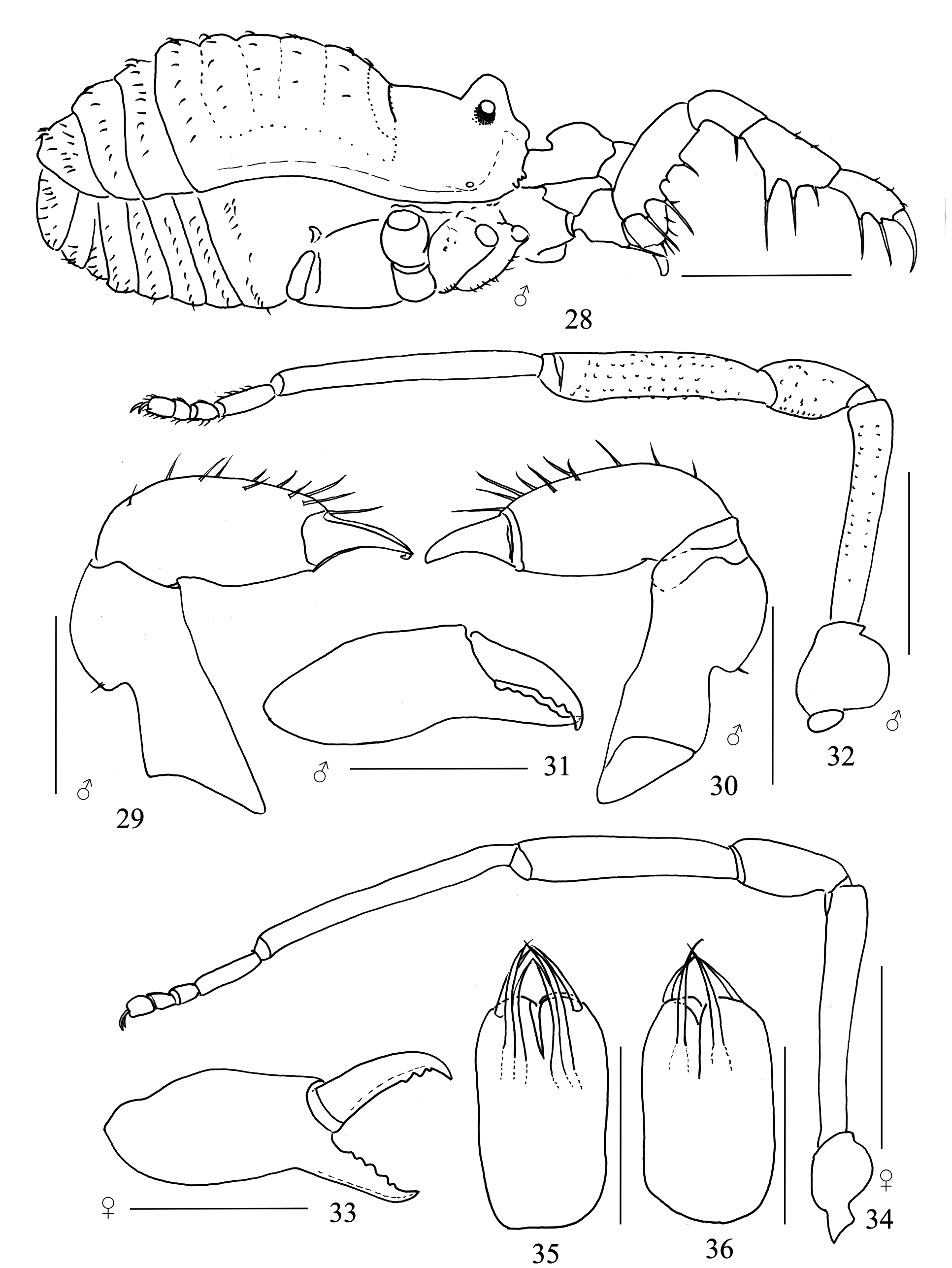
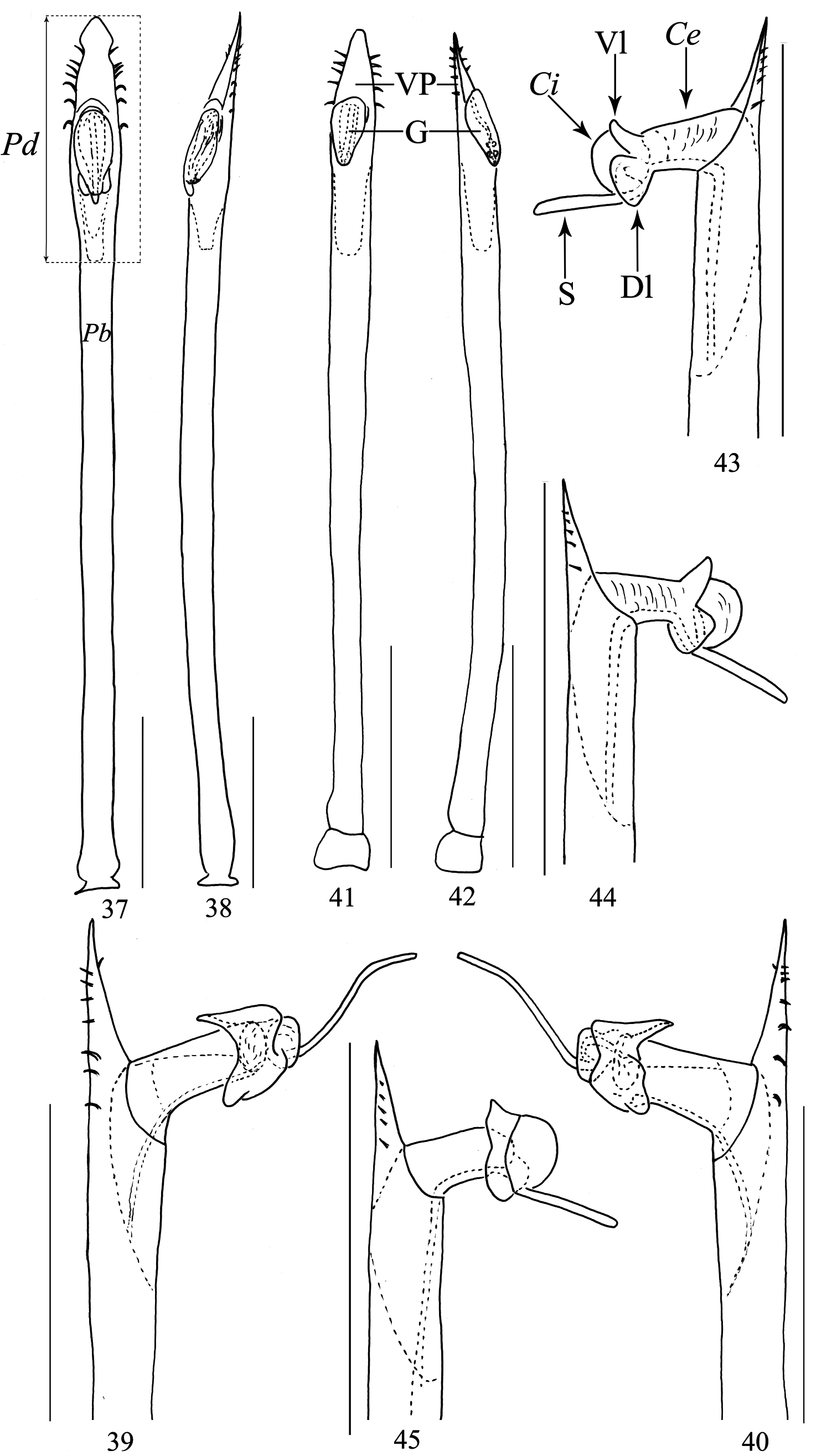
( Figs. 23–36 View FIGURES 23–27 View FIGURES 28–36 , 41–49 View FIGURES 37–45 View FIGURES 46–51 )
Type material. Male holotype, China: Hebei Province, Baoding City, Yi Country, Mt. Yunmeng Shan [ N 39°24´, E 115°15´], alt. 440m, June 12, 2012, C. Zhang leg. (MHBU-Opi-16 ZC1116 ). One female, paratype (MHBU-Opi- 16 ZC1117 ) GoogleMaps , same collecting data as holotype.
Diagnosis. Unique tarsal formula (2:2:4:4); body, including ocularium and legs, unarmed; stylus short and straight.
Etymology. The specific epithet is a noun in apposition, referring to the type locality.
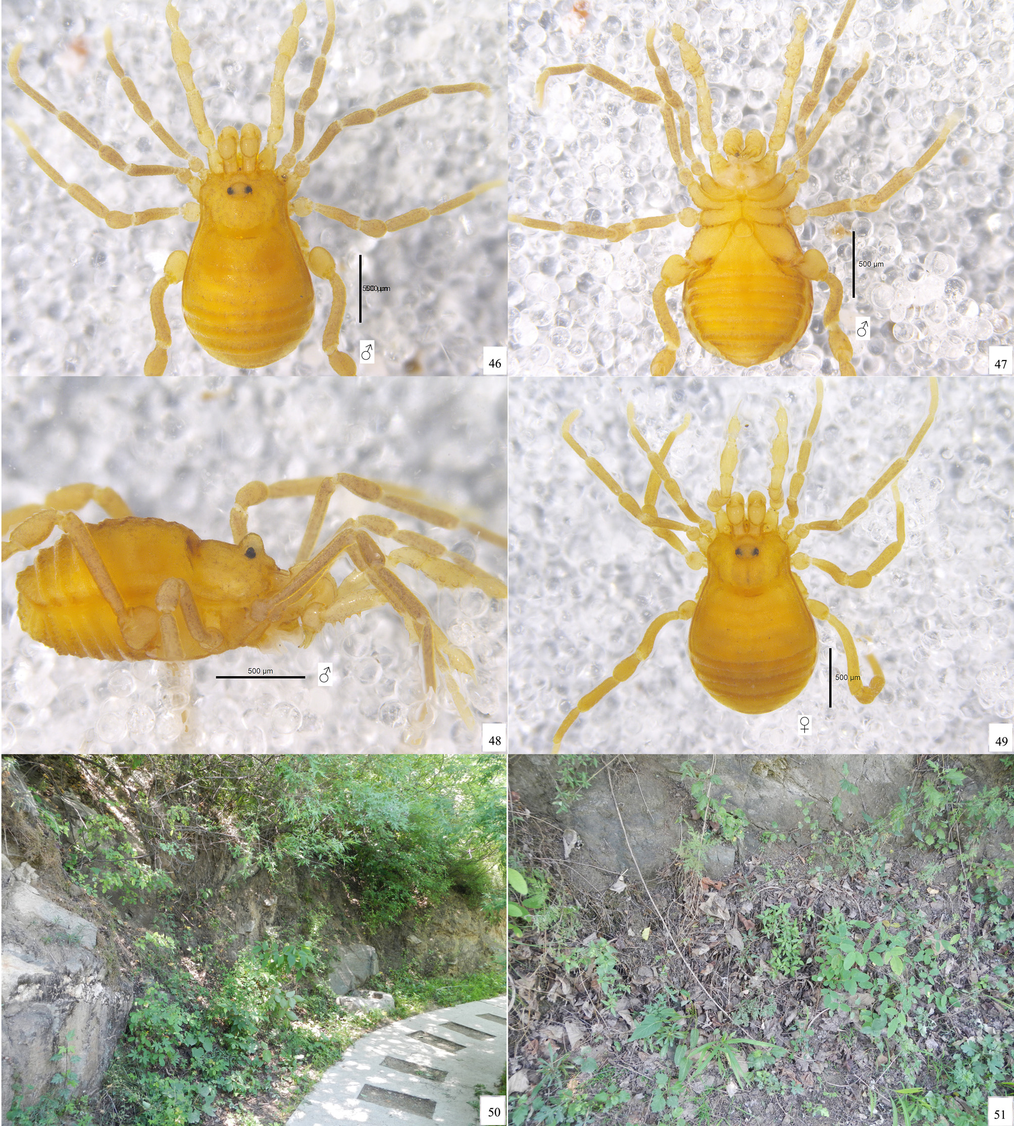
Description. Male habitus as in Figs. 23, 24 View FIGURES 23–27 , 28 View FIGURES 28–36 , 46–48 View FIGURES 46–51 . Coloration ( Figs. 46–48 View FIGURES 46–51 ): entire body yellow; carapace and ocularium with blackish brown reticulations; opisthosomal region of scutum and free tergites with transverse rows of brown stripes; coxae and genital operculum yellow; all free sternites with brown bands; legs yellow to brown as well as basitarsus, remaining tarsomeres whitish yellow.
Dorsum ( Figs. 23 View FIGURES 23–27 , 46 View FIGURES 46–51 ). Dorsal scutum granular and trapezoid in shape, widest portion of body at scutal area V. Carapace with two blunt pegs on each side of anterior margin of carapace near antero-lateral corners. Ocularium unarmed. Scutal sulci of mesotergum indistinct. Scutum and free tergites unarmed.
Venter ( Figs. 24 View FIGURES 23–27 , 47 View FIGURES 46–51 ). Surface of all coxae granulated. Coxa II with many setose tubercles retrolaterally. Coxa III with prolateral and retrolateral tubercular bridges to adjacent coxae. Coxa IV greatly enlarged, with setose tubercles on anterior margin. Genital operculum sub-triangular. Spiracles not concealed. Opisthosomal free sternites with belts of small regular tubercles.
Chelicera ( Figs. 29–31 View FIGURES 28–36 ). Proximal article with a prominent bulla, but without any conspicuous armament. Second article unarmed, with scattered setae on the prodorsal surface. Fingers relatively short, dentition as illustrated ( Fig. 31 View FIGURES 28–36 ); movable finger with three teeth; fixed finger with five teeth.
Pedipalp ( Figs. 25, 26 View FIGURES 23–27 ). Coxa dorsally with one small blunt tubercle. Trochanter ventrally with one short proximal and one long distal setiferous tubercle. Femur ventrally with a row of four setiferous tubercles, two proximal ones being the longest, the medial one being the shortest; on the medial distal side with one setiferous tubercle. Patella with one setiferous tubercle disto-medially. Tibia with two setiferous tubercles mesally; ectally with one short proximal and two long setiferous tubercles. Tarsus with two setiferous tubercles on both sides of ventral surface. Tarsal claw curved and smooth, approximately the same length as the tarsus.
Legs ( Figs. 32 View FIGURES 28–36 , 46–48 View FIGURES 46–51 ). All segments finely granulated. Trochanter III enlarged. Trochanter IV greatly enlarged, oval in lateral aspect and umarmed ( Fig. 32 View FIGURES 28–36 ). Femora III–IV curved, especially femur IV. Femur IV, Patella IV and tibia IV with enlarged granules ( Fig. 32 View FIGURES 28–36 ). Tarsi III–IV with bare double claws, without scopulae. Tarsal claws smooth. Tarsal formula, 2 (1): 2 (1): 4: 4.
Penis ( Figs. 41–45 View FIGURES 37–45 ). Basal portion of the shaft slender, then distended until apical portion (pars distalis). Apex of ventral plate somewhat triangular. Ventral plate with five pairs of setae on lateral margins. Glans free in apical part, with parastylar lobe extending proximally (not inflatable). Capsula externa cylindrical. Capsula interna globular. Diameter of fully inflatable Capsula interna conspicuously longer than that of the capsula externa. Parastylar lobes including one triangular ventral lobe and two square-shaped dorsal lobes. Stylus short, straight, similar in length to capsula externa.
Female. ( Figs. 27 View FIGURES 23–27 , 33, 34 View FIGURES 28–36 , 49 View FIGURES 46–51 ). In general appearance similar to the male ( Figs. 27 View FIGURES 23–27 , 49 View FIGURES 46–51 ), with a slight difference in inner edges of cheliceral finger ( Fig. 33 View FIGURES 28–36 ) and leg IV not enlarged ( Fig. 34 View FIGURES 28–36 ).
Ovipositor ( Figs. 35, 36 View FIGURES 28–36 ) composed of two apical lobes, each bearing two dorsal setae, one ventral seta, and two apical setae.
Measurements. Male holotype (female paratype): body 1.51 (1.54) long, prosoma 0.64 (0.66) wide, opisthosoma 0.96 (1.08) wide; length-to-width ratio 1.57 (1.43). Ocularium 0.12 (0.14) long, 0.20 (0.21) wide. Pedipalp claw 0.19 (0.19) long. Penis 0.81 long. Measurements of pedipalp and legs as in Tables 3, 4.
Habitat. The specimens were collected by sifting leaf litter in shrubs close to the trail depicted in Figs. 50, 51 View FIGURES 46–51 .
Distribution. Known only from the type locality ( Fig. 52 View FIGURE 52 ).
Notes. Petrobunus hebei sp. nov. is the northernmost Laniatores species from China.

No known copyright restrictions apply. See Agosti, D., Egloff, W., 2009. Taxonomic information exchange and copyright: the Plazi approach. BMC Research Notes 2009, 2:53 for further explanation.
|
Kingdom |
|
|
Phylum |
|
|
Class |
|
|
Order |
|
|
SubOrder |
Laniatores |
|
Family |
|
|
Genus |