
Pilargis falconae, Ribeiro & Barbosa & Freitas & Zanol & Glasby & Ruta, 2020
|
publication ID |
https://doi.org/10.11646/zootaxa.4878.1.2 |
|
publication LSID |
lsid:zoobank.org:pub:29DB126D-4751-433B-89BC-EFDD421368F7 |
|
DOI |
https://doi.org/10.5281/zenodo.4574131 |
|
persistent identifier |
https://treatment.plazi.org/id/D4F4E539-27F4-4D16-BBF6-574B031AC761 |
|
taxon LSID |
lsid:zoobank.org:act:D4F4E539-27F4-4D16-BBF6-574B031AC761 |
|
treatment provided by |
Plazi |
|
scientific name |
Pilargis falconae |
| status |
sp. nov. |
Pilargis falconae View in CoL sp. nov.
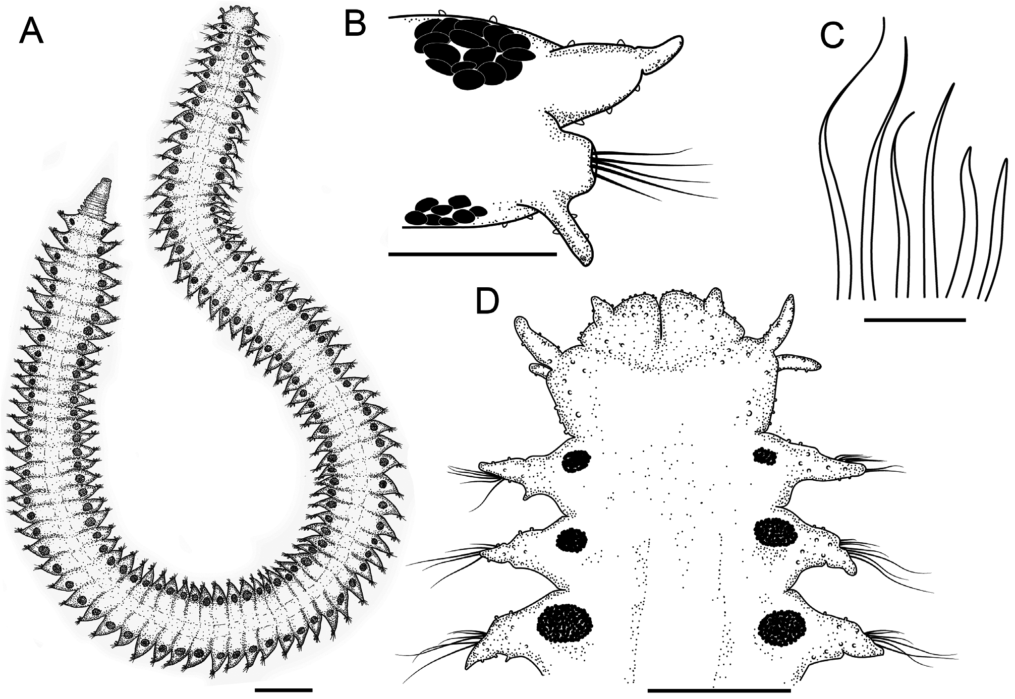
Figures 7 View FIGURE 7 A–C, 8A–D
urn:lsid:zoobank.org:act:
Material examined. BRAZIL: Campos Basin : Holotype: 21º50’2.96”S, 40º5’55.938”W, 476 m, one specimen, 7.ii.2009 (MNRJP-002731). GoogleMaps
Comparative material examined. Pilargis verrucosa Saint-Joseph, 1899 : Northeastern Atlantic Ocean: syntypes, two specimens, females (MNHN-IA-TYPE1234-5); one specimen, male (MNHN-IA-TYPE1236).
Diagnosis. Body surface sparsely papillate. Dorsal tentacular cirri 1.5x longer than ventral ones. All dorsal cirri similar in length. Parapodial glands from first chaetiger on dorsal surface and from the sixth chaetiger on ventral surface. Neurochaetae smooth capillaries, limbate with limbus smooth, unidentate.
Description. Fixed specimen whitish with dark parapodial glands on dorsal and ventral surfaces ( Fig. 7 View FIGURE 7 A–C; 8A, B). The following description is based on the holotype: size 20.89 mm long, 0.90 mm wide at widest point (including parapodia), 92 chaetigers. Body dorsoventrally flattened ( Fig. 7A View FIGURE 7 , 8A View FIGURE 8 ). Body surface with papillae minute and sparse, papillae abundant on cirrophores of tentacular and dorsal cirri, antennae and parapodial cirri ( Fig. 8D View FIGURE 8 ). Prostomium with lateral antennae inserted on posterior margin; median antenna and eyes absent ( Fig. 7B View FIGURE 7 , 8D View FIGURE 8 ). Two biarticulated palps conical ( Fig. 7B View FIGURE 7 , 8D View FIGURE 8 ), palpophore large and palpostyle diminutive. Proboscis not observed. Peristomium dorsally distinct from prostomium with two pairs of tentacular cirri. Dorsal tentacular cirri 1.5x longer than ventral ones and 2.2x longer than lateral antennae. Parapodia sub-biramous and inflated; all parapodia have ventral and dorsal cirri in conical shape ( Fig. 8B View FIGURE 8 ). Dorsal cirri of first chaetiger 1.2x longer than second one, and as long as following cirri ( Fig. 7B View FIGURE 7 ); ventral cirri shorter than dorsal cirri. Cirrophores of dorsal cirri thick and globose, twice as long as cirrostyles. Parapodial glands darkly pigmented, some brownish to reddish, concentrated in elliptical clusters, present on dorsal surfaces of all parapodia, and on ventral surface from the chaetiger 6 and some absent between chaetigers 30 and 35 ( Fig. 7 View FIGURE 7 A–C), ventral glands smaller than dorsal ones. Neuropodial lobes conical, smaller than dorsal cirri. Neurochaetae smooth capillaries variable in length, limbate with limbus smooth and unidentate tips; up to seven neurochaetae per fascicle ( Fig. 8C View FIGURE 8 ). Posterior end abruptly tapering due to posterior regeneration, 12 regenerating segments; parapodial glands barely visible in regenerated segments ( Fig. 7C View FIGURE 7 ). Pygidium with small papillae present on the posterior margin; anal cirri absent ( Fig. 7C View FIGURE 7 ).
Distribution. Atlantic Ocean—Brazil, Campos Basin, specimen examined in this study.
Remarks. There is no doubt that the specimen examined belongs to the genus Pilargis , due to the absence of notochaetae. Among species with papillae small and sparsely distributed, P. falconae sp. resembles P. angeli Salazar-Vallejo & Harris, 2006 , P. berkeleyae Monro, 1933 , P. maculata Hartman, 1947 , and P. modesta Intes & Le Loeuff, 1975 (see Table 2 View TABLE 2 ). However, P. angeli is different in lacking parapodial glands either on the dorsal or ventral surface, and in having dorsal tentacular cirri 1.2x longer than ventral ones, while P. falconae sp. nov. has dorsal and ventral parapodial glands, and dorsal tentacular cirri 1.5x longer than ventral ones. Pilargis berkeleyae differs from P. falconae sp. nov. in having parapodial glands only in the most posterior segments and limbate chaetae bidendate, while P. falconae sp. nov. has parapodial glands from anterior chaetigers and limbate chaetae unidentate. Both P. maculata and P. modesta have dorsal parapodial glands from the first chaetiger. However, P. maculata differs from P. falconae sp. nov. in having bidentate capillaries, sub-spherical glands on the tentacular segment (peristomium), ventral glands from the first or second chaetigers, while P. falconae has limbate chaetae unidentate, lacks glands in the peristomium, and has ventral glands from the sixth chaetiger. On the other hand, P. modesta differs in having spherical-shaped glands located in the cirrophore, and cirrostyles one-fifth the length of cirrophores, while P. falconae sp. nov. has glands in elliptical clusters on the base of parapodia and cirrostyles half the length of cirrophores.
Etymology. Name is in honour of Ana Paula da Costa Falcão, marine biologist and member of the PETRO- BRAS technical staff who collaborated in the HABITATS Project.

No known copyright restrictions apply. See Agosti, D., Egloff, W., 2009. Taxonomic information exchange and copyright: the Plazi approach. BMC Research Notes 2009, 2:53 for further explanation.
|
Kingdom |
|
|
Phylum |
|
|
Class |
|
|
Order |
|
|
Family |
|
|
SubFamily |
Pilarginae |
|
Genus |