
Ploiaria anak Distant, 1909
|
publication ID |
https://doi.org/ 10.11646/zootaxa.4388.4.7 |
|
publication LSID |
lsid:zoobank.org:pub:68164D01-A985-4B29-AB0E-DDED9F864CB0 |
|
DOI |
https://doi.org/10.5281/zenodo.5974412 |
|
persistent identifier |
https://treatment.plazi.org/id/03CFEC6D-FFFE-2813-51EA-F8E21279FE76 |
|
treatment provided by |
Plazi |
|
scientific name |
Ploiaria anak Distant, 1909 |
| status |
|
Ploiaria anak Distant, 1909 View in CoL
Ploearia anak Distant, 1909: 505 . Syntype (s): India, Lucknow ; BMNH (examined).
Ploearia anak: Distant (1910: 180, Fig. 99) (redescription, figure).
Ploiaria anak: Wygodzinsky (1966: 165, 168) View in CoL (in key, records, distribution), Maldonado Capriles (1995: 109) (catalogue), Ambrose (2006: 2396, supplement 5) (checklist), Rédei (2008: 15) (in key).
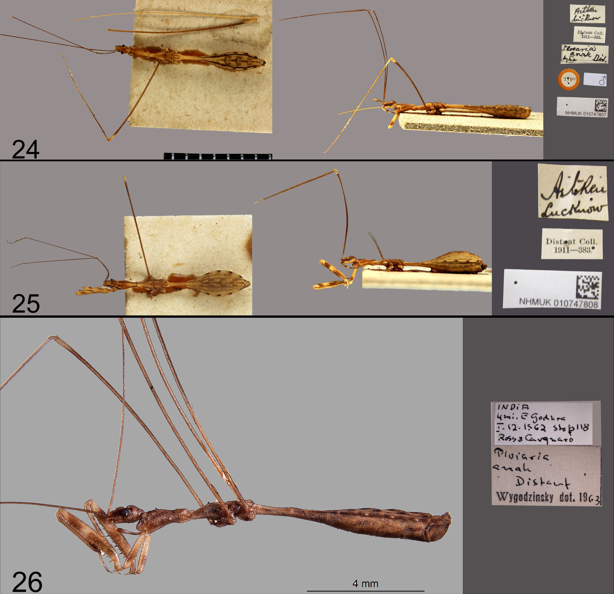
Type material examined. Lectotype (here designated): male, [India, Uttar Pradesh:], “Aitken / Lucknow”, “ Ploearia / anak / Type Dist.”, “Distant Coll. / B.M.1911-383”, “Type” [disc with red margin] ( Fig. 24 View FIGURES 24–26 ). Paralectotype: female, with similar locality and collection label ( Fig. 25 View FIGURES 24–26 ). Both specimens are deposited in BMNH.
Additional specimens examined. India: Pune District, Daund, vi.2016, leg. P. Pansare (1 female, Modern College of Arts, Science & Commerce, Shivajinagar, Pune); same but viii.2016 (1 female, same depository); one male and one female from the same locality but xii.2016, found trapped in spider web (1 male and 1 female, same depository).
Redescription. Female. Colouration: Ground colour of body pale ochraceous, with a pattern of pale brown to dark brown longitudinal bands or patches on head, thorax, legs and abdomen dorsally and laterally. Ventral colouration mostly pale on head and thorax. Fore legs ochraceous, with brown patches on coxa and femur, not extending to ventral side; tarsus pale except extreme apex; spiniform processes of anteroventral row on fore femur completely black including their bases, those of posteroventral row pale with only a narrow black ring at base, set on a pale brown base; mid and hind legs uniformly brownish except femoro-tibial articulations which are pale, femora darker than corresponding tibiae, tarsi very pale. Abdomen dorsally and ventrally partly reddish, with brown patches on tergites; connexivum pale with some brown patches; longitudinal brownish bands present ventrally beyond middle.
Anteocular part of head with a pair of dark lateral bands with black tubercles, median area with colourless fine granules, eyes dark black. Antennae dark brown, base and apex of segment I pale, segment II darker except pale apex, segments III and IV pale. Postocular part of head with a median ochraceous band and lateral brownish areas with dark tubercles. Head beneath pale, smooth and shining. Labium with second visible segment laterally brown, third visible segment pale brown.
Pronotum with a broad, median, ochraceous band, lateral area pale brown with dark brown granules at few places; with a pair of ochraceous spots laterally on each side near anterior margin; prosternum pale cream with a few brown specks laterally and in front of fore coxae. Meso- and metanota with thin, median ochraceous line and one broad line of similar colour on either side, rest area dark brown. Mesosternum pale cream; metasternum (and some part just anterior to mid coxae) pale brown, with median carina.
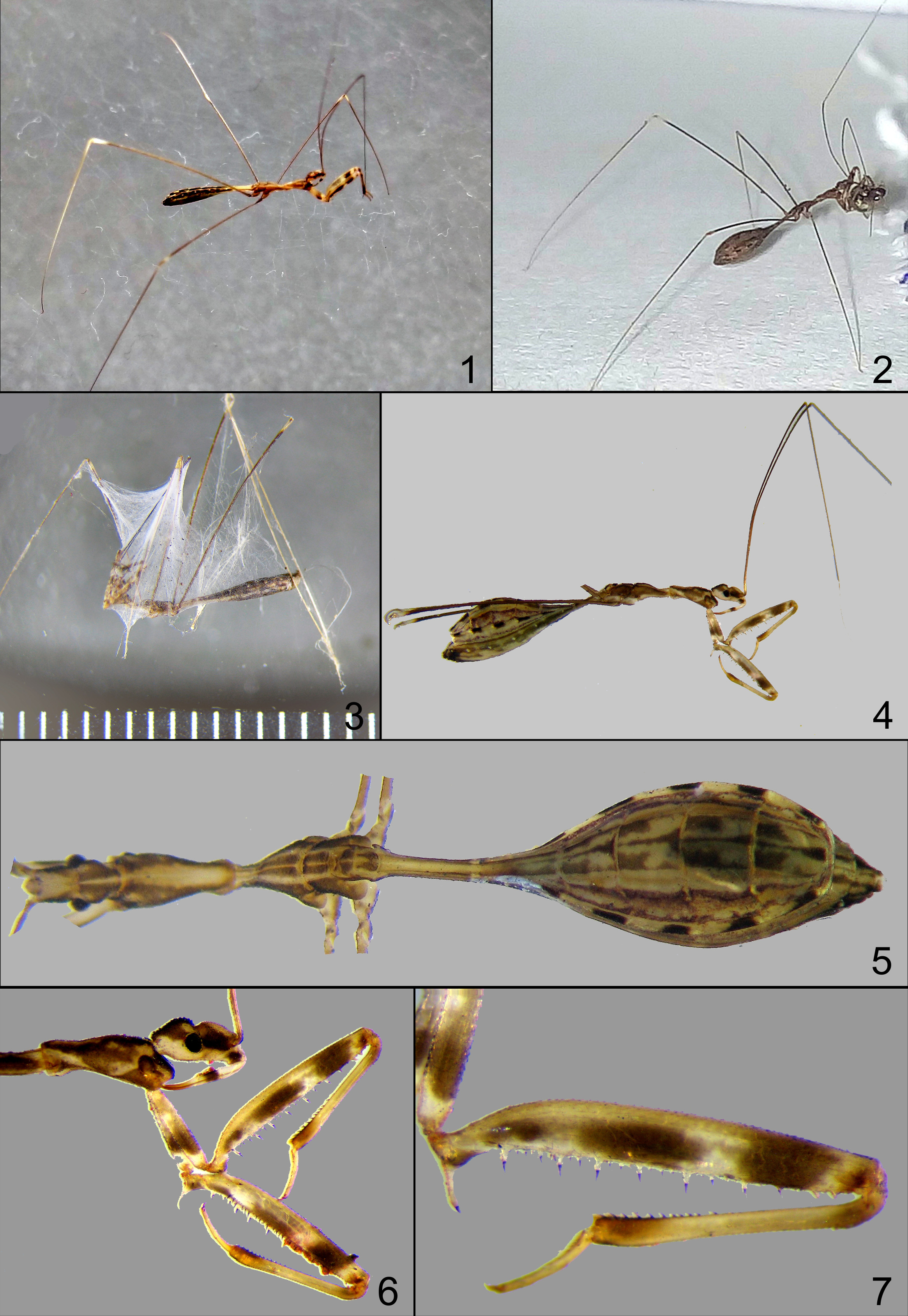
Structure: Head ( Figs. 5–6 View FIGURES 1–7 , 8 View FIGURES 8–12 ) elongate oval, both anteocular and postocular parts distinctly tumescent dorsally, rounded laterally, granulate. Antenniferous tubercles situated dorsally; clypeus and mandibular plates sloping anteriad and projecting beyond antenniferous tubercles, clypeus slightly more prominent than mandibular plates. Clypeus and mandibular plates finely granular, granules on disc of anteocular area small and colourless, those on lateral area coarser and darker. Transverse interocular sulcus prominent, eyes large. Posteriormost part of head forming a short, almost parallel-sided neck. Antennae thin and long, first and second joints subequal in length, third and fourth considerably shorter. Ventral surface of head smooth and shining. Labium with first two visible segments moderately swollen, third visible segment narrowed, slightly curved, sharp at tip ( Fig. 8 View FIGURES 8–12 ), extending up to base of fore coxae, resting on prosternal groove which has fine transverse stria.
Thorax ( Figs. 5–6 View FIGURES 1–7 , 9–10 View FIGURES 8–12 ): Pronotum broad anteriorly, gradually narrowed posteriorly, posterior fourth almost parallel-sided, median area very finely, lateral part more coarsely granulate. Anterior margin straight behind head, anterior corner slightly elevated and rounded. Mesonotum narrowed anteriorly, gradually dilated posteriorly, dorsomedian part finely granulate, with a fine median sulcus which is distinct in posterior third of segment. Dorsolateral part demarcated by a distinct carina or raised margin along its entire length, area laterad to this carina with large granules. Metanotum subquadrate with fine median longitudinal sulcus, finely granulate dorsomedially, lateral area with larger granules ( Figs. 9–10 View FIGURES 8–12 ). Pro- and mesosterna medially almost smooth, shining, with very scattered fine granules; metasternum medially finely granulate and with a distinct carina between mid and hind coxae; mid and hind coxae oblong, shining and smooth. Prothoracic acetabula directed forward; hind coxae closer to each other than mid coxae.
Legs: Fore legs ( Figs. 6–7 View FIGURES 1–7 , 11 View FIGURES 8–12 ) robust. Fore coxae moderately long, shorter than fore femur, unarmed; fore trochanter thick, armed with a long process (4–5 times as long as its width at base) provided with a small apical spine and also with a very small spine on its inner face. Fore femur slightly broader in proximal half than distally, longer than coxae, with an anteroventral and a posteroventral row of spiniform processes; posteroventral row with about 6 longer, elevated and several minute, wart-like processes, each with a short apical spine ( Fig. 11 View FIGURES 8–12 ). Fore tibia shorter than fore femur, more or less straight, its apical portion slightly dilated and bent, with few small setae and fine tubercles in proximal half and short spiniform processes in distal half, apex with a tuft of stiff setae ventrally; tarsus three-segmented, segment I longest, segment III shortest.
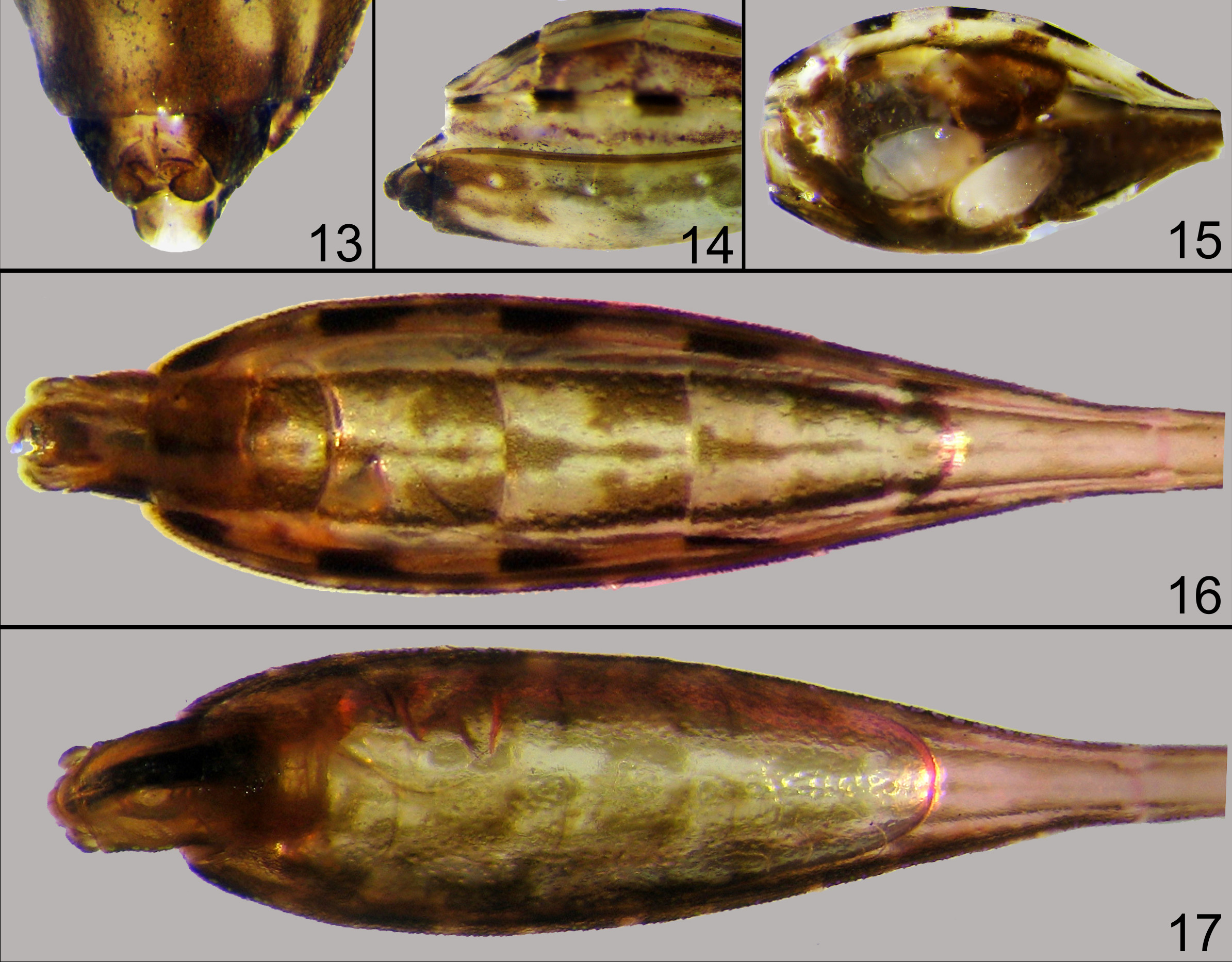
Abdomen ( Figs. 5 View FIGURES 1–7 , 12–15 View FIGURES 8–12 View FIGURES 13–17 ) with basal fifth narrow and parallel-sided, beyond which laterally dilated, broadly oval and dorsally convex, and again narrowed towards posterior tip; abdomen six times broader at its widest place than basally. Segment I with lateral angular projection on either side; segment II gradually dilated posteriorly; segments III–V successively broader, again narrowed from segment VI onwards. Entire dorsal surface very finely granulate, ventral surface finely granulate and sculptured; ventral intersegmental sutures indistinct; spiracles very small, situated on ventral laterotergites, surrounded by fine white border ( Fig 12 View FIGURES 8–12 , 14 View FIGURES 13–17 ).
Female terminalia as in Figs. 13–14 View FIGURES 13–17 .
Male. Colouration. Colour only slightly different to female, especially ventrally (see Figs 16–17 View FIGURES 13–17 ).
Structure. Abdomen slightly narrower than in female ( Figs. 16–17 View FIGURES 13–17 ).
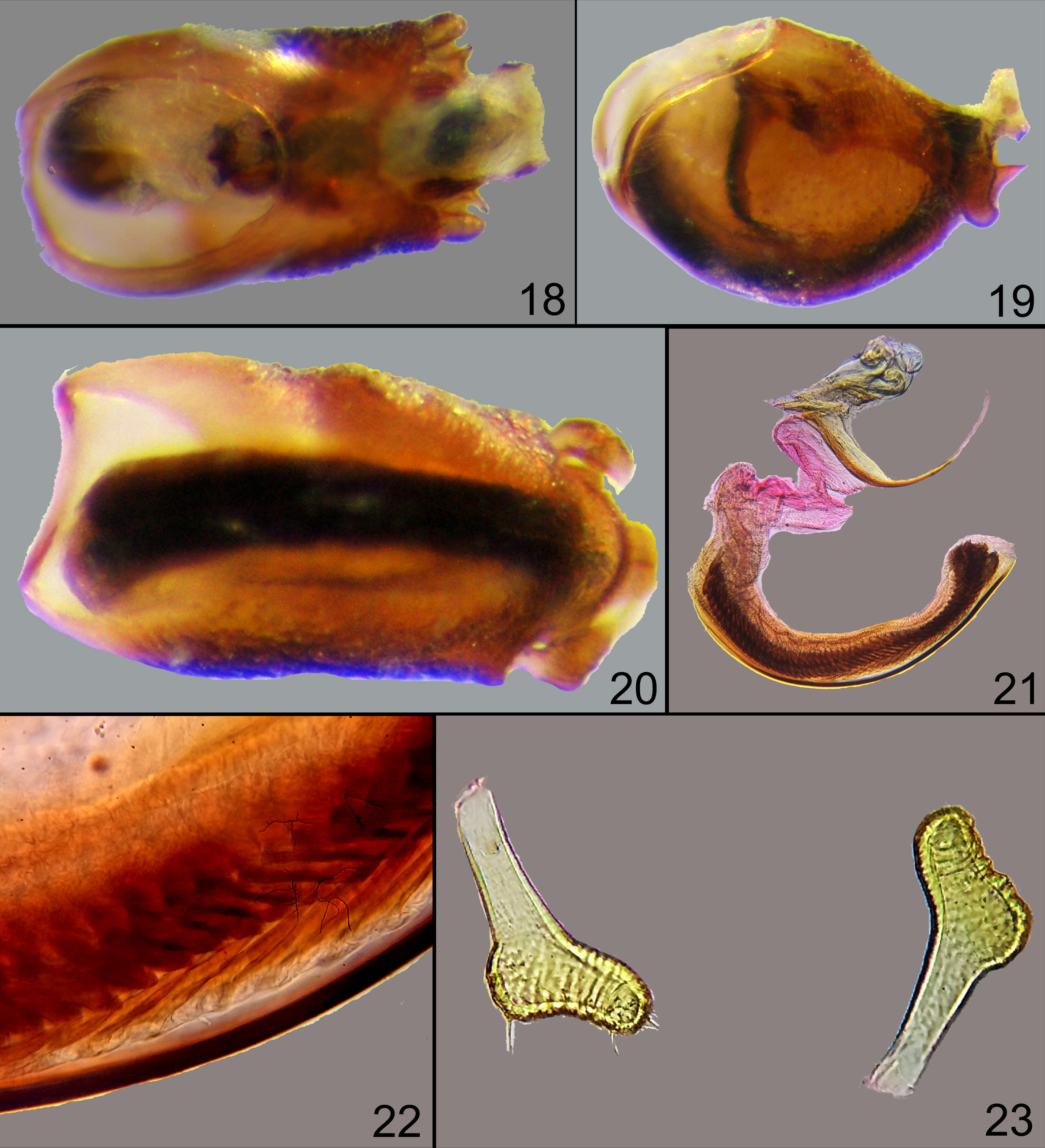
Male terminalia. Pygophore 0.8 mm long, slightly elongate oval, moderately sclerotized, ventrally distinctly convex, dorsally flat, with a sharp posterosuperior spine preceded by rounded protuberance; with a pair of small spines, directed vertically towards dorsum, situated laterally mesad to paramere, visible in dorsal ( Fig. 18 View FIGURES 18–23 ) and lateral views ( Fig. 19 View FIGURES 18–23 ); anterior (basal) opening oval in dorsal view, outline of phallus visible in lateral view, endosoma visible in ventral view ( Fig. 20 View FIGURES 18–23 ) in macerated preparatums. Parameres symmetrical, widely separated, small, sock-like ( Fig. 23 View FIGURES 18–23 ). Phallus ( Figs. 21–22 View FIGURES 18–23 ) more or less similar to the condition found in Ploiaria huangorum Rédei & Tsai, 2010 .
Measurements (in mm; male / female). Total length 10.5 / 9.75, length of anteocular region of head 0.50 / 0.60, length of postocular region of head 0.43 / 0.50, width across eyes 0.63 / 0.60, interocular distance 0.35 / 0.30; length of first visible labial segment 0.35 / 0.30, length of second visible labial segment 0.38 / 0.40, length of third visible labial segment 0.38 / 0.40; maximum width of pronotum 0.65 / 0.68, width of pronotum at base of head 0.45 / 0.40, length of pronotum 1.35 / 1.40; length of mesonotum 1.13 / 1.00; length of metanotum 0.63 / 0.90; minimum width of abdomen (at base) 0.25 / 0.25, maximum width of abdomen (at middle) 1.75 / 1.65; fore leg: length of coxa 1.13 / 1.00, length of femur 2.55 / 2.35, length of tibia 1.75 / 1.60, length of tarsus with claw 0.75 / 0.65, length of first tarsal segment 0.28 / 0.31, length of second tarsal segment 0.23 / 0.25, length of third tarsal segment 0.38 / 0.35; mid leg: length of coxa 0.50 / 0.50, length of femur 7.70 / 6.50, length of tibia 10.0 / 8.50, length of tarsus 0.30 / 0.30; hind leg: length of coxa 0.50 / 0.50, length of femur 10.5 / 9.00, length of tibia 13.75 / 13.0, length of tarsus with claw 0.30 / 0.30; length of prosternum 1.50 / 1.50, length of mesosternum 1.10 / 1.20, length of metasternum 0.60 / 0.50; length of first antennal segment 6.75 / 6.00, length of second antennal segment 6.00 / 5.50, length of third antennal segment 0.75 / 0.80, length of fourth antennal segment 1.10 / 1.10.
Distribution. Pakistan: Punjab ( Wygodzinsky 1966); India: Uttar Pradesh ( Distant 1909), Tamil Nadu ( Ravichandran 1988, Ambrose 2006), Rajasthan: Mahwah, Gujarat: Godhra ( Wygodzinsky 1966), Maharashtra: Pune District (new record).
Remarks. P. anak was described from an unspecified number of specimens (syntypes) from ‘Lucknow (Aitken)’. A male and a female syntype ( Figs. 24, 25 View FIGURES 24–26 ) are present in the BMNH, from which the male is selected as lectotype. Photo of an additional non-type male from the environs of Godhra, Gujarat, deposited in the CASC, identified and listed by Wygodzinsky (1966), has been examined as well ( Fig. 26 View FIGURES 24–26 ). One male studied in detail was found dead, trapped in a spider web, and many parts of the body became detached during cleaning. Subsequently, in November 2017, a fresh live male was collected, and the measurements of male were taken from this individual.
Bionomics. Both live females of P. anak were observed moving slowly on the wall where small spiders were also detected. When brought to the laboratory, we observed that the bug could move on a spider web without getting entangled ( Fig. 1 View FIGURES 1–7 ). Knowing that some emesine bugs feed on spiders ( Wygodzinsky 1966, Wignall & Taylor 2010, Soley et al. 2011) and Eugubinus araneus Distant (1904: 207) was reported “living in nest of spider”, we provided one of the females of P. anak females with a small moth, a small dipteran, a lygaeid and a mirid bug, and a small spider; within a short time the spider was aggressively caught and its content sucked. Two more spiders offered were treated similarly ( Fig. 2 View FIGURES 1–7 ). This species is apparently a specialist predator of spiders. Nevertheless, we also found one male and one female of P. anak and a specimen of another emesine bug ( Stenolemus sp.) dead and trapped in spider web; this indicates that these emesines sometimes get trapped in spider webs ( Fig. 3 View FIGURES 1–7 ).
The female collected in August 2016, with distended abdomen ( Figs. 4–5 View FIGURES 1–7 ), was found to carry three large, nearly fully developed eggs, each about 1.5 mm long ( Fig. 15 View FIGURES 13–17 , one egg removed). This indicates that the breeding season of this species is during July and August and that the female can deposit at least three eggs at a time.

No known copyright restrictions apply. See Agosti, D., Egloff, W., 2009. Taxonomic information exchange and copyright: the Plazi approach. BMC Research Notes 2009, 2:53 for further explanation.
|
Kingdom |
|
|
Phylum |
|
|
Class |
|
|
Order |
|
|
Family |
|
|
Genus |
Ploiaria anak Distant, 1909
| Pansare, Pratik P., Ghate, Hemant V. & Webb, Mick 2018 |
Ploearia anak
| Distant, 1909 : 505 |