
Rhyacophila miyanoura, Ito, 2021
|
publication ID |
https://doi.org/ 10.11646/zootaxa.5023.1.2 |
|
publication LSID |
lsid:zoobank.org:pub:F6CB0172-C3B9-4E9F-ACBF-2BC006F588F5 |
|
DOI |
https://doi.org/10.5281/zenodo.5225416 |
|
persistent identifier |
https://treatment.plazi.org/id/03CF878F-4F2A-FFFE-68A8-1FCFFD2EFE10 |
|
treatment provided by |
Plazi |
|
scientific name |
Rhyacophila miyanoura |
| status |
sp. nov. |
Rhyacophila miyanoura sp. nov.
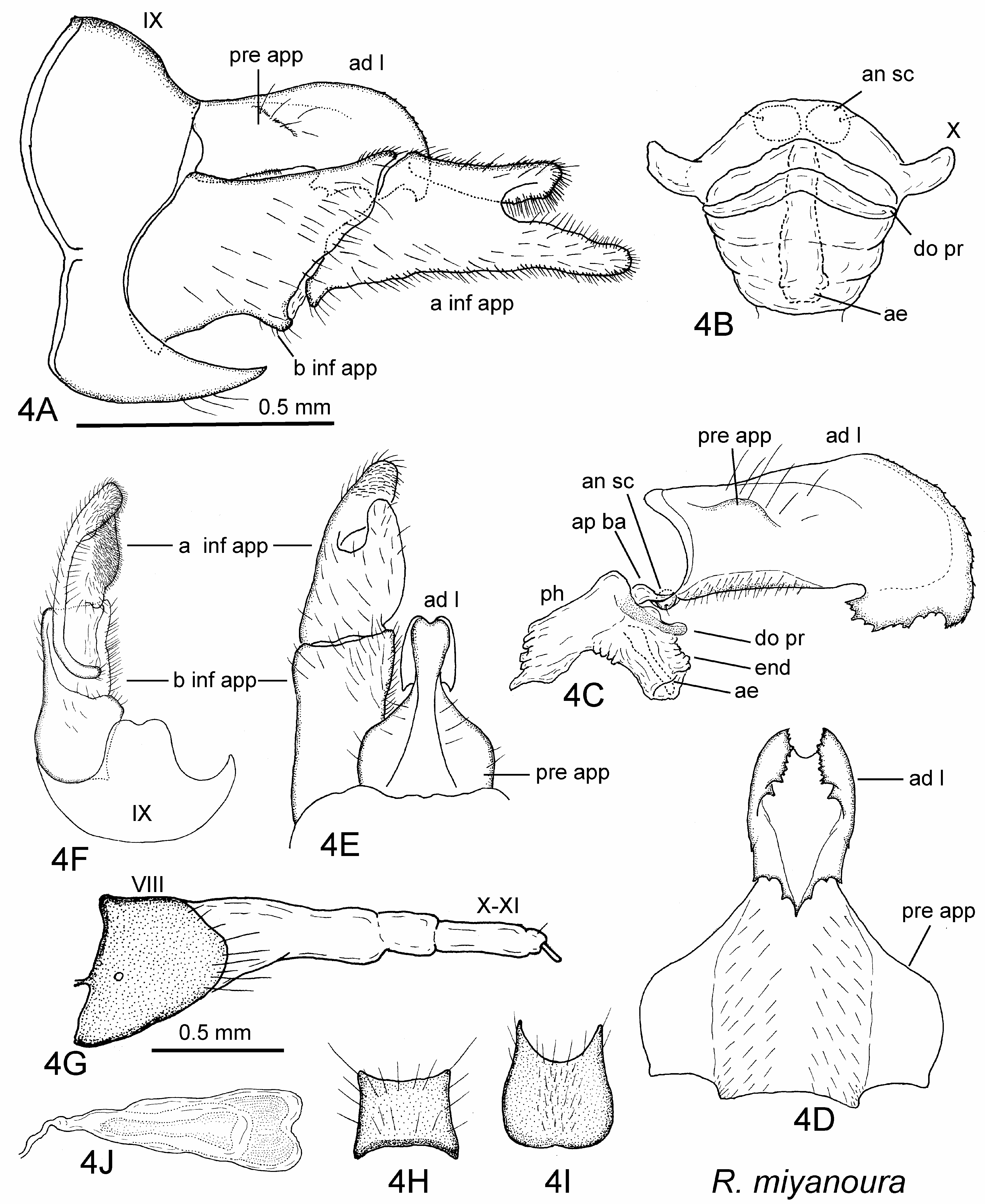
( Figs 4 View FIGURE 4 , 6C View FIGURE 6 )
Diagnosis. This new species belongs to the R. yosiiana Species Group (see Discussion), and the male resembles those of R. satoi Kuranishi 1997 , inhabiting central Ryukyu, southern Japan, and R. yosiiana Tsuda 1940 , found on Honshu, central Japan ( Taira & Nozaki 2021), in having a scoop-shaped apicodorsal lobe of segment IX and a very small phallic apparatus without parameres ( Kuranishi 1997; Hattori 2005). The male of R. miyanoura is clearly discriminated from those of R. satoi and R. yosiiana as follows: The venter of segment IX has a large bilobed projection in the middle, the ventroposterior margin of the apicodorsal lobe of segment IX is irregularly serrated, and the apical segment of each inferior appendage is deeply bifurcate in R. miyanoura ; whereas in R. satoi and R. yosiiana the venter of segment IX is short, the ventroposterior margin of the apicodorsal lobe of segment IX (‘segment X’ of Kuranishi 1997) is round, and the apical segment of each inferior appendage is not bifurcate. The female of R. miyanoura also resembles that of R. satoi in the shape of the short segment VIII and the vaginal apparatus having a bilobed posterior margin ( Kuranishi 1997); they cannot be distinguished from each other easily.
Adult. Lengths of forewings and hind wings: Each forewing 6.2–10.0 mm long (mean 7.2 mm, n = 18) and each hind wing 5.6–8.9 mm (mean 6.5 mm, n = 18) in males, 7.0–9.0 mm (mean 7.6 mm, n = 9) and 6.6–8.0 mm (mean 7.0 mm, n = 9) in females, respectively. Head dark brown, warts light brown with brown setae, antennae brown, scapes thicker and longer than other segments; palpi brown. Thorax dark brown dorsally. Legs brown with brown spurs. Wings brown with darker veins and fulvous pterostigma, few whitish dots on forewings of both sexes. Abdomen with brown tergites and light brown sternites, dark pigments scattered dorsally; scent glands of sternite V opening on antero-angle mounds; small mid ventral process on sternite VII (or VI and VII) in male and on sternite VI in female, process on sternite VI of male often very small or completely absent.
Male genitalia ( Figs 4A–4F View FIGURE 4 ). Segment IX (IX) in lateral view slightly convex dorsally with large apicodorsal lobe (ad l), venter with pair of large posteroventral projections; apicodorsal lobe scoop-shaped, in ventral view with apicomesal incision posteriorly, convex lateral margins, irregularly serrate posteroventrally. Preanal appendages (pre app) fused with basal half of apicodorsal lobe of segment IX, broadened laterally in dorsal and ventral views. Segment X (X) near basoventral edges of preanal appendages, semi-membranous, invisible in lateral view, short and wide in caudal view. Anal sclerites (an sc) small, round, weakly sclerotized; apical band (ap ba) short, semimembranous.
Phallic apparatus very small, not exceeding basal 1/4 of basal segment of inferior appendage (b inf app). Phallotheca (ph) membranous with semi-membranous dorsal process (do pr); retracted aedeagus (ae) slender, surrounded by endotheca (end); parameres absent.
Inferior appendages large, basal segment (b inf app) almost parallel sided with oblique caudal margins in lateral and dorsal views, apical segment (a inf app) bifurcate at apical 1/3, dorsal branch shorter than ventral one, both branches round and spiny apicomesally.
Female genitalia ( Figs 4G–4J View FIGURE 4 ). Segment VIII (VIII) short, length 0.8 times as long as basal width, posterior margins slightly convex in lateral view, subquadrate with anterior and posterior margins slightly incised in dorsal view, in ventral view roundish with large posterior incision. Vaginal apparatus slightly sclerotized, slender anteriorly, gradually thickened posteriorly with bilobed apical margin.
Holotype. Male , Japan, Yakushima Island, Miyanoura-gawa, small tributary (30.41˚N, 130.53˚E, 40–80 m a.s.l.), 10.v.2006, TI, S ( CBM-ZI 0159998 ).
Paratypes ( SPMN-IS). Yakushima Island : 5 males, 2 females, same data as holotype ; 1 male, Shirataniunsuikyo, 24.iv.2003, A. Ishizuka; 5 males, 2 females, same locality, 9.v.2006, TI , S; 1 male, 1 female, Yakusugiland, Ara-kawa, Seiryu-bashi, 12.vi.2007, TI , S; 2 males, 3 females, Yakusugi-land, Ara-kawa, small stream, 12.vi.2007, TI , S, L; 1 female, Miyanoura, Kue-gawa, Biwatsubo-bashi, 8.v.2006, TI , L; 1 male, Onoaida, Onoaidagyoko, 17.iv.2003, A. Ishizuka.
Other specimens. Yakushima Island: 1 male, Ara-kawa, 1200 m, 25.ix–23.x.2006, T. Yamauchi et al., M ( T. Ito) ; 1 male, same locality, 4–7.vi.2007, T. Yamauchi et al., M ( T. Ito) .
Distribution. Japan (Yakushima).
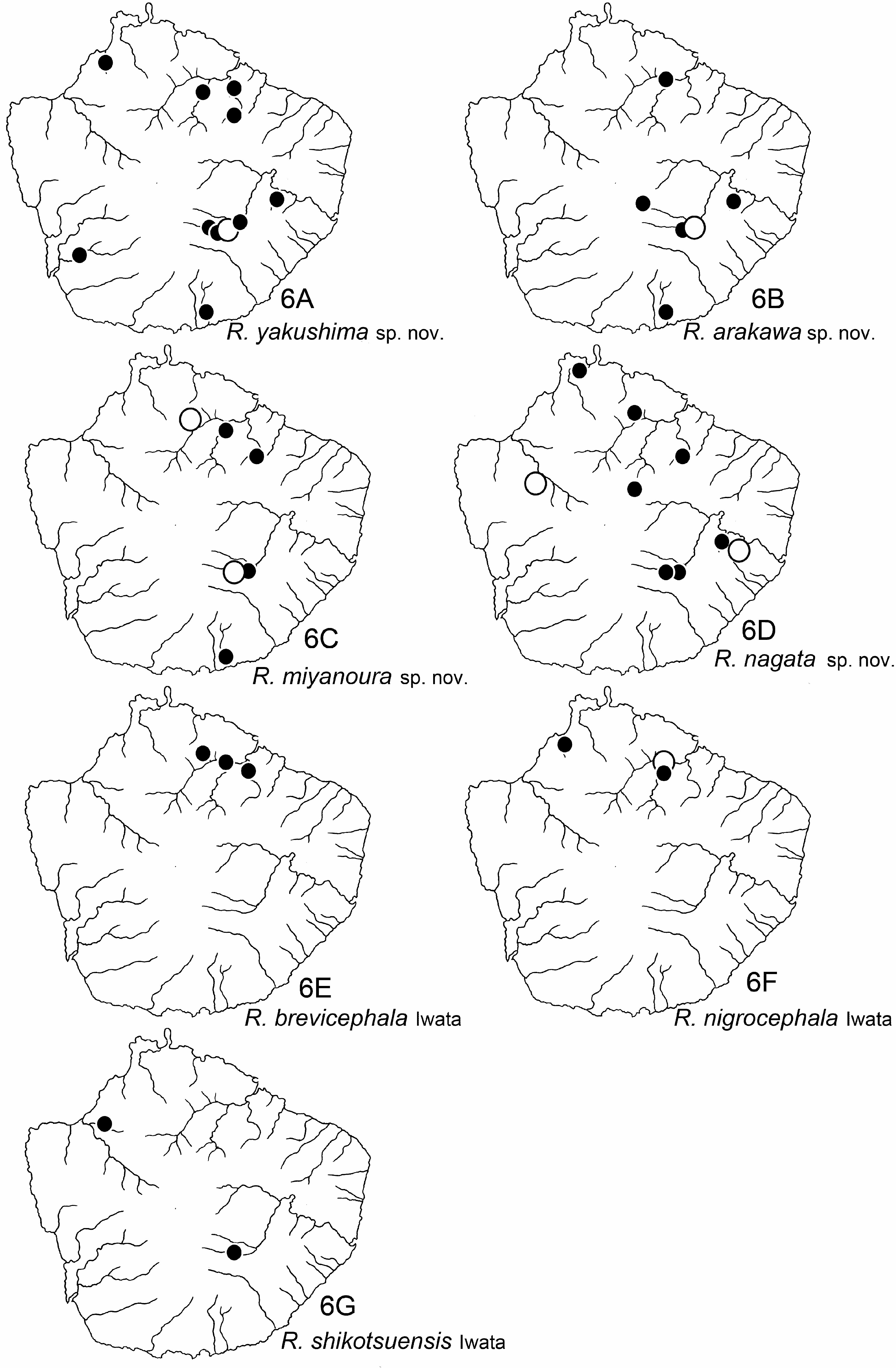
Habitat. Adults of this species were collected beside fast streams with stony bottoms ranging in elevation from 20 to 1100 m a.s.l. ( Fig. 6C View FIGURE 6 ).
Etymology. The name “ miyanoura ” is a noun in apposition, coined from the name of the type locality.
Japanese name. Miyanoura-nagare-tobikera.
| TI |
Herbarium of the Department of Botany, University of Tokyo |
| T |
Tavera, Department of Geology and Geophysics |
No known copyright restrictions apply. See Agosti, D., Egloff, W., 2009. Taxonomic information exchange and copyright: the Plazi approach. BMC Research Notes 2009, 2:53 for further explanation.