
Enhydrosoma kosmetron, Karanovic, Tomislav, Kim, Kichoon & Lee, Wonchoel, 2015
|
publication ID |
https://doi.org/ 10.11646/zootaxa.3990.4.1 |
|
publication LSID |
lsid:zoobank.org:pub:90BE4EAF-8594-4164-AE26-4F47E04A4A5D |
|
DOI |
https://doi.org/10.5281/zenodo.5616528 |
|
persistent identifier |
https://treatment.plazi.org/id/03CE8781-FFCA-E672-6180-2F28FAF1E2D5 |
|
treatment provided by |
Plazi |
|
scientific name |
Enhydrosoma kosmetron |
| status |
sp. nov. |
Enhydrosoma kosmetron sp. nov.
( Figs. 16–21 View FIGURE 16 View FIGURE 17 View FIGURE 18 View FIGURE 19 View FIGURE 20 View FIGURE 21 )
Type locality. South Korea, South Sea, Gwangyang Bay, sampling station 3 (see Karanovic et al. 2014), muddy sediments, 3453'03.9" N 12739 View Materials '50.5"E.
Specimens examined. Holotype female (NIBRIV 0000287224) dissected and mounted on one slide, collected from the type locality, 17 February 2013, collected by K. Kim. Allotype male (NIBRIV 0000287225) dissected and mounted on one slide, collected from Korea, South Sea, Gwangyang Bay, sampling station 10 (see Karanovic et al. 2014), 34°55'15.4"N 127°47'07.9"E, 18 February 2012, collected by K. Kim. Additional paratypes: one dissected female (NIBRIV 0000287226) mounted on one slide; four males and two females together on one SEM stub (NIBRIV 0000287227); three males and three females together on another SEM stub (NIBRIV 0000287228); all from Korea, South Sea, Gwangyang Bay, sampling station 10 (see Karanovic et al. 2014), 34°55'15.4"N 127°47'07.9"E, 18 February 2012, collected by K. Kim. One female used for molecular analysis collected by K. Kim from the type locality on 26 July 2012 (see Table 1 View TABLE 1 ).
Etymology. This species is named after the Greek noun kosmetron , meaning “broom” and referring to the broom-like ornamentation of the male antennulae. The specific name is to be treated as a noun (gender masculine) in apposition to the generic name.
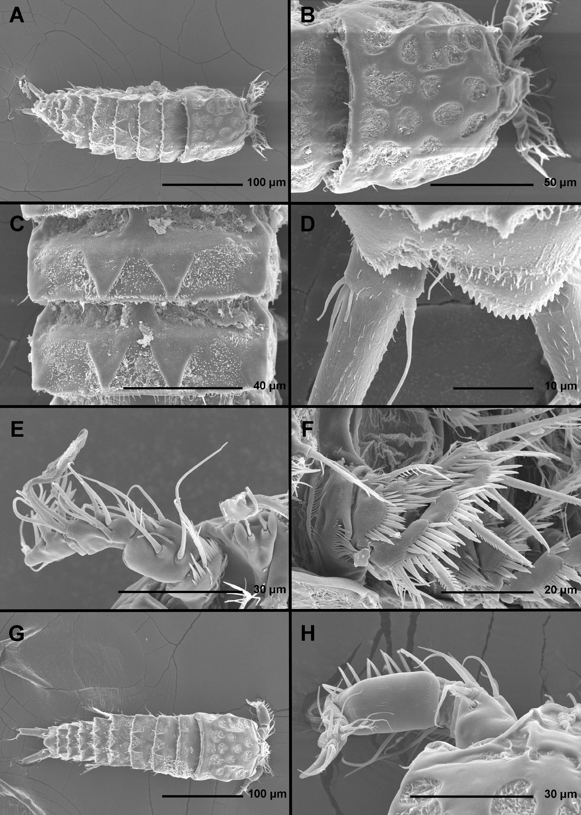
Description of female. Based on holotype and several paratypes. Total body length from 441 to 495 µm (mean = 483 µm, n = 7). Body segmentation, colour, nauplius eye, hyaline fringes, general integument thickness, angle between telescoped and non-telescoped parts of pleurons on most free somites, and most somite ornamentation as in Enhydrosoma apimelon sp. nov., however, surface relief on most somites slightly different. Habitus ( Figs. 16 View FIGURE 16 A) generally cylindrical in dorsal view, widest at posterior end of cephalothorax and tapering posteriorly, boundary between prosome and urosome inconspicuous; prosome/urosome length ratio about 1.1, and prosome only slightly more voluminous than urosome. Body length/width ratio about 3.7 in dorsal view; cephalothorax about 1.2 times as wide as genital double-somite. Free pedigerous somites without lateral or dorsal expansions, heavily sculptured, pleurons only partly covering coxae of swimming legs in lateral view. Integumental relief more defined by ridges than depressions in dorsal and lateral view; ventro-lateral ridges on urosomites as in E. apimelon , but ventral surface with additional (mostly internal) ridges. Most cuticular depressions and posterior margin of prosomites without hair-like spinules but full of bacterial growth and detritus, making observation of cuticular pores and sensilla in those regions very difficult; posterior margin of urosomites with hair-like spinules, especially on ventral surface. Hyaline fringe of all somites narrow and rough, in some places nearly serrated, and in all but last two somites also with conical mound-looking protuberances with sensilla on tip, as in E. apimelon . In addition to hairlike spinules, surface ornamentation of somites and caudal rami consists of at least two different types of sensilla (slender and bottle-shaped; see Fig. 16 View FIGURE 16 C), simple cuticular pores, and few stronger spinules; exact number of pores and spinules difficult to establish.
Rostrum ( Fig. 16 View FIGURE 16 B, E) as in E. apimelon , except for space between sensilla smaller than width of one horn-like projection.
Cephalothorax ( Fig. 16 View FIGURE 16 B) as in E. apimelon , but with completely different relief in dorsal view, without oval depression and with more pronounced longitudinal ridges (compare Figs. 3 View FIGURE 3 B and 16B), without comb of long setules in anterior part along inner surface of lateral margin (as observed in E. apimelon ) or ventral flaps that protect first three pairs of mouth appendages (as observed in E. robustum sp. nov.); lateral relief similar to that in E. apimelon , except for posterior depression larger and more triangular; most sensilla easy to homologize.
Pleuron of free prosomites ( Fig. 16 View FIGURE 16 A) very similar to each other, without triangular dorsal plates and posterior hair-like spinules, but with posterior conical mound-looking protuberances at base of sensilla, as well as dorsal and dorso-lateral ridges similar to those in E. apimelon but slightly smaller; all sensilla homologous to those in E. apimelon ; angle between telescoped and non-telescoped parts of pleuron relatively wide as in E. apimelon .
First urosomite ( Fig. 16 View FIGURE 16 A) slightly narrower and significantly longer than fourth pedigerous somite and pleuron without free lateral margin, but relief very similar to that of other pedigerous somites, with pronounced dorso-lateral ridges resulting in angular shape in cross-section.
Genital double-somite ( Figs. 16 View FIGURE 16 A, 19A) 1.6 times as wide as long in ventral view; completely fused ventrally but with deep suture indicating original segmentation between genital and third urosomites dorsally, thus dividing double-somite into equally long and similarly wide halves; general shape and ornamentation as in E. apimelon , except for more pronounced ridges on ventral surface and additional pair of ventral pores. Female genital complex ( Fig. 19 View FIGURE 19 A) weakly sclerotized and hardly distinguishable from internal sutures and soft tissue, except for large copulatory pore near midlength of somite and wide copulatory duct; genital operculum as in E. apimelon but with longer armature elements and shorter spinules.
Third urosomite ( Figs. 16 View FIGURE 16 A, 19A) very similar in shape and ornamentation to posterior part of genital doublesomite, but without ventral pores and with three or four large spinules on ventral surface at base of each ventral sensillum.
Fourth urosomite ( Figs. 16 View FIGURE 16 A, D, 19A) slightly shorter and narrower than third urosomite and without any sensilla and large spinules, but with well-developed dorsal, dorso-lateral, and ventro-lateral ridges (latter finely serrated), with posterior row of hair-like spinules, and with ventro-lateral cuticular pores.
Anal somite ( Figs. 16 View FIGURE 16 A, D, 19A, B) only slightly clefted medially, with one pair of large dorsal sensilla at base of anal operculum, several minute spinules, small group of large ventral spinules on medial corners, and one pair of ventral simple pores; ventro-lateral corners produced as in preanal somite, but slightly longer and more flared out, also serrated; dorso-lateral corner also produced into small serrated flaps; anal operculum ( Figs. 16 View FIGURE 16 D, 19B) semicircular, short, not reaching posterior margin of somite (shorter than in E. apimelon but longer than in E. robustum ), serrated, representing 39% of somite's width; anal sinus widely open, with three rows of hair-like spinules.
Caudal rami ( Figs. 16 View FIGURE 16 A, D, 19A, B) spindle-shaped, widest at about proximal quarter of their length, about 1.4 times as long as anal somite, about 2.2 times as long as wide (ventral view), strongly divergent, with strong dorsal ridge, and with space between them about one ramus’ width. Armature as in E. apimelon but dorsal seta longer and inserted more posteriorly (at about midlength); ornamentation as in E. robustum ; no tubular pore at base of anterior lateral setae; outer distal process carrying posterior lateral seta even more produced than in E. robustum ; proportion of setae other than dorsal similar to that in E. apimelon ; principal apical seta 1.3 times as long as ramus.
Antennula ( Figs. 16 View FIGURE 16 F, 20A) as in E. apimelon , except for second segment with one seta less (thus armature formula being: 1.8.7+ae.1.11+ae), and five additional spiniform setae and with strong spinules (three setae on second segment, one on third segment, and stronger apical seta on fifth segment). Lnegth and ornamentation of segments as in E. apimelon ; as in E. apimelon and E. robustum one unipinnate slender seta on second segment inserted into cone-shaped depression.
Antenna ( Figs. 19 View FIGURE 19 C), labrum, paragnaths, general shape and armature of maxilla ( Fig. 20 View FIGURE 20 B), maxilliped ( Fig. 20 View FIGURE 20 C), endopod of first swimming leg ( Fig. 20 View FIGURE 20 D), second swimming leg ( Fig. 20 View FIGURE 20 F), exopod of third swimming leg, and exopod of fourth swimming leg as in E. apimelon and E. robustum , except for small differences in proportion of certain segments and armature elements and very minor differences in ornamentation.
Mandibula ( Fig. 19 View FIGURE 19 D) size and segmentation as in E. apimelon , but palp with four setae and seta on cutting edge of coxal gnathobase much stronger and longer.
Maxillula ( Fig. 19 View FIGURE 19 E) size and segmentation as in E. apimelon , but both basis and praecoxal arthrite with one additional inner seta.
Maxilliped ( Fig. 19 View FIGURE 19 C) as in E. apimelon , except endopodal strong spine shorter and slender endopodal seta proportionately longer; coxa ornamented with three rows of spinules.
Exopod of first swimming leg ( Fig. 20 View FIGURE 20 E) with five elements on third segment.
Endopod of third swimming leg ( Fig. 20 View FIGURE 20 G) and endopod of fourth swimming leg ( Fig. 20 View FIGURE 20 H) both with very short outer spines in addition to two slender and long terminal setae on second segment.
Fifth leg ( Fig. 20 View FIGURE 20 I) biramous as in E. apimelon , comprising conical exopod and baseoendopod, but exopod much more robust and endopodal lobe shorter; exopod also armed with three elements, but lateral elements inserted closer to distal margin (as in E. robustum ), with distal lateral element almost apical and difficult to homologize, and apical element very small and bare. Outer margin of exopod with numerous long hair-like spinules, inner margin smooth, without tubular pores; baseoendopod as in E. robustum with robust distal seta, but with only one cuticular spiniform process on inner margin covered by small spinules; distal exopodal element about 0.3 times as long as proximal endopodal spine, 0.2 times as long as distal endopodal spine, 0.75 times as long as proximal exopodal element, 0.25 times as long as middle exopodal element, 0.3 times as long as distal endopodal element, and less than 0.2 times as long as entire exopod; exopod about 3.3 times as long as wide. Endopodal lobe without tubular pores, but with two simple pores at base of inner spines.
Sixth legs ( Fig. 19 View FIGURE 19 A) as in E. apimelon , but with proportionately smaller basal spinules and larger armature; inner smooth seta about 1.7 times as long as outer unipinnate spine.
Description of male. Based on allotype and four other paratypes. Body length ranging from 279 to 312 Μm (mean = 302 µm, n = 5). Genital somite and third urosomite not fused ( Fig. 19 View FIGURE 19 F). Habitus ( Figs. 16 View FIGURE 16 G, 17A, 18A), colour, rostrum, shape and ornamentation of cephalothorax ( Fig. 18 View FIGURE 18 B, C, D), shape and ornamentation of free prosomites ( Fig. 16 View FIGURE 16 G, 18A), shape and ornamentation of last three urosomites ( Figs. 16 View FIGURE 16 H, 17C, H, 18H, 19F), general shape, armature and ornamentation of caudal rami ( Figs. 16 View FIGURE 16 H, 17C, 18H, 19F), antenna ( Fig. 17 View FIGURE 17 E), labrum ( Fig. 17 View FIGURE 17 F), paragnaths ( Fig. 17 View FIGURE 17 F), mandibula ( Figs. 17 View FIGURE 17 F, 21C, D), maxillula ( Fig. 17 View FIGURE 17 F, 21E), maxilla ( Fig. 17 View FIGURE 17 B, F), maxilliped ( Fig. 17 View FIGURE 17 F), first swimming leg ( Fig. 17 View FIGURE 17 G), second swimming leg ( Fig. 17 View FIGURE 17 A), exopod of third swimming leg ( Fig. 17 View FIGURE 17 A), and fourth swimming leg ( Fig. 21 View FIGURE 21 G) as in female. Prosome/urosome ratio about 1.2, greatest width at posterior end of cephalothorax, body length/width ratio about 3.3; cephalothorax 1.6 times as wide as genital somite in dorsal view.
Genital somite ( Figs. 17 View FIGURE 17 H, 19F) 1.8 times as wide as long in ventral view, similar in ornamentation to that in E. robustum , and also with left sixth leg functioning as genital operculum; no spermatophore visible inside any observed specimens.
Third urosomite ( Figs. 17 View FIGURE 17 H, 19F) as posterior part of genital double-somite in female but proportionately narrower, and with four or five large ventro-lateral spinules.
Caudal rami ( Figs. 16 View FIGURE 16 H, 17C, 19F) very similar to those in female but slightly longer in proportion to anal somite and more robust.
Antennula ( Figs. 17 View FIGURE 17 D, 18E, F, G, 21A, B) as in E. apimelon , except for second segment with two spiniform setae, third segment with one spiniform seta, fourth segment with large broom-like structure instead of brush and with only six or seven large spinules, fifth segment with large spiniform process and three slender setae, and sixth segment with large characteristically shaped tube pore ( Fig. 18 View FIGURE 18 G); second segment as in female with slender plumose seta inserted into cone-like depression ( Fig. 18 View FIGURE 18 F).
Endopod of third swimming leg ( Fig. 21 View FIGURE 21 F) transformed and apparently three-segmented, with outer spine fused to segment and enlarged, secondary segmentation resulting in very small third segment bearing two long and slender apical setae.
Fifth leg ( Figs. 17 View FIGURE 17 H, 21H) somewhat smaller than in female but with similar basic structure; exopod about 2.3 times as long as wide, with all three elements present and of similar proportions as in female, except for apical one bipinnate; endopodal lobe without distal armature element or inner spiniform processes, its distal tip with bunch of hair-like spinules; proximal endopodal inner spine about 1.6 times as long as distal endopodal inner spine and 1.2 times as long as longest element on exopod.
Sixth legs ( Figs. 17 View FIGURE 17 H, 19F) as in E. robustum , withsimple cuticular plates, unarmed, with transverse row of slender spinules.
Variability. Despite numerous examined specimens and detailed examination using SEM (see Figs. 16 View FIGURE 16 A, G, 17A, 18A), we are not able to report on any significant morphological variability. The number of ventro-lateral spinules on fourth male and female urosomite and third male urosomite varies between three and five.


No known copyright restrictions apply. See Agosti, D., Egloff, W., 2009. Taxonomic information exchange and copyright: the Plazi approach. BMC Research Notes 2009, 2:53 for further explanation.