
Phortica Schiner, 1862
|
publication ID |
https://doi.org/ 10.5281/zenodo.5333042 |
|
persistent identifier |
https://treatment.plazi.org/id/03CC87B2-9819-006E-BBED-6ED63BF5F8CD |
|
treatment provided by |
Diego |
|
scientific name |
Phortica Schiner, 1862 |
| status |
|
Phortica Schiner, 1862 View in CoL (sensu stricto)
Phortica Schiner, 1862: 433 View in CoL ; Máca, 2003: 251. Type species: Drosophila variegata Fallén, 1823 .
Amiota (Phortica) : Wheeler, 1952: 167.
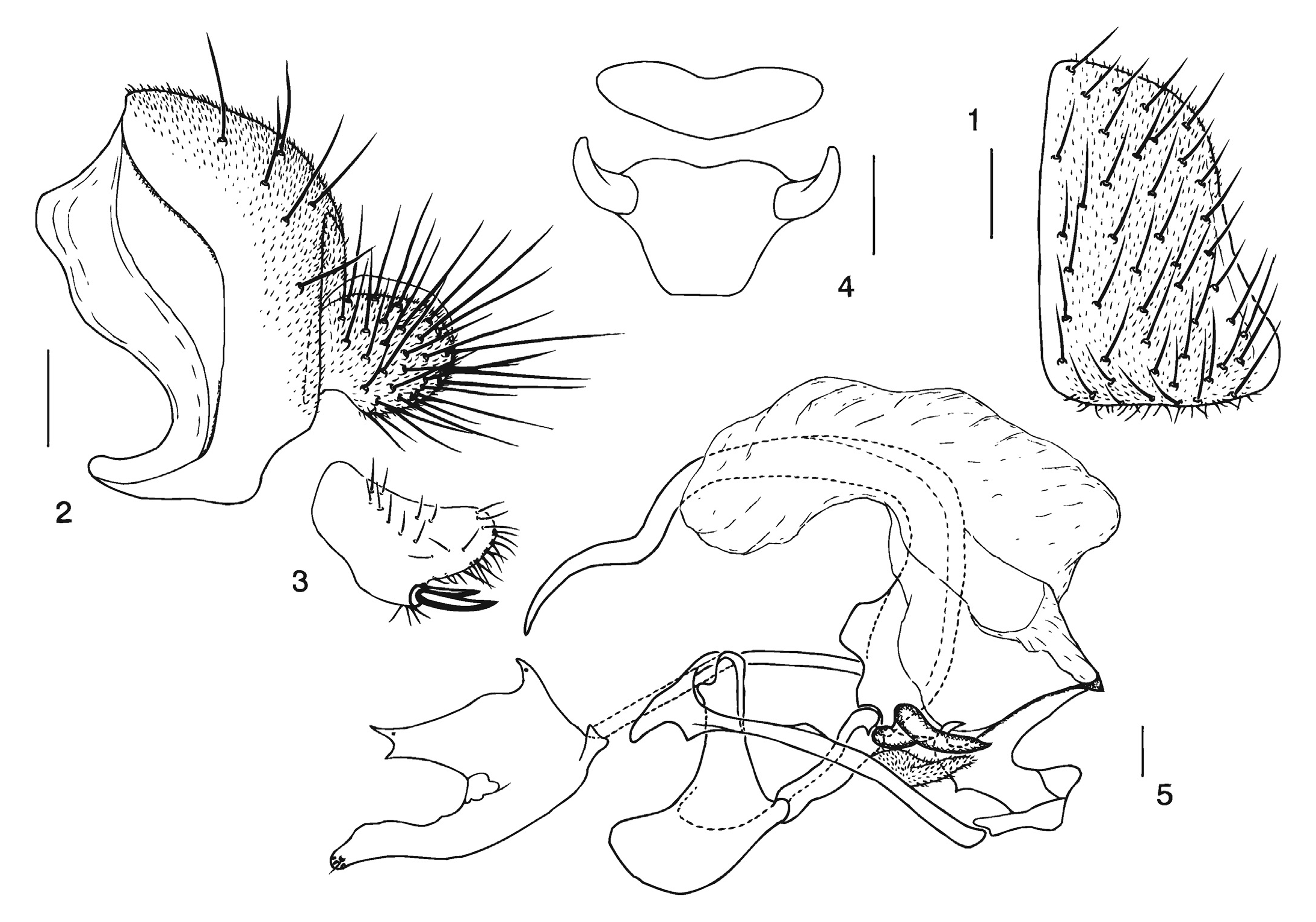
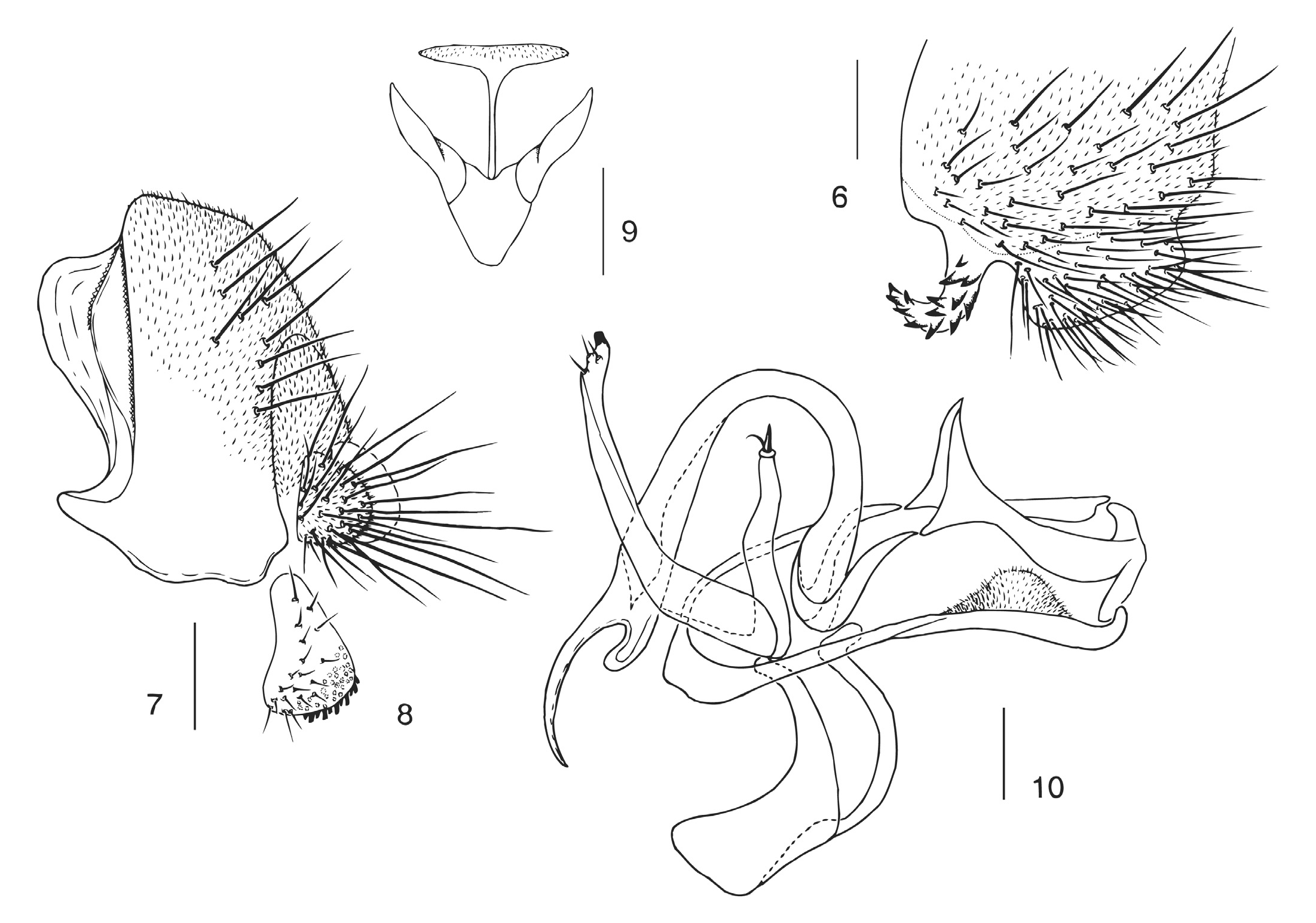
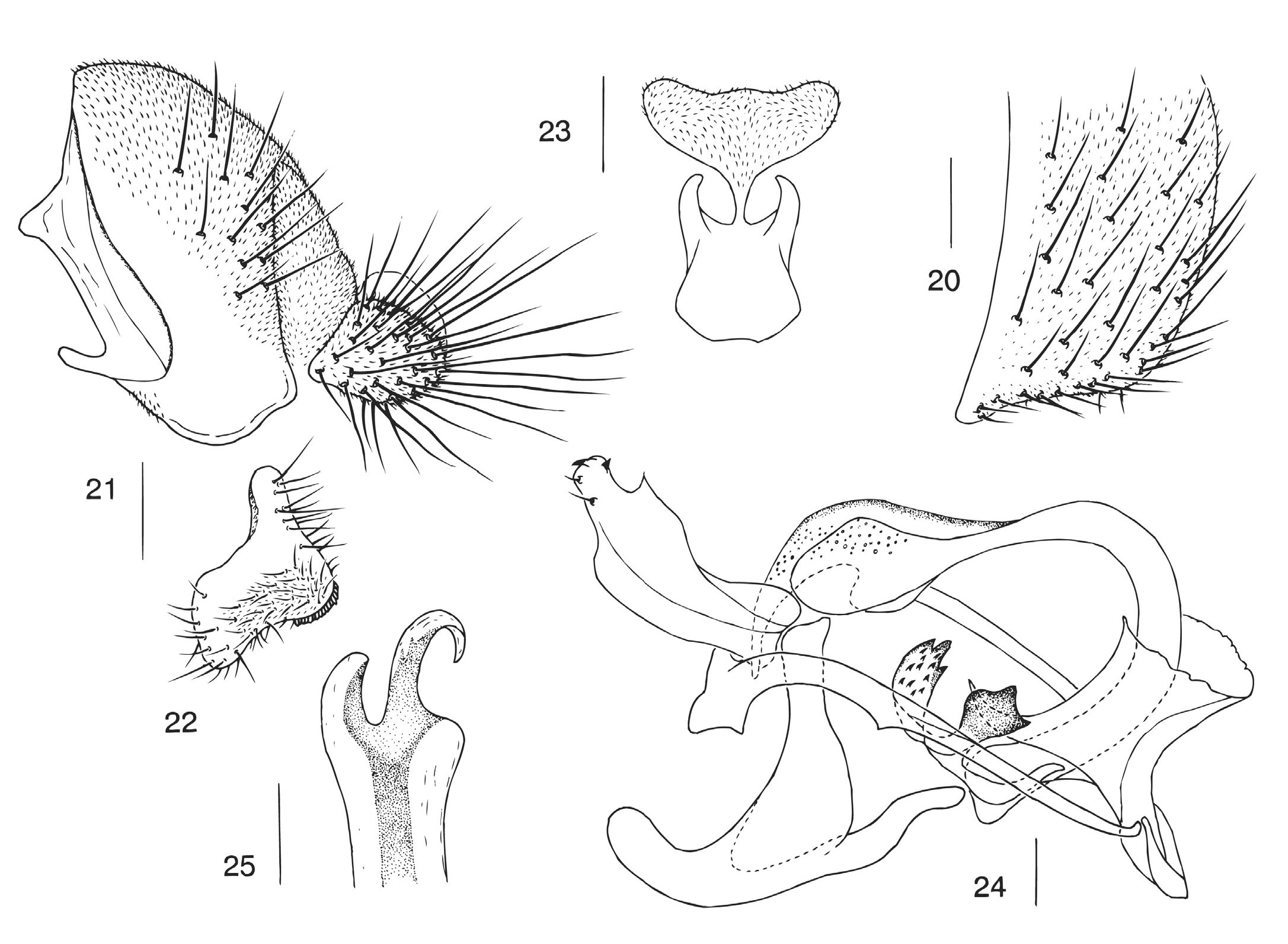
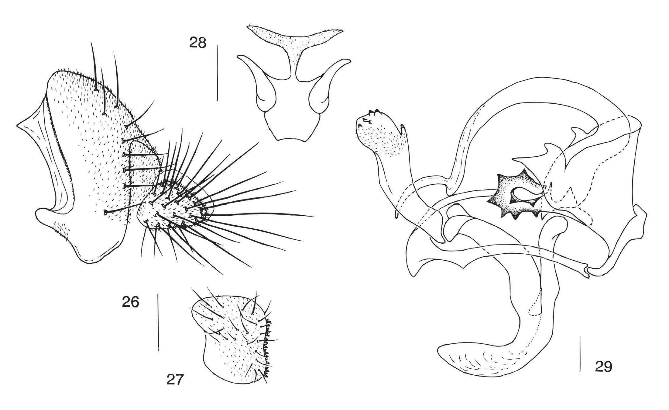
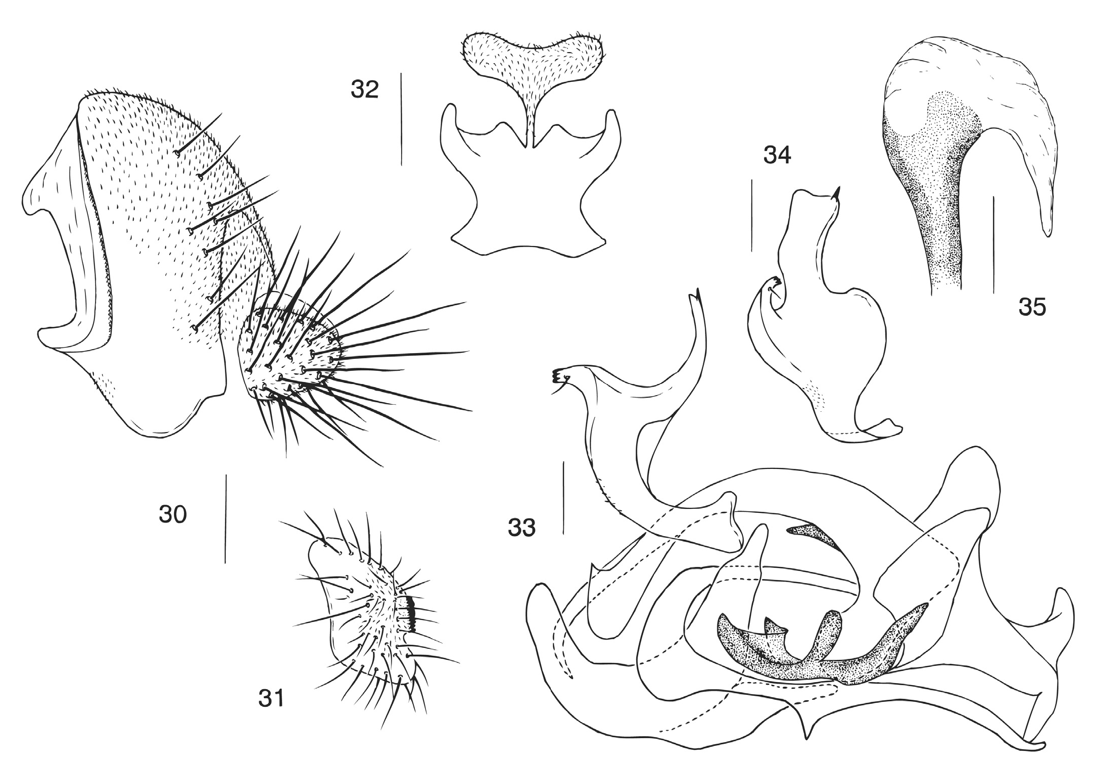
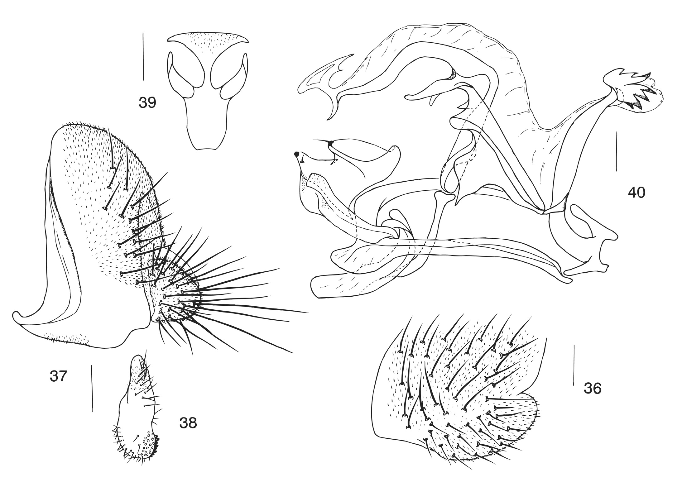
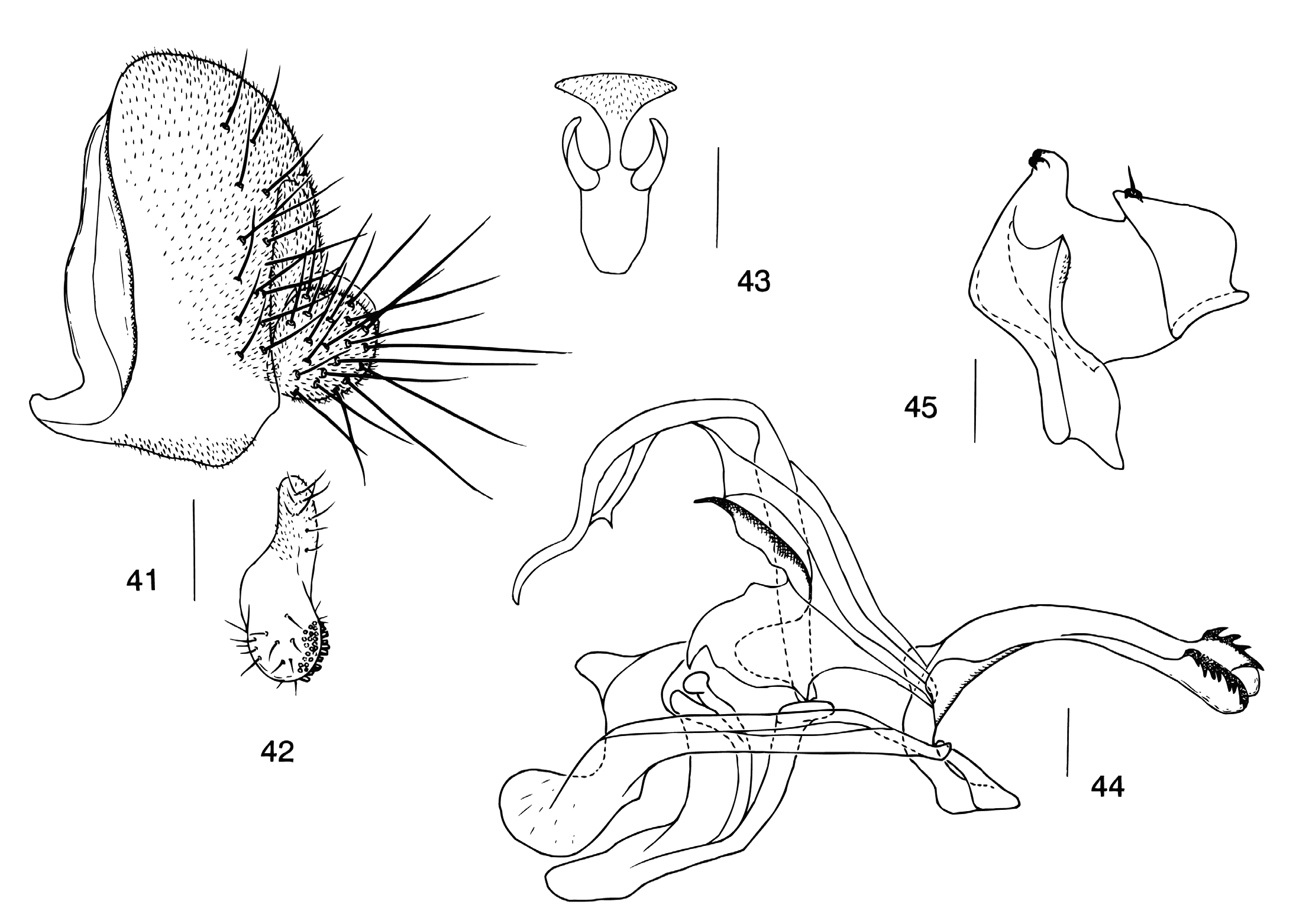
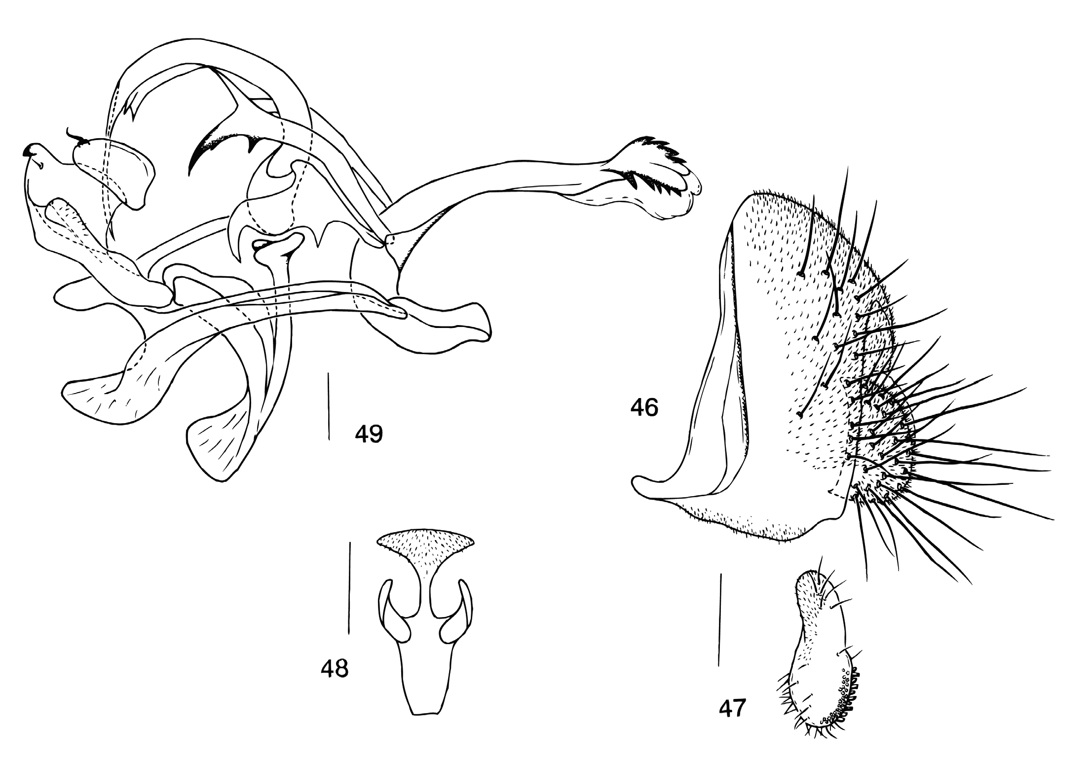
Diagnosis. – Leg tibiae with 3 dark rings; additional plate present between cerci and 10th sternite ( Figs. 4 View Figs , 9 View Figs , 14 View Figs , 18 View Figs , 23 View Figs , 28 View Figs , 32 View Figs , 39 View Figs , 43 View Figs , 48 View Figs , 52 View Figs , 56 View Figs ); aedeagal median rod developed, basally and/or medially connected to basal corners of gonopods by 1 or 2 pair(s) of bridges ( Figs. 5 View Figs , 10 View Figs , 15 View Figs , 19 View Figs , 24 View Figs , 29 View Figs , 33 View Figs , 40 View Figs , 44 View Figs , 49 View Figs , 53 View Figs , 57 View Figs ).
Description. – Eyes brownish red. Ocellar triangle brown to black, with 1 pair of small setae below ocellar setae. Frons brown to black, medially with a few interfrontal setulae. Fronto-orbital plate pale, often silvery white; proclinate orbital seta nearer to inner vertical seta than to ptilinal fissure. Pedicel and first flagellomere grayish yellow. Face yellow to brown, with yellowish white patches on lower corners. Clypeus medially white to yellow, laterally dark brown to black. Gena grayish brown; postgena dark brown. Palpus somewhat triangular, grayish yellow distally, brown basally, with 1 hollow sense organ and a few setae distally. Vibrissa prominent; other orals small. Occiput glossy, brownish black.
Thorax yellow to orange brown, usually with brownish to black patches and pollinose pattern. Postpronotal lobe pale yellow, with 1 long and a few short setae. Acrostichal setulae in ca. 10 irregular rows. Prescutellar setae usually 1 pair. Anepisternum usually lacking setulae. Scutellum usually concolorous with thorax, with dark brown to black patch. Basal scutellar setae divergent; apicals cruciate.
Wing hyaline, sometimes smoky; veins grayish yellow. Basal medial-cubital crossvein present; C 1 setae 2, indistinctly differentiated. Costal vein with spinules on ventral surface between R 2+3 and R 4+5. R 2+3 slightly curved to costa at tip; R 4+5 distally convergent with M 1. Halters white.
Legs yellow; femora usually brown to black except for apical portions. Foreleg femur with 2-3 irregular rows of long setae on posterior surface. Preapical dorsal setae present on all tibiae. Midleg tarsus ventrally with 2 rows of minute cuneiform setulae on inner and outer sides; hindleg tarsus with 1 row of minute cuneiform setulae on underside; fore- and hindleg first tarsomeres each as long as three succeeding tarsomeres together; midleg first tarsomere as long as other tarsomeres combined.
Abdominal tergites yellow to orange yellow; second to fifth tergites with broad brownish to black bands on posterior margins. Sternites usually grayish yellow.
Male terminalia: Epandrium almost not constricted middorsally, with pubescence and setae; apodeme developed along anterior margins. Cercus almost oval, but elongate in P. hani (Zhang & Shi, 1997) and P. varipes Duda, 1926 , separated from epandrium, entirely pubescent and setigerous. Surstylus with numerous setae on outer surface. Membrane between epandrium and cercus pubescent. Hypandrium arched, usually with 1 pair of apodeme processes on anterior portion; posterior ends contiguous to lateral corners of gonopods and anteroventral corners of epandrium. Gonopods fused to each other, forming posteromedian plate, anteriorly forming vertical process. Parameres usually basally contiguous to anterior portion of hypandrium and tips of distally bifurcated ventral branch of aedeagal apodeme. Aedeagus composed of outer membranous tube and more or less sclerotized median rod; outer membrane posteriorly connected to vertical process of gonopod; median rod basally contiguous to dorsal branch of aedeagal apodeme; basal bridge sometimes with sclerotized branch ( Figs. 5 View Figs , 24 View Figs , 29 View Figs , 33 View Figs ); ventral bridge (termed inner paraphysis by Bächli et al., 2004) usually contiguous to medial process of aedeagus ( Figs. 40 View Figs , 44 View Figs , 49 View Figs , 53 View Figs ), sometimes elongated and dilated apically ( Fig. 57 View Figs ). Aedeagal apodeme mostly laterally flattened. Ejaculatory apodeme rudimentary (in Asian species).
Classification. – Supraspecific division within the genus Phortica has not been fully settled yet, although the following species-group or complexes have been erected: the variegata complex by Máca (1977), the foliiseta complex by Tsacas & Okada (1983), the magna complex by Chen & Toda (1997), the omega complex by Chen & Toda (1998) and the varipes group by Máca (2003). According to Chen’s (2001) phylogenetic analysis, the classification within the genus Phortica will be fully revised in the near future. For the present we follow the previous classification system within Phortica , only assigning some species to known speciescomplexes, except for the variegata complex of which the diagnosis is insufficient in the light of recent phylogenetic analysis by Chen (2001).


No known copyright restrictions apply. See Agosti, D., Egloff, W., 2009. Taxonomic information exchange and copyright: the Plazi approach. BMC Research Notes 2009, 2:53 for further explanation.
|
Kingdom |
|
|
Phylum |
|
|
Class |
|
|
Order |
|
|
Family |
Phortica Schiner, 1862
| Chen, Hong-Wei, Toda, Masanori J., Lakim, Maklarin B. & Mohamed, Maryati B. 2007 |
Amiota (Phortica)
| Wheeler, M 1952: 167 |
Phortica
| Maca, J 2003: 251 |
| Schiner, I. R 1862: 433 |