
Aplysia (Varria) inca d’Orbigny, 1837
|
publication ID |
https://doi.org/10.11646/zootaxa.5222.3.1 |
|
publication LSID |
lsid:zoobank.org:pub:582B99D3-0BE3-4FE0-B9FC-D23555B66E77 |
|
DOI |
https://doi.org/10.5281/zenodo.7467209 |
|
persistent identifier |
https://treatment.plazi.org/id/03CA87ED-FFA4-FF84-69A0-F91ACA02FE3D |
|
treatment provided by |
Plazi |
|
scientific name |
Aplysia (Varria) inca d’Orbigny, 1837 |
| status |
|
Aplysia (Varria) inca d’Orbigny, 1837 View in CoL
( Figures 18–32 View FIGURE 18 View FIGURE 19 View FIGURE 20 View FIGURE 21 View FIGURE 22 View FIGURE 23 View FIGURE 24 View FIGURE 25 View FIGURE 26 View FIGURE 27 View FIGURE 28 View FIGURE 29 View FIGURE 30 View FIGURE 31 View FIGURE 32 )
Aplysia inca d´Orbigny, 1837 View in CoL : 207–209, pl. 19, figs. 1–3; Alamo & Valdivieso 1997: 84; Uribe et al. 2013a: 47.
? Aplysia chierchiana Mazzarelli & Zuccardi, 1889: 52 ; Mazzarelli & Zuccardi, 1890: 52; Alamo & Valdivieso 1997: 83; Nakamura 2006: 79; Uribe et al. 2013a: 47.
Tethys inca — Pilsbry, 1895: 87, pl. 19, figs. 29–31.
? Tethys chierchiana — Pilsbry, 1895: 87.
Aplysia (Varria) inca View in CoL — Eales, 1960: 321; Paredes et al. 1999b: 31; Ramírez et al. 2003: 264; Nakamura 2006: 79.
Type material. Syntype, 1 dissected individual, deposited at MNHN, Paris ( Valdés & Héros 1998).
Type locality. Between Callao and San Lorenzo Island, Peru ( d’Orbigny 1837) .
Material examined. PERU, Piura: Sechura ( Matacaballo ), 1 specimen 60 mm length, 17.I.2004 (LaBSIM 15.06-0013) . PERU, La Libertad: Huanchaco , 1 specimen 110 mm length, 21.III.2018, J. Esplana col. (LaBSIM 15.06-0034) . PERU, Ancash: Samanco , 3 specimens 113–149 mm length, 1992, V.H. Vera col. (LaBSIM 15.06- 0006), 3 specimens 70–80 mm length, 6.III.1996 (LaBSIM 15.06-0009) . PERU, Lima: Ancón , 1 specimen 70 mm length, 9.X.1977, C. Paredes col. (LaBSIM 15.06-0002.1), 2 specimens 63–106 mm length, 5.II.1993, C. Paredes col. (LaBSIM 15.06-0007), 3 specimens 58–80 mm length, 10.IV.1995, J. Quesada col. (LaBSIM 15.06-0008), 1 specimen 96 mm length, 27.IX.1997, C. Paredes col. (LaBSIM 15.06-0010), 1 specimen 79 mm length, 26.IX.1999, C. Paredes col. (LaBSIM 15.06-0011), 1 specimen, 83 mm length; 8.XII.2000, F. Cardoso col. (LaBSIM 15.06- 0042), 1 specimen 91 mm length, 8.XII.2000, F. Cardoso col. (LaBSIM 15.06-0012), 1 specimen 101 mm length, 2. VI.2017, F. Cardoso col. (LaBSIM 15.06-0020.1), 2 specimens 66–102 mm length; 17. VI.2017, F. Cardoso col. (LaBSIM 15.06-0022), 3 specimens, 19.XI.2017, P. Guardales & J. Leandro col. (LaBSIM 15.06-0027.1-3), 1 specimen 185 mm length; 7.IX.2018 (LaBSIM 15.06-0038). PERU, Callao: La Punta ( La Arenilla ), 2 specimens 161–177 mm length, 6.XII.2000, F. Cardoso col. (LaBSIM 15.06-0044) . PERU, Lima: Barranco , 16 specimens 87–186 mm length, 15.I.2018, A. Mendivil col. (LaBSIM 15.06-0032) . PERU, Lima: Chorrillos , 4 specimens 136– 146 mm length, 16.IX.2017, A. Mendivil col. (LaBSIM 15.06-0025), 6 specimens 115–186 mm length, 5.XI.2017, A. Mendivil col. (LaBSIM 15.06-0026), 5 specimens 136–199 mm length, 9.XII.2017, A. Mendivil col. (LaBSIM 15.06-0028), 1 specimen 84 mm length, 10.I.2018, A. Mendivil col. (LaBSIM 15.06-0031) . PERU, Lima: Pucusana , 2 specimens 149–151 mm length, 11.XII.1977 (LaBSIM 15.06-0004), 1 specimen 40 mm length, 1983 ( IMARPE 04-001902 View Materials ), 1 specimen 179 mm length, 2017, C. Cisneros col. (LaBSIM 15.06-0017), 3 specimens 147–205 mm length, 16.XII.2017, A. Mendivil & A. Bravo col. (LaBSIM 15.06-0029.1-3), 2 specimens 222–233 mm length, 6.I.2018, A. Mendivil col. (LaBSIM 15.06-0030.1-2) . PERU, Ica: Paracas , 1 specimen 64 mm length, IV.2017, P. Carbajal col. ( IMARPE 04-001901 View Materials ) . PERU, Ica: Laguna Grande , 1 specimen 77 mm length, 6. V.1984, C. Paredes col. (LaBSIM 15.06-0005), 1 specimen 108 mm length, 24.XII.1998, C. Paredes col. (LaBSIM 15.06-0043.1).
Description
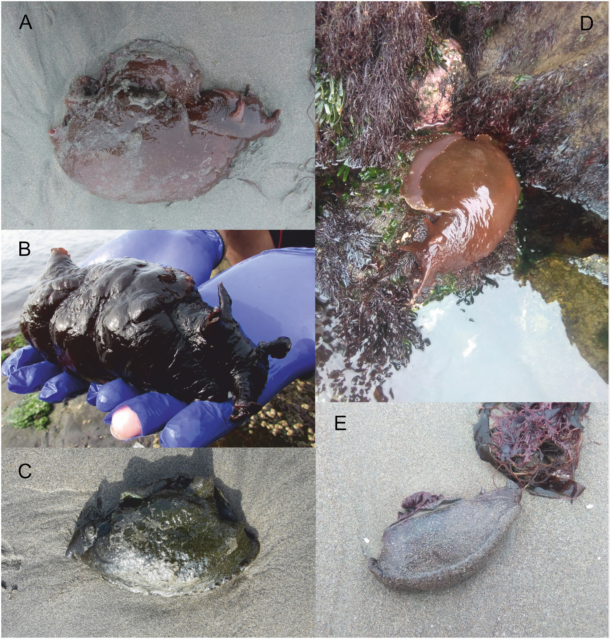
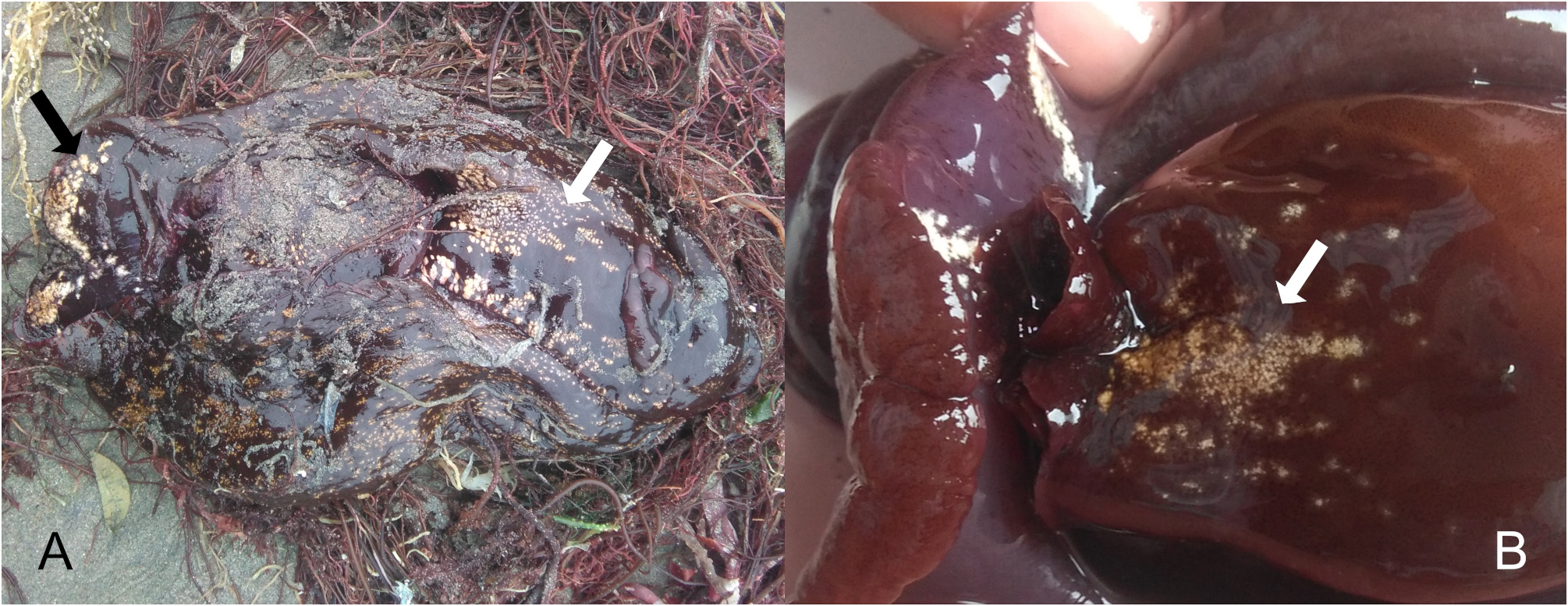
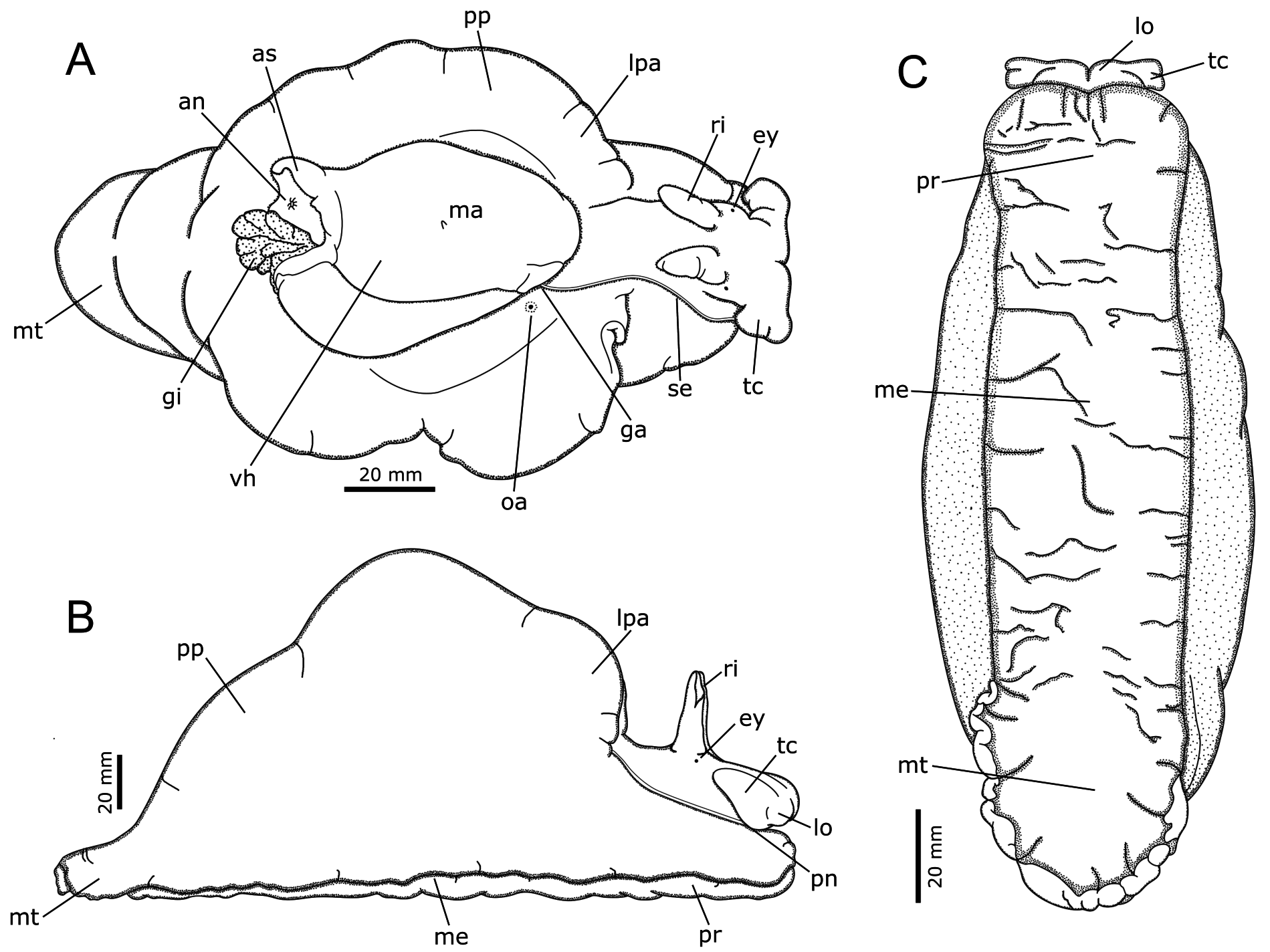
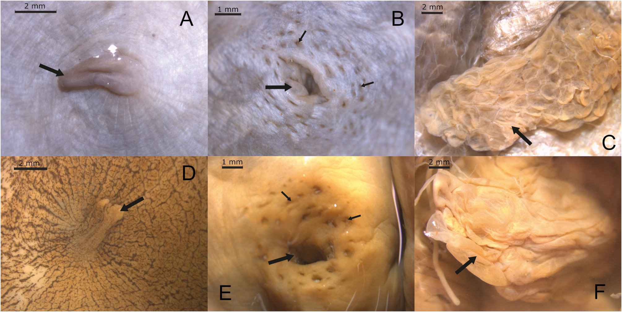
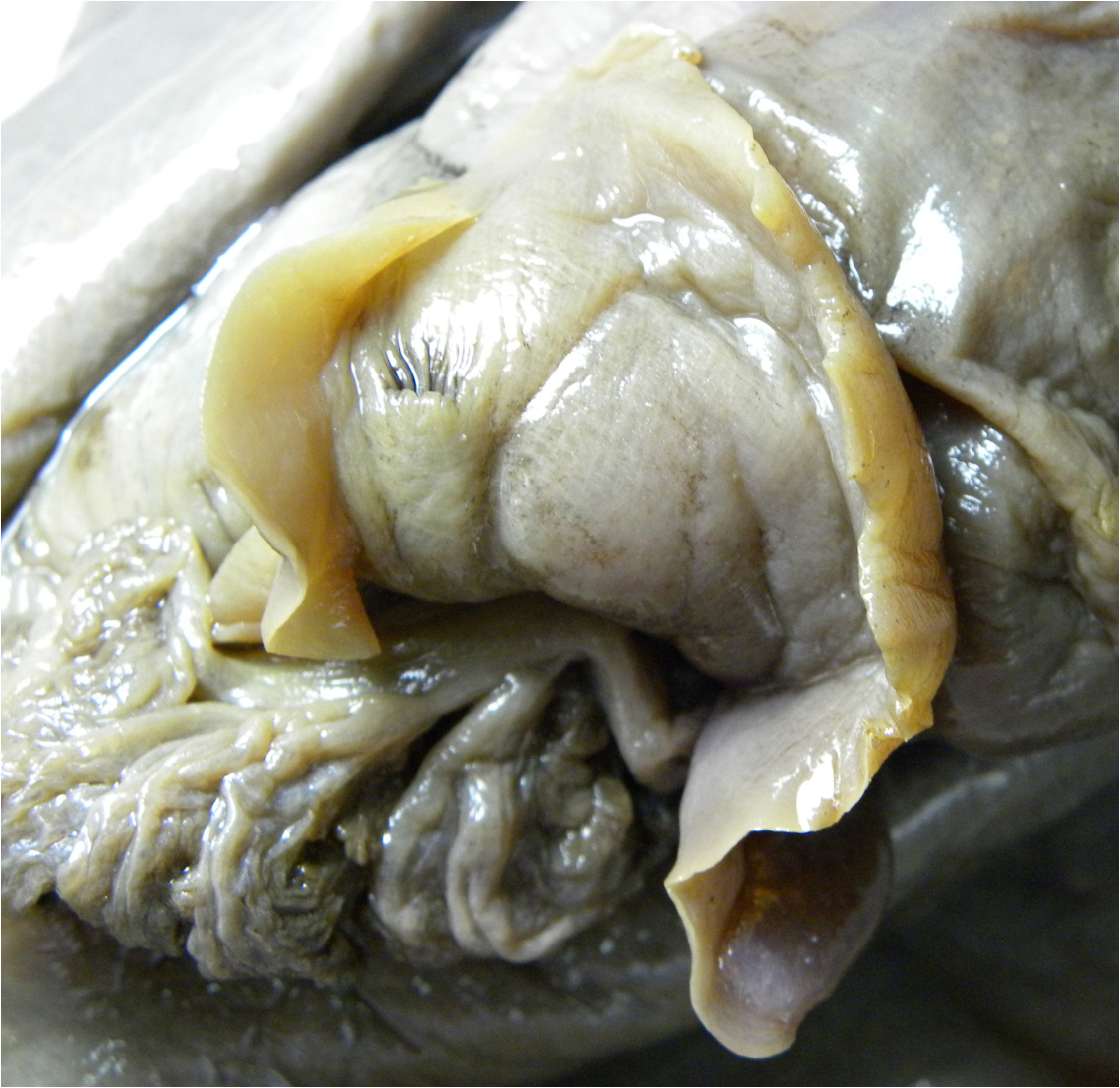
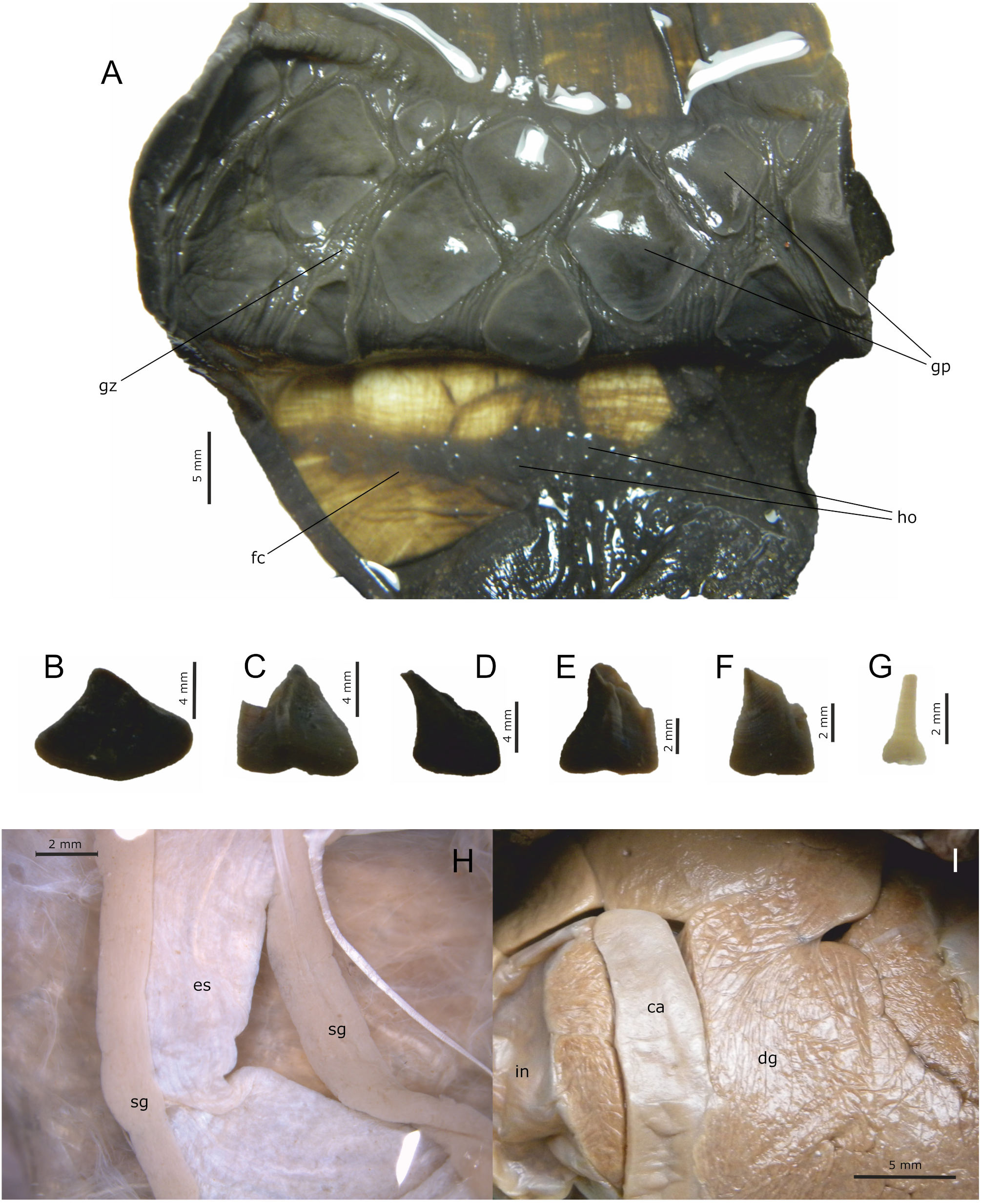
External morphology. Size ~ 230 mm in length. Color variable, purple, reddish, or dark green, with or without white or yellow spots, sometimes aggregated in dense concentrations, specially between anterior end of parapodia and rhinophores, posterior internal margin of parapodia and dorsal mantle ( Figs. 18 View FIGURE 18 , 19 View FIGURE 19 ); parapodia edge sometimes lighter than body color; mantle papilla sometimes with fine radial lines ( Fig. 21 View FIGURE 21 ). Body soft, elongated, relatively high, narrowing gradually towards cephalic region ( Fig. 20A View FIGURE 20 ). Cephalic tentacles elongated, thick, enrolled, with narrower bases, forming oral veil ( Fig. 20A View FIGURE 20 : tc). Oral lobes located on ventral surface of oral veil ( Figs. 20A–B View FIGURE 20 : lo). Rhinophores conical, thick, close together. Foot sole relatively narrow, thick, with pair of lobes poorly differentiated in propodium, with narrow and elongated metapodium, sometimes developed in pointed tail ( Fig. 20C View FIGURE 20 ). Parapodia broad, widely open, joined low down posteriorly, joined anteriorly with lobes slightly developed in anterior margin of parapodia ( Figs. 20A–B View FIGURE 20 : lpa). Visceral hump relatively large, oval, elevated, well differentiated, occupying more than 1/3 of body length ( Fig. 20A View FIGURE 20 ). Mantle foramen reduced to elevated papilla, located on center of mantle ( Fig. 20A View FIGURE 20 , 21A, 21D View FIGURE 21 : arrow). Anal siphon wide, elongated, tubular, occupying ~1/3 of mantle length, freely exposed within parapodial cavity ( Fig. 22 View FIGURE 22 ). Opaline gland resembling a bunch of grapes, with large, rounded main opening surrounded by many very small secondary openings, occupying small area on pallial cavity floor ( Figs. 20A View FIGURE 20 : oa, 21B–C, 21E–F: arrows). Opaline gland secretion milky white. Ink gland secretion purple, copious.
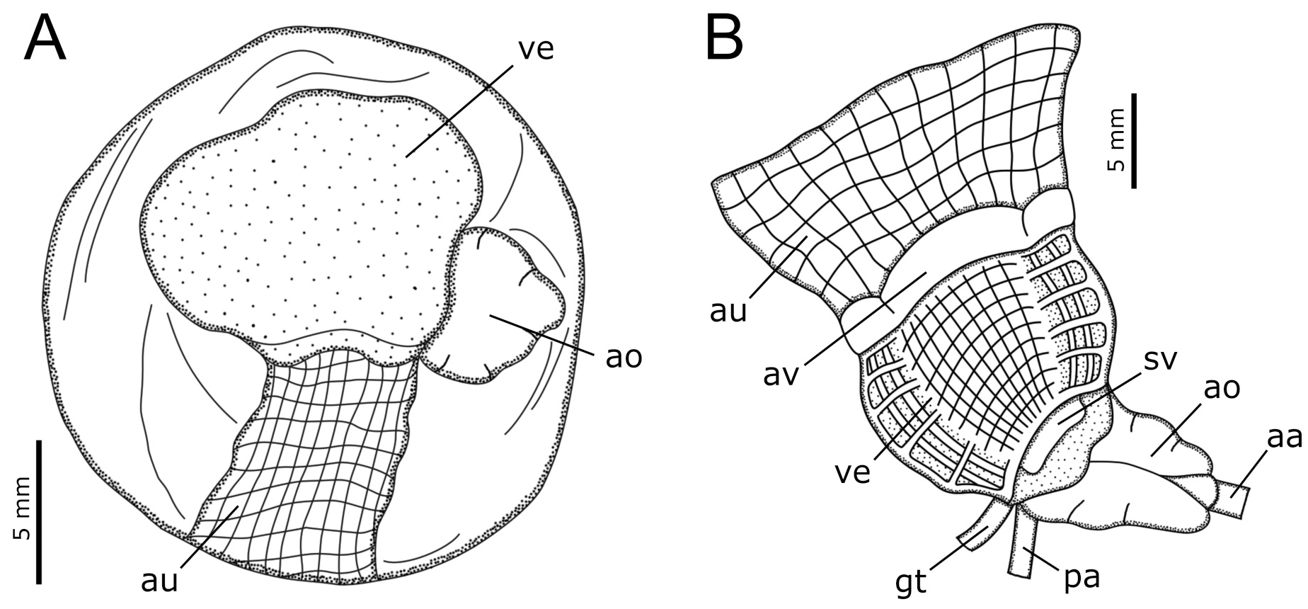
Shell. Relatively large, shell length more than 1/3 of body length, width/length ratio about 3:4, arched, rounded to oval, relatively broad, wider close to anal sinus; on apertural view left margin slightly convex; right margin convex, narrowing gradually towards apex, anal sinus relatively narrow, usually concave ( Fig. 23 View FIGURE 23 ). Protoconch in apex of calcified layer. Sculpture formed by very fine concentric lines.
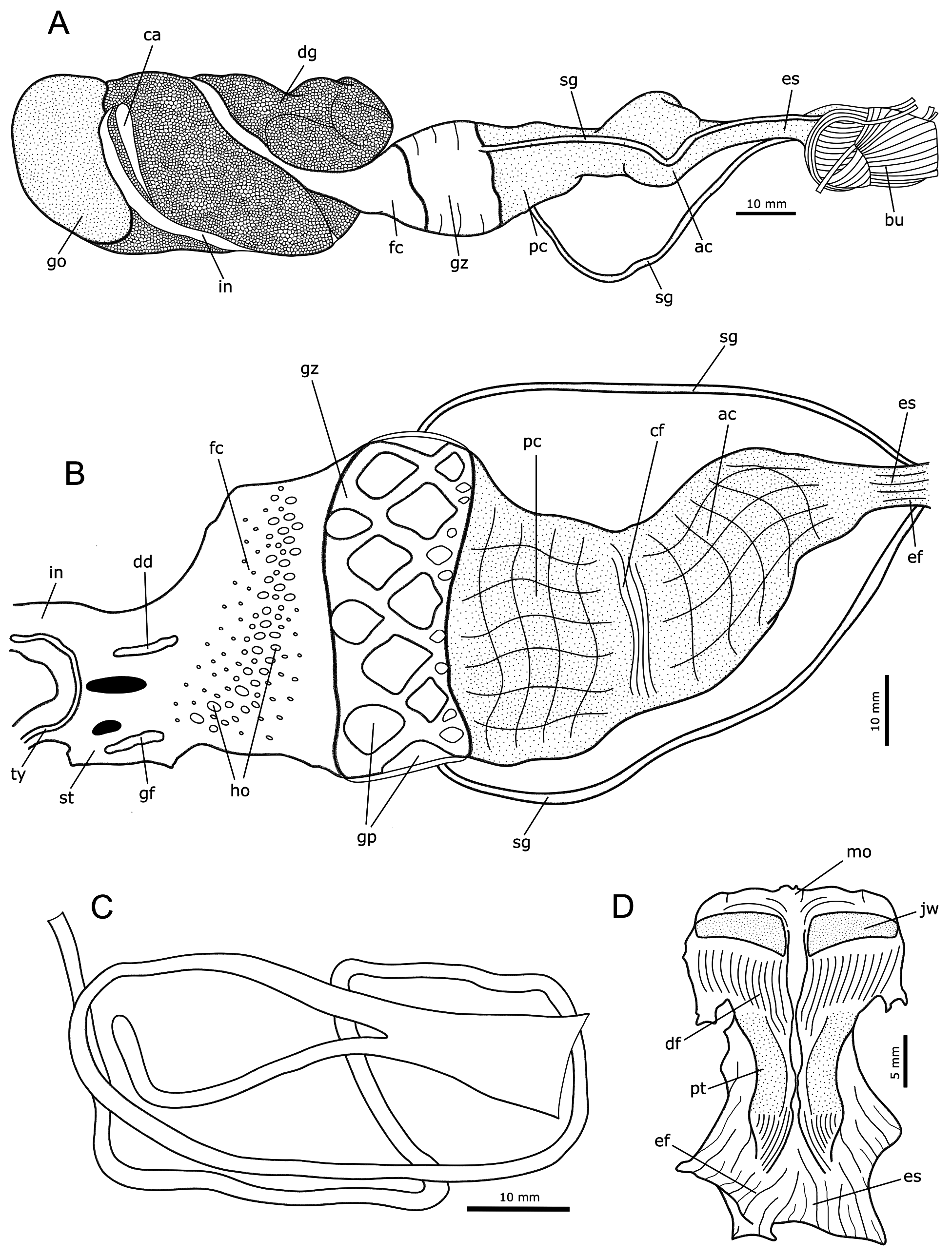
Haemocoel organs. Haemocoel of A. inca very similar to A. nigra ( Fig. 24 View FIGURE 24 ), except for the grape-shaped opaline gland on dorsal wall of haemocoel, occupying a very small fraction of haemocoel volume ( Figs. 21C, 21F View FIGURE 21 : arrow).
Circulatory and excretory systems. Arrangement of circulatory and excretory systems very similar to A. nigra ( Fig. 25 View FIGURE 25 ).
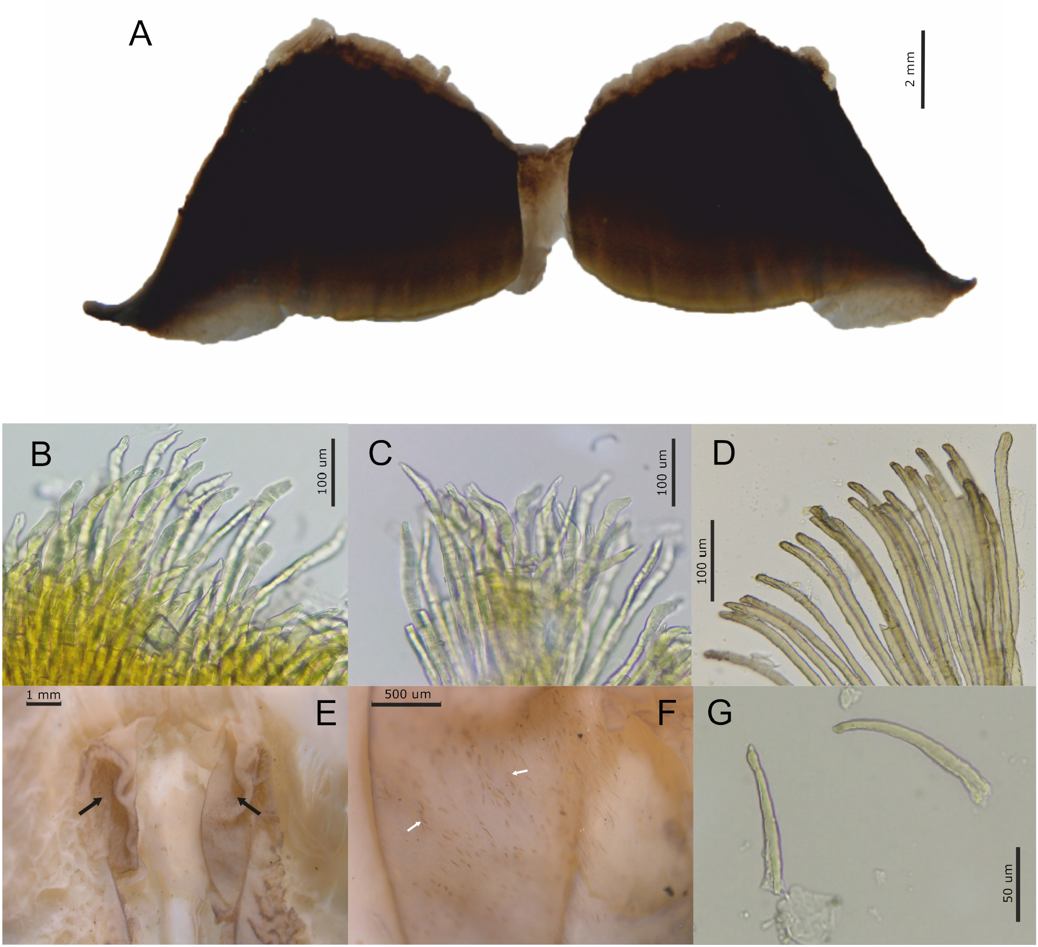
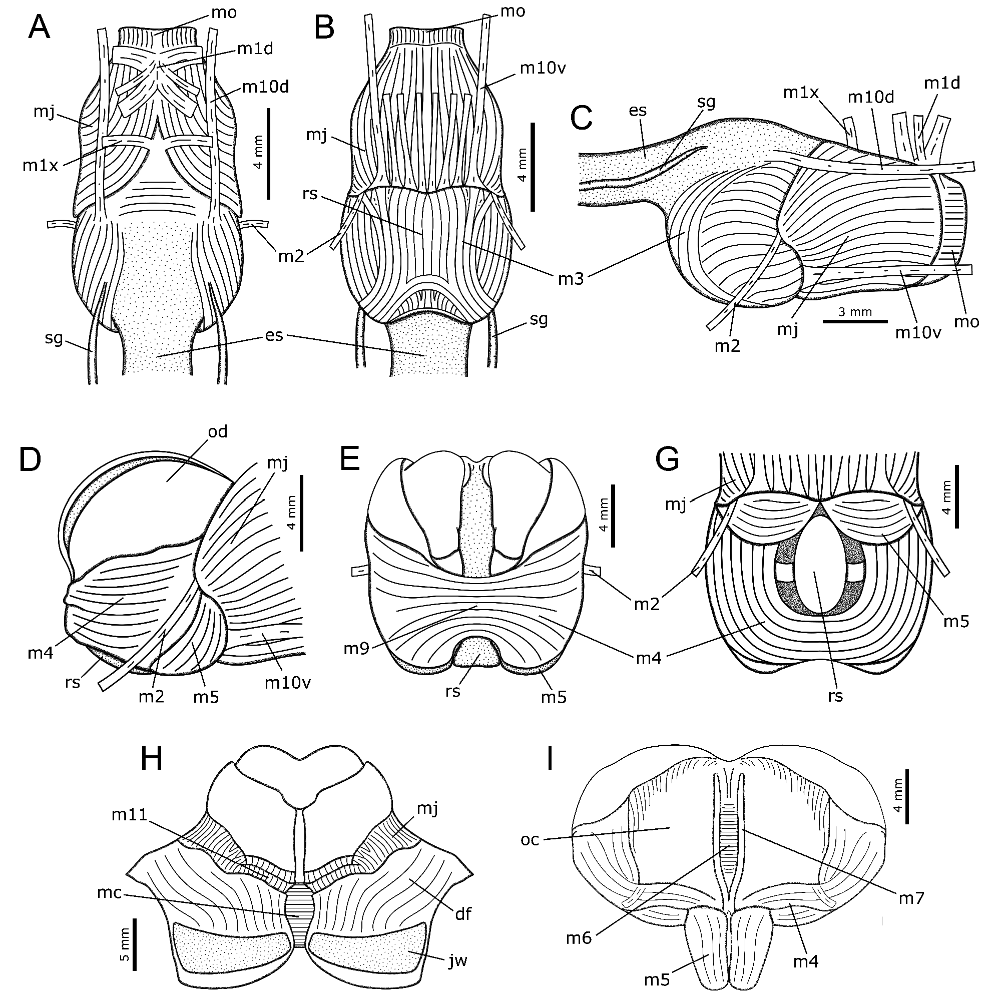
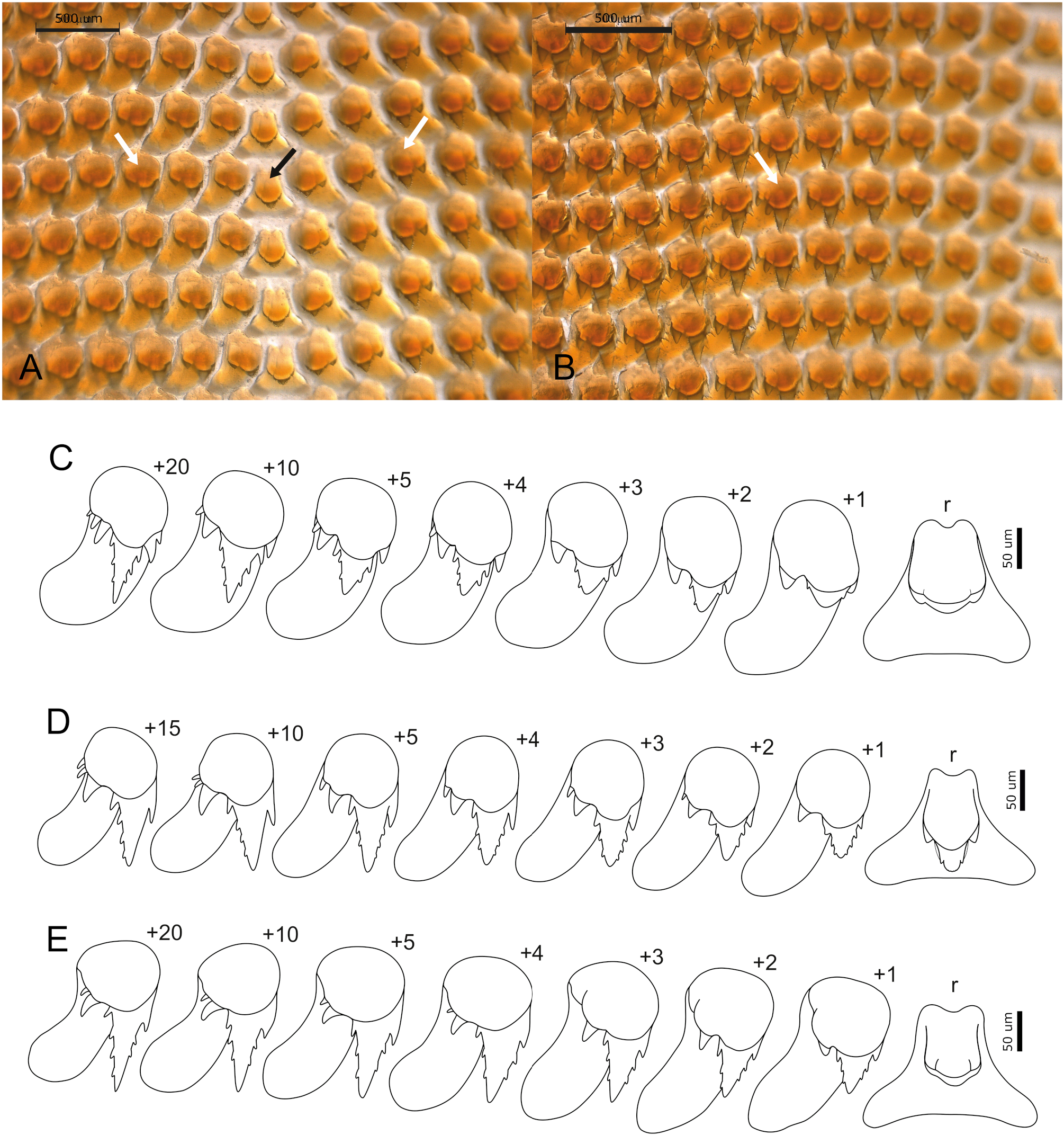
Digestive system. Digestive system of A. inca similar to A. nigra , as described above, except for the following: Jaws thin, without longitudinal ridges, ~2 times wider than long, anterior and posterior borders smooth ( Fig. 26A View FIGURE 26 ), with elongated jaw elements, sometimes knobbed, slightly curved toward distal end, tip narrower with a longitudinal slit ( Fig. 26B–D View FIGURE 26 ). Dorsal folds less developed than in A. nigra ( Fig. 26E View FIGURE 26 : arrows), with elongated palatal elements, curved toward distal end, tip narrower ( Figs. 26F–G View FIGURE 26 ). Odontophore muscles: m1x, pair of auxiliary jugal muscles, ~10 times longer than wide ( Figs. 27A, 27C View FIGURE 27 ); m2, pair of strong retractor muscles of buccal mass, relatively thin, ~15 times longer than wide ( Figs. 27A–C View FIGURE 27 ); m7, pair of very narrow and thin muscles, ~20 times longer than wide ( Fig. 27I View FIGURE 27 ). Pair of odontophore cartilages forming radular bolsters narrow and slightly elevated ( Fig. 27I View FIGURE 27 : oc). Radular formula 48 x 1.19.1.17.1 (specimen length: 66 mm, LaBSIM 15.06-0022.2), to 67 x 2.40.1.40.2 (specimen length: 130 mm, LaBSIM 15.06-0032.5). Rachidian tooth bilaterally symmetrical, wider om base, with small, triangular central cusp with 2 small denticles on each side, 1 small secondary cusp on each side of central cusp, lacking secondary cusps ( Fig. 28 View FIGURE 28 : black arrow). Lateral teeth asymmetrical, elongated, with large and triangular main cusp with up to 4 denticles on each side, 1 secondary cusp well developed with up to 2 additional smaller cusps distally ( Fig. 28 View FIGURE 28 : white arrow). Marginal teeth small, with vestigial cusp. Pair of salivary glands ducts thin, cylindrical, equal in width along their length ( Figs. 29 View FIGURE 29 A-B, 30H: sg). Crop subdivided in two chambers of similar volume ( Figs. 29 View FIGURE 29 A-B). Gizzard presenting 10-11 large, pyramidal chitinous plates of rhomboid base, distributed in 3 transverse rows occupying posterior region of gizzard ( Figs. 29B View FIGURE 29 , 30A–D View FIGURE 30 ); presenting up to 20 small, conical chitinous plates of oval to circular base, usually distributed in a single row occupying anterior region of gizzard ( Figs. 29B View FIGURE 29 , 30A, 30E–F View FIGURE 30 ). Filter chamber occupying 1/3 of gizzard volume, presenting many small, conical gastric hooks distributed in several irregular transverse rows forming a relatively narrow band ( Figs. 29B View FIGURE 29 , 30A, 30G View FIGURE 30 ). Digestive caecum running embedded within digestive gland along most of its length, tip wide and flattened ( Figs. 29A, 29C View FIGURE 29 , 30I View FIGURE 30 : ca); typhlosole continuing into intestine for about 1/3 of stomach length. Intestine slightly convoluted ( Fig. 29C View FIGURE 29 ).
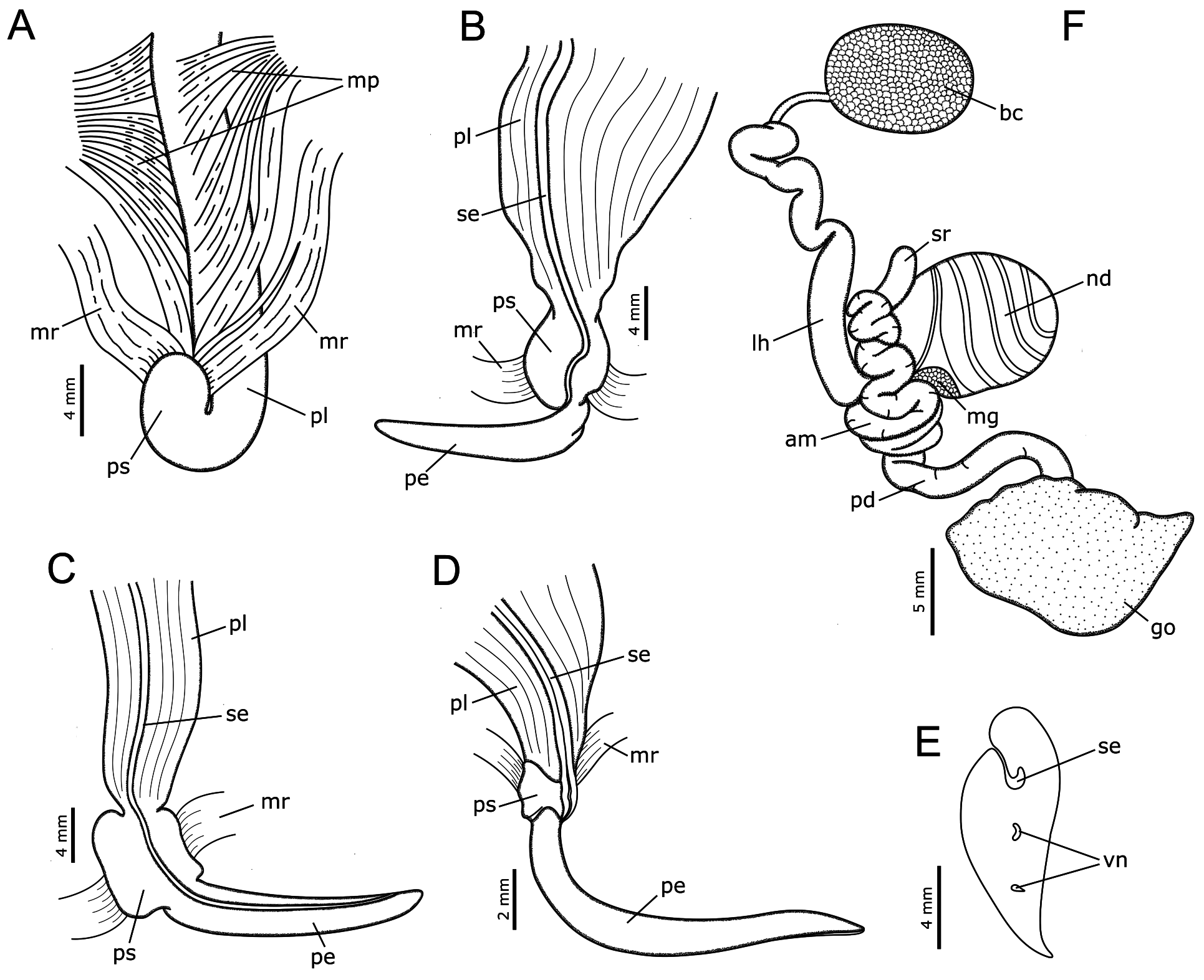
Reproductive system. Penial sheath well differentiated into proximal penial canal and distal penial sac ( Fig. 31A View FIGURE 31 ). Penial canal elongated, with muscular, thick wall, highly folded ( Figs. 31B–D View FIGURE 31 : pl). Penial sac bulbous, with thin wall, unarmed ( Figs. 31B–D View FIGURE 31 : ps). Penis thin, elongated, ~12 times longer than wide, unarmed; with triangular, rounded tip ( Fig. 31E View FIGURE 31 ), seminal groove ending on longer side of tip, opposite side thinner ( Figs. 31B–D View FIGURE 31 ). Pair of retractor muscles of penis thick, ~10 times longer than wide ( Fig. 31A View FIGURE 31 : mr). Protractor muscles of penis formed by many thin fibers ( Fig. 31A View FIGURE 31 : mp).
Hermaphrodite reproductive system of A. inca like A. nigra , except for the following: Preampullary duct wide, about same width as ampulla, slightly curved. Ampulla thick, strongly convoluted, leading to very narrow postampullary duct ( Fig. 31F View FIGURE 31 : am). Large hermaphroditic duct relatively narrow, cylindrical, curved, but not convoluted as ampulla, ~2 times wider than ampulla, slightly attached to dorsal inner surface of haemocoel ( Fig. 31F View FIGURE 31 : lh).
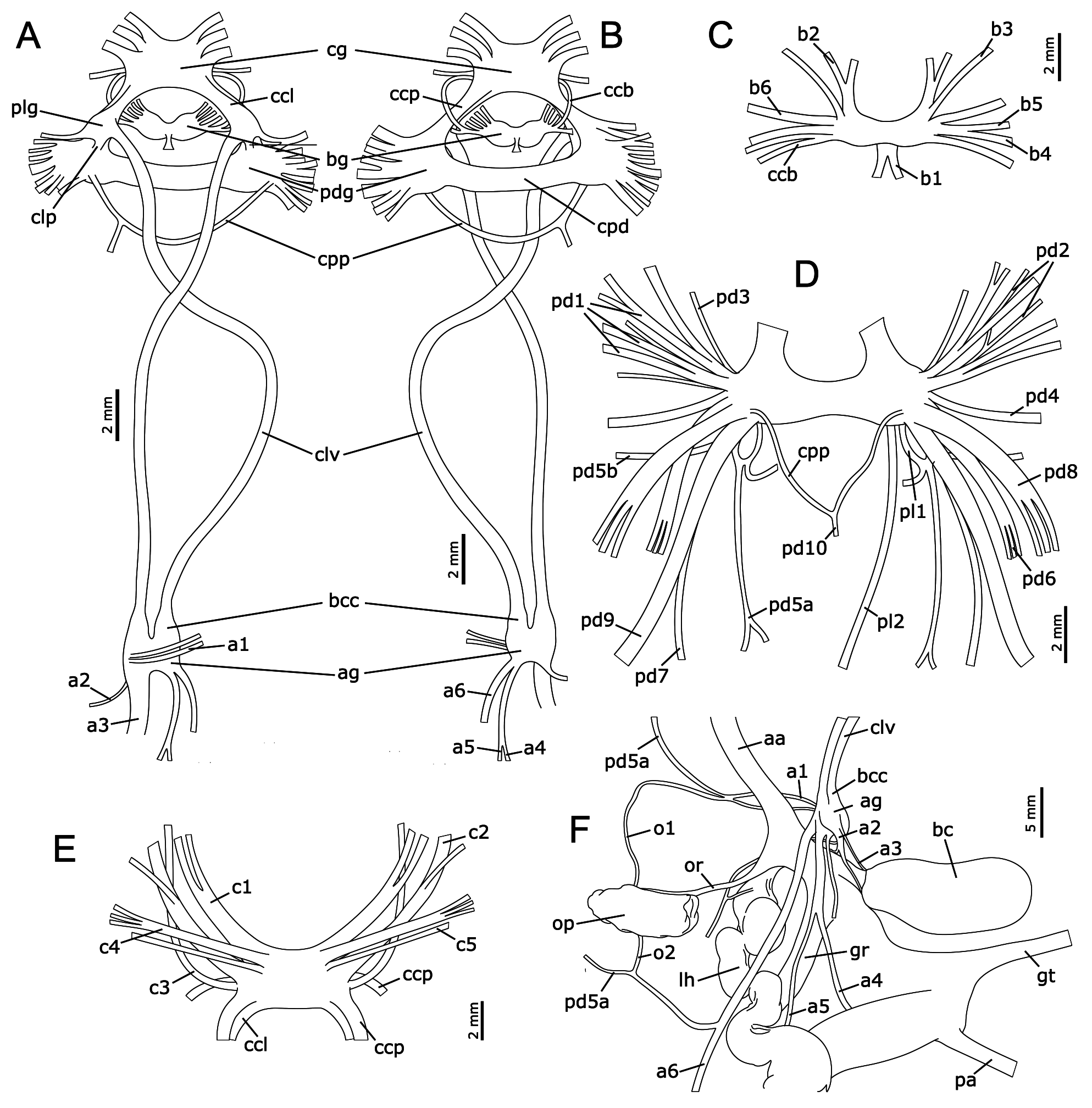
Nervous system. Nervous system of A. inca very similar to A. nigra ( Fig. 32 View FIGURE 32 ), except for the following: Cerebral ganglia fused forming single mass, slightly wider than pedal ganglion. Nerves leaving pedal ganglia ( Fig. 32D View FIGURE 32 ): pd5, slender, bifurcating into 2 branches: pd5a, arrangement very similar to A. nigra ; pd5b, inserting into lateral anterior body; pd5c not observed. Cerebropleural connectives slightly asymmetric, right connective slightly longer than left one. Pleural ganglia almost same size. Pleurovisceral connectives ~1 1/2 times longer than nerve ring length.
Geographic distribution. Sechura, Piura ( 5° S) to Laguna Grande, Ica ( 14° S), Peru .
Habitat and ecology. Sandy beaches, tidal pools, rocky shores with algae; intertidal to subtidal. Mass mortality events seem to be more common than in A. nigra . Apparently more abundant than A. nigra .
Remarks. D’Orbigny (1837) described A. inca as a moderately elongated, tall animal; color purple, covered with white rounded spots; with parapodia well developed joined low down posteriorly, and anal siphon elongated and wide ( Fig. 1B View FIGURE 1 ). Eales (1957, 1960) examined the type material of A. inca adding to the original description a penial sheath unarmed; penis broad, short, pointed at the tip; and opaline gland partly compound with a group of openings arranged in a circle. The specimens examined here are consistent with the original description of A. inca and the observations of Eales (1957, 1960), except for the shape of the penis which is generally elongated rather than broad and short as stated by Eales (1957, 1960). Although A. inca has been the most reported species of the subgenus Varria in Peru ( Eales 1960, Paredes et al. 1988, Paredes et al. 1999a, Ramírez et al. 2015), during the nineteenth century two other species were also described for the Peruvian coast: Aplysia lessonii Rang 1828 and Aplysia chierchiana Mazzarelli & Zuccardi, 1889 ; however, currently only A. inca is considered a valid species (MolluscaBase 2021).
Aplysia lessonii was described from Paita, northern Peru, as rounded, tall animals, grayish pink with very fine red markings, parapodia joined posteriorly and wide anal siphon ( Rang 1828, fig. 1A). Mazzarelli & Zuccardi (1890) reported A. lessonii for Honolulu, Hawaii as animals with a large mantle foramen, feature not mentioned by Rang (1828). Kay (1964) reported five species of Aplysia for the Hawaiian Islands, but there was no mention of A. lessonii . Since none of the species described by Kay (1964) nor those reported later for Hawaii ( Golestani et al. 2019: A. elongata ) are consistent with the original description of A. lessonii , and we did not find a subsequent record of this species for the northern Pacific, the record of A. lessonii for the Hawaiian Islands should be considered probably erroneous. D’Orbigny (1837) distinguished A. inca and A. lessonii using the external coloration and morphological traits such as parapodia, mantle and cephalic tentacles; however, as Rang (1828) described A. lessonii on the basis of preserved specimens, the differences noted by d’Orbigny (1837) between the two species are questionable.
Eales (1960) could not examine the type material of A. lessonii deposited at MNHN, Paris ( 1 syntype dissected, see Valdés & Héros 1998); but she concluded that by similitudes in body shape, foot, parapodia, mantle and shell, it could be a junior synonym of A. keraudreni Rang (1828) from the southwest Pacific. Aplysia keraudreni is a rather large dark brown animal, covered with white spots and a fine black reticulum ( Eales 1960, Willan & Morton 1984); originally described from the Society Islands in the Polynesia, but also reported from New Zealand ( Eales 1960, Willan 1979, Willan & Morton 1984, Morley & Hayward 2015) and Australia ( Nimbs 2017a). Since the external coloration of A. keraudreni is not consistent with the description of A. lessonii ( Rang 1828) nor with that reported in another species from the Eastern Pacific ( Pilsbry 1895, Eales 1960, Beeman 1968, Bebbington 1977, Behrens & Hermosillo 2005, Camacho-García et al. 2005, Valdés 2019), A. keraudreni is very likely restricted to the southwest Pacific. Therefore, the records of A. keraudreni for Peru ( Eales 1960, Bebbington 1977, Paredes et al. 1999 b, Ramírez et al. 2003, Nakamura 2006, Uribe et al. 2013a) should be considered erroneous. Until the examination of the material type of A. lessonii can corroborate the similitudes between both species, we decide to retain the name of A. inca for the typical species of the subgenus Varria in Peru.
Mazzarelli & Zuccardi (1889) introduced the name of A. chierchiana for a dark animal, with black rounded spots and small white dots, wide parapodia, mantle foramen reduced to a small papilla, and opaline gland compound and multiporous; suggesting the mantle papilla as the diagnosis feature of this species ( Fig. 1E View FIGURE 1 ). Although Mazzarelli & Zuccardi (1890) considered A. chierchiana very distinct from A. inca , with both having the same type locality, Eales (1960) did not compare them and rejected the name A. chierchiana , without specifying whether due to lack of type material, or by an inadequate description. In this work, we found that the most characteristic feature of A. chierchiana are the black rounded spots that cover the body of the living animals, not described in A. inca ( d’Orbigny 1837, Eales 1960). Specimens that can be identified as A. chierchiana were found in Sechura, Samanco and San Lorenzo Island; where A. inca specimens have also been observed, with both species similar in body shape, foot, anal siphon, mantle papilla, shell, radula, jaws, and penis. Although A. chierchiana is included here as a probable synonym of A. inca , these specimens are not included in the anatomical redescription of A. inca until their taxonomic status is clarified using molecular data.
Uribe et al. (2013a, b) recorded A. juliana for Máncora, Isla Santa (Samanco), Ancón and Paracas. However, these animals have parapodia joined low down posteriorly and release purple ink, characteristics not reported in A. juliana but quite common among the species of the subgenus Varria ( Eales 1960) , suggesting that these records are likely A. inca . Although Máncora might be the northernmost record of A. inca , it is not included here until it can be corroborated with the revision of additional material from northern Peru. Aplysia inca has been previously recorded for Pisco ( Paredes et al. 1988), Ancón and Barranca ( Paredes et al. 1999a), and Lobos de Tierra ( Ramírez et al. 2015), although the last record needs to be verified and is not included in the distribution of the species.


| MNHN |
Museum National d'Histoire Naturelle |
| VI |
Mykotektet, National Veterinary Institute |
| V |
Royal British Columbia Museum - Herbarium |
No known copyright restrictions apply. See Agosti, D., Egloff, W., 2009. Taxonomic information exchange and copyright: the Plazi approach. BMC Research Notes 2009, 2:53 for further explanation.
|
Kingdom |
|
|
Phylum |
|
|
Class |
|
|
SubClass |
Heterobranchia |
|
Order |
|
|
Family |
|
|
Genus |
|
|
SubGenus |
Varria |
Aplysia (Varria) inca d’Orbigny, 1837
| Mendivil, Alejandro & Cardoso, Franz 2022 |
Aplysia inca d´Orbigny, 1837
| Uribe, R. & Nakamura, K. & Indacochea, A. & Pacheco, A. S. & Hooker, Y. 2013: 47 |
| Alamo, V. & Valdivieso, V. 1997: 84 |
Aplysia (Varria) inca
| Nakamura, K. 2006: 79 |
| Ramirez, R. & Paredes, C. & Arenas, J. 2003: 264 |
| Paredes C. & Huaman, P. & Cardoso, F. & Vivar, R. & Vera, V. 1999: 31 |
| Eales, N. B. 1960: 321 |
Tethys inca
| Pilsbry, H. A. 1895: 87 |
Tethys chierchiana
| Pilsbry, H. A. 1895: 87 |
Aplysia chierchiana
| Uribe, R. & Nakamura, K. & Indacochea, A. & Pacheco, A. S. & Hooker, Y. 2013: 47 |
| Nakamura, K. 2006: 79 |
| Alamo, V. & Valdivieso, V. 1997: 83 |
| Mazzarelli, G. F. & Zuccardi, R. 1890: 52 |
| Mazzarelli, G. F. & Zuccardi, R. 1889: 52 |