
Ansonia longidigita
|
publication ID |
https://doi.org/10.5281/zenodo.191764 |
|
DOI |
https://doi.org/10.5281/zenodo.6226155 |
|
persistent identifier |
https://treatment.plazi.org/id/03CA87A2-FFB1-FFE2-69D2-FD67F9D0F835 |
|
treatment provided by |
Plazi |
|
scientific name |
Ansonia longidigita |
| status |
|
Ansonia longidigita View in CoL
Colour in life ( Fig. 3 View FIGURE 3 ). The background colour of the body is beige and translucent. On this background welldefined symmetric black markings are arranged in a specific pattern: A rostral band begins around each of the nostrils and runs anteriorly to the snout; both bands diverge slightly laterally ( Fig. 3 View FIGURE 3 a). These nostril bands are isolated from other markings. A black orbital band extends from one cheek (region ventrolateral to eye) to the other, embedding both eyes in the course. Perpendicular to and connected to the orbital cross-band lies a sagittal, longitudinal marking. It covers the braincase dorsally. Posterior to it, a pair of broad transverse bands begin close to the vertebral column and extend laterally onto the flanks. At the flanks this epidermal pigmentation is underlain by deeper pigmentation of the abdominal cavity, giving the impression of one dark band wrapping around the flanks onto the lateral parts of the venter ( Fig. 3 View FIGURE 3 b–c). Posterior to these trunk markings, two longitudinal black, broad bands begin at the posterior end of the trunk. They run laterally along the sides of the upper parts of the tail musculature, but leave the dorsal face and ventral parts of the tail musculature uncovered.
Between the black markings, the dorsum and dorsal face of the tail muscle are pigmented with loosely scattered melanocytes. Epidermal melanocytes are small and circular in shape.
The ventral side (venter, oral disc) of the tadpole is mostly unpigmented and translucent, except for the ventral extensions of the transverse trunk markings. The gut coil and developing front limbs are visible in ventral view. The gills and the heart shine through the ventral skin in red. The pericard is partially pigmented. The iris is black. The skin of the oral sucker is without pigmentation. The blood vessels of the tail are not sheathed by melanophores; thus, the vena caudalis ventralis is inconspicuous.
The colour in preservation shows the same markings as living specimens, however, markings usually turn lighter.
External morphological features. A small tadpole, the largest specimen in our sample measuring 13.2 mm in total length at stage 33 ( Fig. 4 View FIGURE 4 a; Tab. 2), with moderately long tail (58–62% of total length). The body contour in dorsal view is moderately tear drop shaped. In dorsal view, the body is widest approximately at eye level, without any pronounced constriction of the contour. The body is moderately depressed dorsoventrally. The snout is expanded corresponding to the enlarged ventral sucker. In life and adhering to the substrate, the snout profile is straight ( Fig. 3 View FIGURE 3 c). The eyes are located dorsally, at clear distance from the body contour in dorsal view.
The external nares are much closer to the eyes than to the snout ( Fig. 3 View FIGURE 3 a, 4a). The interorbital distance is 1.45±0.13 (mean±SD) times the internarinal distance. The spiracle is sinistral and the spiracular orifice is free from the body wall. The spiracle is directed posterodorsally, opening ventrally close to the substratum when the tadpoles is attached by its sucker. The gut is arranged in a transverse coils.
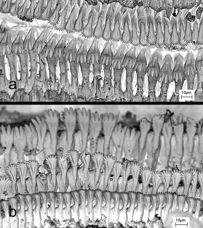
The oral disc is ventral and slightly wider than the snout in adhesion state ( Fig 3 View FIGURE 3 b, 4a). The marginal papillation of the oral disc is only present on the lower lip; the margin of the upper lip is fleshy and devoid of papillae ( Fig. 4 View FIGURE 4 a). Marginal papillae are arranged mostly uniserially; some papillae may stand outside that row in the middle third of the labium. The oral disc margins possess inconspicuous lateral indentations at the end of the papillae row. Papillae are short, blunt and adjoining ( Fig. 4 View FIGURE 4 a). Submarginal papillae are absent. The labial ridges bear uniserial keratodont rows. The Labial Tooth Row Formula (LTRF) is 2/3. The upper lip keratodont rows do not extend caudally beyond lower lip keratodont rows but end anterior to the latter ( Fig. 3 View FIGURE 3 b). The keratodonts are spoon-shaped with incisions along their edges ( Fig. 5 View FIGURE 5 ). Keratodont shape is differentiated according to rows: keratodonts are smaller and more finely serrated on the distal row than on the proximal upper lip row ( Fig. 5 View FIGURE 5 a); a similar proximodistal gradient (finely serrated on distal row) applies to the keratodonts of the three lower lip rows ( Fig. 5 View FIGURE 5 b). The upper beak comprises two short, clearly spaced keratinized edges. The gap between the two edges equals clearly less than twice the edges’s lengths. The lower beak is shallow V-shaped and serrated along its edge.
The dorsal tail fin starts shortly posterior to the trunk-tail junction. Dorsal and ventral tail fins are of equal heights. At approximately mid-tail position the tail reaches its maximum height. The fins’ edges are only slightly convex, almost parallel in lateral view ( Fig. 3 View FIGURE 3 c, 4a), tapering rapidly in a rounded tip at the posterior end.
The anal siphon is located medially, embedded in a flap of skin extending from the posterior end of the body between the limb anlagen. The tail musculature is strong and high (lateral view) proximally and reduced in height distally with a smooth but noticeable reduction in height at about mid-tail length.
Variation. The described pattern of black bands is characteristic and does not vary among the specimens examined or localities. Only the degree to which the posterior bands fuse was found variable: unlike Fig. 3 View FIGURE 3 , there was a solid black area in the dorsal trunk above the vertebral columns (bands fused) in some specimens ( Fig. 4 View FIGURE 4 a); rostral bands, however, are never fused. Transverse gut coils were present in most specimens, but less well organized in the transverse plane in some. For metric variation, see Tab. 2.
Ecological notes. We collected Ansonia longidigita tadpoles from small streams (< 2 m wide), from rocky side pools and rock faces in moderate, non-foaming current (= low energy) during the day. They were observed on flat, inclined, or vertical rock faces. Ansonia longidigita tadpoles occurred in syntopy with A. platysoma tadpoles in the same river (at the Camp 2 site of Gunung Mulu), but with clearly different microhabitat preferences.


No known copyright restrictions apply. See Agosti, D., Egloff, W., 2009. Taxonomic information exchange and copyright: the Plazi approach. BMC Research Notes 2009, 2:53 for further explanation.