
Coronaproctus castanopsis Li, Xu & Wu, 2023
|
publication ID |
https://doi.org/ 10.11646/zootaxa.5254.3.9 |
|
publication LSID |
lsid:zoobank.org:pub:20849690-9237-4D95-ACBA-2C56D7D4DDB1 |
|
DOI |
https://doi.org/10.5281/zenodo.7727811 |
|
persistent identifier |
https://treatment.plazi.org/id/03C78795-D05C-7B32-BBF1-FCBCFEFE06B4 |
|
treatment provided by |
Plazi |
|
scientific name |
Coronaproctus castanopsis Li, Xu & Wu |
| status |
sp. nov. |
Coronaproctus castanopsis Li, Xu & Wu sp. nov.
Material examined. Holotype: adult female. CHINA / Zhejiang province / Kaihua county / Qianjiangyuan National Park (29º 40´N, 118º 35´E) / on the twigs of Castanopsis eyrei ( Fagaceae ) / April 19, 2022 / collected by S-A Wu ( NFUC). GoogleMaps
Paratypes: same data as holotype, 3 adult females mounted singly on 3 slides GoogleMaps ; same locality and host plant as holotype, 10.ix.2020, 3 adult females on 4 slides GoogleMaps ; same locality and host species as holotype, 20.xii.2020, 7 adult males mounted singly on 7 slides ( NFUC) GoogleMaps .
Adult female
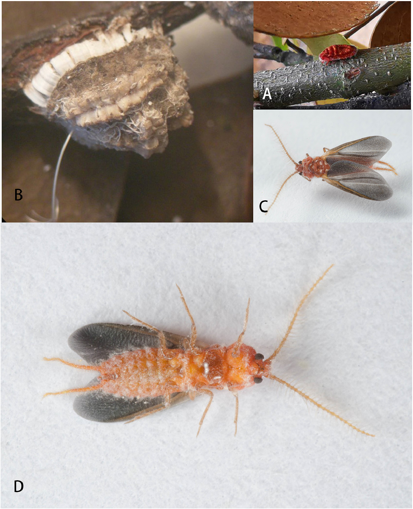
Unmounted material ( Fig. 1A, B View FIGURE 1 ). Body of adult female oblong oval, convex dorsally, orange yellow to red; naked just after the final moult ( Fig. 1A View FIGURE 1 ), mature adult female covered dorsally by a solid waxy test ( Fig. 1B View FIGURE 1 ). Test also oblong oval, about 7.5–8.5 mm long, 6.0–7.0 mm wide and 5.5–6.5 mm high, strongly convex above, sides compressed and slightly concave, bearing marginal and dorsal tufts of wax processes, but with little secretion between the tufts: grayish white, processes paler. Dorsum of thorax with 5 truncate-conical processes, distributed from head in 2-2-1, each process flower-like at apex. Dorsum of abdomen without processes but with a fine white wax tube arising from anal tube; dorsum of abdomen also with a dorsolateral wax ridge and a complete marginal row of processes. Dorsolateral ridge widely separated from marginal ridge except at the two extremities, where they approach each other. Each dorsolateral ridge with a longitudinal furrow along its midline. Body margin with a row of about 20 white wax processes, each with a wax flake bent downwards. Abdominal venter with a deep median cleft, covered by a white layer of felted wax.
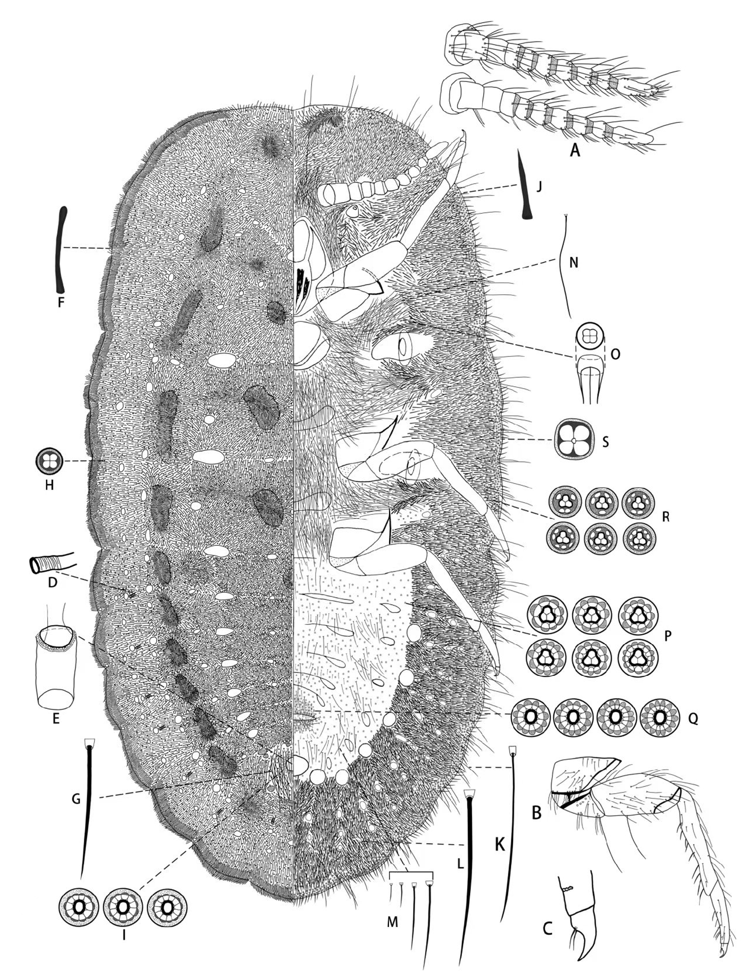
Slide-mounted material ( Fig. 2 View FIGURE 2 ). Body of adult female oval, 5.1–7.8 mm long, 2.6–4.0 mm wide. Derm lightly sclerotized except ventromedial abdomen membranous and with corrugated folds. Antennae ( Fig. 2A View FIGURE 2 ) each 10- segmented and 0.83–1.0 mm long, with basal segment broadest, tapering distinctly to apex; segment lengths (µm): I, 45–75; II, 88–125; III, 70–105; IV, 63–88; V, 63–88; VI, 70–100; VII, 75–110; VIII, 68–110; IX, 63–95 and X, 175–220; each segment with different lengths of setae, segments II–IX normally each with a sensory seta, but sometimes sensory setae of some segments absent; apical segment with 2–6 sensory setae and 2 long slender setae. Mouthparts well developed; labium 1-segmented, conical. Eyes sclerotized, each hemispherical, situated laterad of scape. Thoracic spiracles large, each 275–450 µm wide, without wax pores within atrium. Legs ( Fig. 2B View FIGURE 2 ) well developed, subequal in length; hind leg segment lengths (µm): coxa 150–250, trochanter + femur 600–800, tibia 480–600, tarsus 300–330, claw 60–88; hind tibia + tarsus about 1.3 times longer than trochanter + femur, tarsus about 0.55–0.63 times as long as tibia; trochanter with 4 campaniform sensilla on each surface, a short thin seta (22–41 µm long) and a long seta (300–375 µm long); femur with a long seta (275–375 µm long) on inner side; tibia with spine-like setae on inner side; tarsus with a sensillum near apex ( Fig. 2C View FIGURE 2 ); claw ( Fig. 2C View FIGURE 2 ) without denticle, with a pair of digitules with pointed apices, these shorter than the claw. Abdominal spiracles ( Fig. 2D View FIGURE 2 ) numbering 7 pairs, situated on dorsum, each about 20 µm wide and without wax pores within atrium. Vulva forming a transverse opening, 320–400 µm wide. Anal tube ( Fig. 2E View FIGURE 2 ) sclerotized, 175–250 µm long, 100–150 µm wide, with sclerotized ring at inner end, comprised of polygonal pores of unclear structure; anal opening simple, without sclerotization. Ventral cicatrices numbering 13 (rarely 12 or 14), arranged in a U-shape along the lateral and posterior ventromedial areas of abdomen; middle cicatrix largest, oblong, others smaller and each subcircular. Marsupium present.
Dorsum. Spines with apices slightly swollen and blunt ( Fig. 2F View FIGURE 2 ), each 44–89 µm long, abundant throughout dorsum except around anal tube, forming clusters with denser spines on submarginal and midline areas, also more numerous along margin but apparently not forming distinct spine clusters. Long flagellate setae ( Fig. 2G View FIGURE 2 ), each 225– 305 µm long, present densely in anal area and scarcely among spines. Disc pores of 2 types: (i) small quadrilocular pores ( Fig. 2H View FIGURE 2 ), each 7–8 µm in diameter, distributed throughout dorsum, scattered among spines but scarce within spine clusters, and often forming a single row around each spine cluster; (ii) multilocular pores ( Fig. 2I View FIGURE 2 ), each 9–12 µm in diameter, with an elliptical center (sometimes trilocular or quadrilocular) and 10–12 outer loculi, present around anal opening.
Venter. Lanceolate spines with pointed apices ( Fig. 2J View FIGURE 2 ), each 40–95 µm long, densely present on marginal and submarginal areas of venter, more numerous at apex of head. Flagellate setae of 2 types: (i) long setae, each 200–430 µm long ( Figs 2K, L View FIGURE 2 ), distributed along margins of body, usually thin, but 3 pairs on last segment and 1 pair on penultimate segment thick; and (i) short setae ( Fig. 2M View FIGURE 2 ), each about 30.0–50.0 µm long, scattered on median area of abdomen, but densely distributed around vulva. Hair-like setae ( Fig. 2N View FIGURE 2 ), each 125–250 µm long, densely distributed on median area of cephalothorax. Disc pores of 5 types present: (i) quadrilocular pores ( Fig. 2O View FIGURE 2 ), each 7–8 µm in diameter, rather deeply invaginated and 11–13 µm high in profile, distributed among long hair-like setae in cluster on median area of cephalothorax; (ii) multilocular pores ( Fig. 2P View FIGURE 2 ) each 10–12 µm in diameter, with a trilocular centre and 8–13 outer loculi, scattered on median area of abdomen except around vulva; (iii) multilocular pores ( Fig. 2Q View FIGURE 2 ) each 10–12 µm in diameter, with an elliptical centre and 12–15 outer loculi, densely distributed around vulva; (iv) multilocular pores ( Fig. 2R View FIGURE 2 ) each 8–10 µm in diameter, with a trilocular centre (sometimes quadrilocular) and 4–9 outer loculi, scattered in marginal areas of venter and submarginal areas of cephalothorax; and (v) large quadrilocular pores ( Fig. 2S View FIGURE 2 ) each 10–12 µm wide, nearly square in shape, abundant along margin of body and scattered in submarginal and marginal areas of abdomen.
Adult male
Unmounted material ( Fig. 1C, D View FIGURE 1 ). Body about 4–6 mm long, cylindrical with abdomen slightly depressed. Wingspan about 9.5 mm. Membranous parts of body yellow, sclerotized parts brown. Antennae each long and filiform, 10-segmented; basal 2 segments short and stout, segments III–X each cylindrical and trinodose, each with 3 whorls of long setae. Compound eyes large and dark. Fore wings ( Fig. 1C, D View FIGURE 1 ) each large and broad with membrane smoky in colour and costal complex brown. Legs slender. Abdomen 8 segmented, segment VIII with a pair of long caudal extensions ( Fig. 1C, D View FIGURE 1 ).
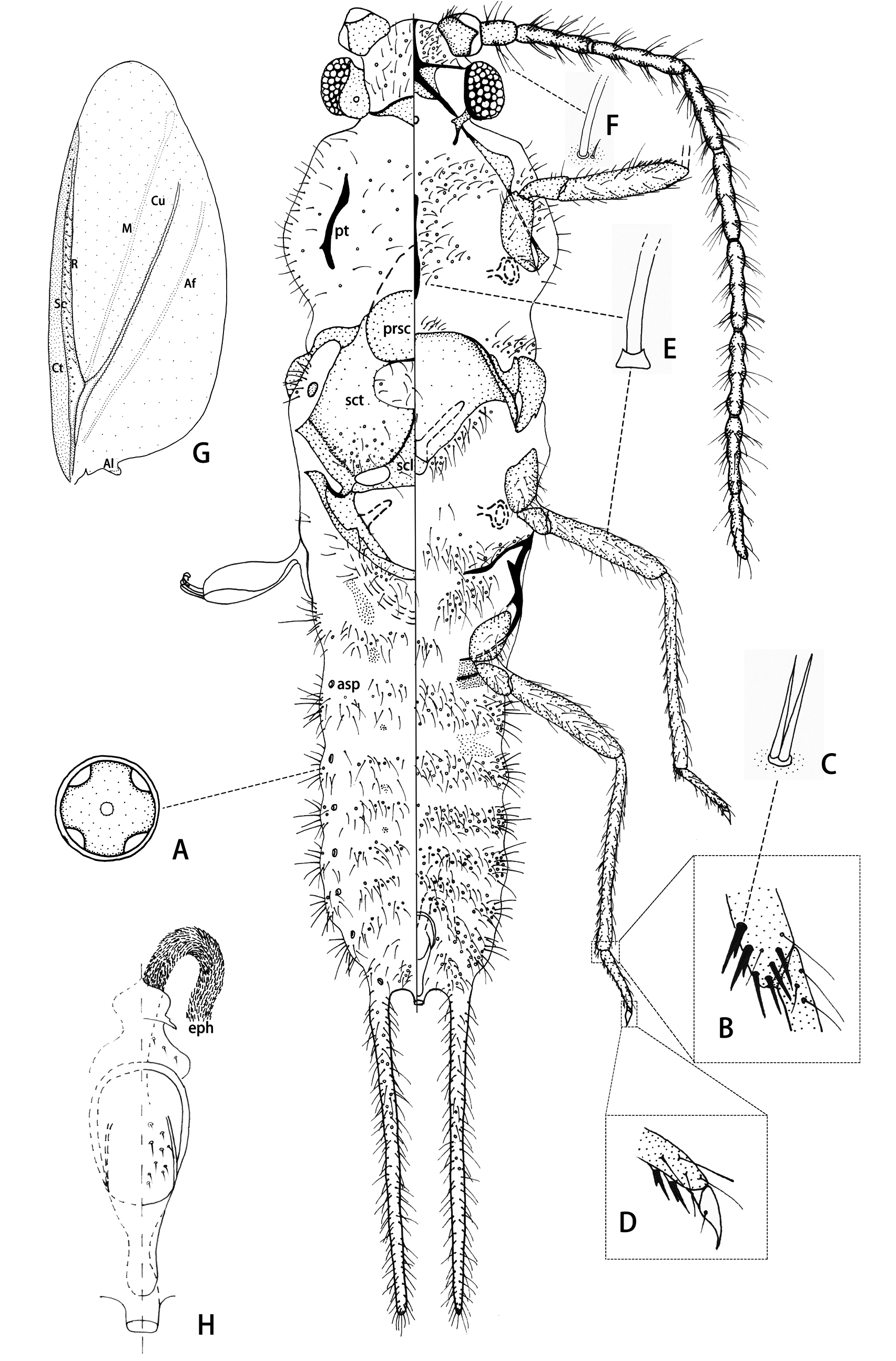
Slide-mounted material ( Fig. 3 View FIGURE 3 ) (n = 7). Body of adult male about 4–7 mm long, 1.05 mm wide across prealare. Loculated pores ( Fig 3A View FIGURE 3 ) each about 20 μm in diameter, with 4 loculi, present on head, thorax, and abdomen. Sclerotized areas of body without nodulations. Legs well developed and setose, with many setae ( Fig. 3B, C View FIGURE 3 ) bifurcated; tarsi each 2-segmented; claw ( Fig. 3D View FIGURE 3 ) without any denticles; claw digitules acute, much shorter than claw. Abdominal segment I not visible ventrally; abdomen with 1 pair of long caudal extensions on segment VIII only, each about 1/3 body length; abdomen without tubular ducts.
Head triangular in dorsal view, length about 500 μm, width across compound eyes 850–950 μm. Dorsum: postoccipital suture well developed, extending across posterior part of epicranium, and posteriorly with a strongly sclerotized postocciput. Dorsal epicranium covered in numerous collared setae ( Fig. 3E View FIGURE 3 ). Lateral view: compound eyes each about 250 mm long, with about 102–112 ommatidia. A wide, sclerotized ocular sclerite present along lateral margin of each compound eye with a single ocellus dorsally, each ocellus about 40 μm in diameter. Venter with a strong sclerotized series of ridges forming a five-armed star, composed of: (i) ventral mid-cranial ridge (about 180–200 μm long), fusing with (ii) a pair of preocular ridges laterally between each antenna and compound eye, and (iii) a pair of preoral ridges. Rest of ventral part of epicranium sclerotized, apart from a membranous area between ventral mid-cranial ridge and anterior margin of preocular ridges. With 18–20 collared setae ( Fig. 3E View FIGURE 3 ) between mid-cranial ridge and antenna base, plus 6–10 quadrilocular pores ( Fig. 3A View FIGURE 3 ); lateral area between preocular and preoral ridge without setae or pores; area posterior to preoral ridges with 26–28 collared setae ( Fig. 3E View FIGURE 3 ) and 7 or 8 quadrilocular pores ( Fig. 3A View FIGURE 3 ).
Antennae filamentous, 3.25–3.5 mm long (ratio of total body length to antennal length 1: 0.5–0.81), each with 10 segments. Scape sclerotized, about 150 μm long, 200 μm wide, with 7–21 setae (each 15–85 μm long); pedicel 185 μm long, 100 μm wide, with 8–9 long setae (125–400 μm long) ( Fig. 3F View FIGURE 3 ), 0–3 satellite setae, and 10–21 short setae (each 50–95 μm long); segments III–X each trinodose, with 3 whorls of long setae ( Fig. 3E View FIGURE 3 ), segment lengths (μm): III, 340–350; IV, 390–400; V, 360–400; VI, 400–420; VII, 360–380; VIII, 330–370; IX, 280–350; and X, 350–370.
Thorax. Prothorax: neck distinct. Dorsum: pronotum and pronotal sclerite absent. A pair of diagonal posttergites (pt) present, each about 350–420 μm long. Lateral view with a pair of strong cervical sclerites, each articulating anteriorly with ventral projection from ocular sclerite and preoral ridges; each cervical sclerite with a broad proepimeron. Pleural apophysis distinct. Venter: prosternum with a well-sclerotized median ridge, 460–520 μm long, which broadens posteriorly but is without obvious sternal apophyses. Most membranous areas covered with collared setae (each 120–410 μm long) and quadrilocular pores.
Mesothorax: Dorsum: prescutum (prsc) large and approximately oval (about 270 µm long and 670 µm wide); mesoprephragma large and nearly triangular; prescutal ridges short; prescutal sutures sclerotized. Scutum (sct) with an approximately rectangular membranous area medially posterior to prescutum, 180 µm long, 500 µm wide; each posterior part with 10–27 hair-like setae ( Fig. 3E View FIGURE 3 ) and 8–17 quadrilocular pores ( Fig. 3A View FIGURE 3 ). Scutellum (scl) approximately triangular, 400 µm long and 600 µm wide, with 0–4 hair-like setae ( Fig. 3E View FIGURE 3 ) but without pores; each outer angle with an approximately oval membranous area. Mesopostnotum well sclerotized and U-shaped, posteriorly extending internally under the metathoracic metapostnotum to form a large mesopostphragma; mesopostphragma antero-laterally with a pair of slender sclerotized mesopostnotal apophyses. Lateral view: prealare elongate; tegula well-developed and circular, with 9 setae. Mesothoracic spiracles large, outer part of peritreme about 150–160 μm wide, without pores in atrium. Venter: basisternum large, about 600 μm long, 820 μm wide, with a thin median ridge; bounded anteriorly by well-developed marginal ridge and posteriorly by well-developed precoxal ridges; basisternum with an anterior group of 9–15 setae ( Fig. 3E View FIGURE 3 ), 2 small lateral groups of 11–17 setae ( Fig. 3E View FIGURE 3 ) and 4–8 loculated pores ( Fig. 3A View FIGURE 3 ), and with 2 posterior groups of 17–33 setae ( Fig. 3E View FIGURE 3 ) and 4–7 loculated pores ( Fig. 3A View FIGURE 3 ); furca slightly waisted, with long arms strongly divergent.
Metathorax: Dorsum: metapostnotum present as a pair of long oval sclerites dorsolaterally. Lateral view: posterior spiracles similar in structure to anterior spiracles, each about 170 μm wide. Metapleural wing process heavily sclerotized and extending anterodorsally from anterior end of pleural ridge; precoxal ridge well developed, extending medio-ventrally, about 380 μm long. Metepimeron represented by a large sclerotization extending posteriorly around metacoxae. Venter: metasternum absent; anteriorly with 32 metasternal setae and 11 loculated pores, and posteriorly with 25 metasternal setae and 16 loculated pores.
Wings ( Fig. 3G View FIGURE 3 ) large and well developed, each 3.5–4.15 mm long and 1.23–1.75 mm wide; ratio of length to width 1: 0.35–0.42; ratio of total body length to wing length 1: 0.59–0.88. Costal thickening (Ct) well developed and sclerotized; subcostal vein (Sc) present along Ct from the base toward the apex. Radius ( R) present, with a line of 33–38 circular sensoria and 12–20 fine setae before. Cubitus (Cu) extending close to edge of wings. Rest of wing membranous; media (M) and anal fold (Af) in clear lines; alar lobe (Al) well developed and sclerotized. Hamulohalteres mainly sclerotized, broadest about middle of length, 490–500 μm long, about 150 μm wide; each with 3 or 4 hamuli, each hamulus hooked with a small apical knob.
Legs long and slender. Three pairs of legs almost equal in length (measurements in μm): each coxa 325–400, trochanter + femur 750–825, tibia 800–950, tarsus 300, and claw 75–100. Trochanter with about 12–16 shortish setae and 1 long flagellate seta (230–325 μm long), and with 3–5 circular campaniform sensilla on each side. Profemur with numerous shortish setae, ventrally with many bifurcated setae ( Fig. 3C View FIGURE 3 ) on anterior surface and 1 long, basal flagellate seta (about 151 μm long); mesofemur with 72 shortish setae (50–100 μm long) and 2 long ventral setae (250–310 μm long); metafemur with about 70–72 shortish setae (50–130 μm long) and 1 long ventral seta (about 290 μm long). Protibia with numerous bifurcated setae and a few spur-like setae, distal 3/4th of meso- and metatibiae with bifurcated setae ( Fig. 3C, D View FIGURE 3 ) ventrally, these replaced by spur-like setae and flagellate setae laterally and dorsally. Tarsus 2 segmented, proximal segment short and triangular, distal segment with a few bifurcated setae on ventral surface, and with flagellate setae dorsally and laterally. Claw with a pair of short setose digitules, but without denticles.
Abdomen with 1 pair of caudal extensions arising from margins of segment VIII, each about 1.40–1.55 mm long, with numerous long setae. Pairs of small tergites present on segments I–IV. Venter with small sternites mediolaterally on anterior borders of segments III and IV. Dorsal abdominal setae ( Fig. 3E View FIGURE 3 ) frequently fewer than ventral abdominal setae and arranged in bands across each segment; segments I–IV each with a band containing about 39–46 setae and 10–14 loculated pores, segments V –VIII each with a band containing 21–28 setae and 7–12 loculated pores. Pleural setae present in groups, each group containing 2–6 long setae (200–420 μm long) and 22–34 shortish setae. Ventral abdominal setae also arranged in segmental bands: segment I with 13 setae and 7 loculated pores ( Fig. 3A View FIGURE 3 ); segments II– VI each with 58–106 setae and 25–37 loculated pores; and segment VIII with 18–25 setae and 12–15 loculated pores. Abdominal spiracles (asp) numbering 7 pairs, present on dorsal submargins of segments II–VIII; spiracle on segment VIII distinct, with a sclerotized opening about 40 µm wide. Anus present between caudal extensions on segment VIII, with a tubular opening, connected to penial sheath by a sclerotized band. Penial sheath ( Fig. 3H View FIGURE 3 ) heavily sclerotized, positioned anteroventrally to anus; penial sheath about 420 μm long, 130 μm wide at broadest point, narrowing distally, with many small setae at base and in middle. Aedeagus sclerotized, with a long, strongly setiferous, eversible endophallus (eph).
Etymology. The specific name castanopsis is based on the genus name of the insect’s host plant; it is a noun in apposition.
| V |
Royal British Columbia Museum - Herbarium |
| VI |
Mykotektet, National Veterinary Institute |
| R |
Departamento de Geologia, Universidad de Chile |
No known copyright restrictions apply. See Agosti, D., Egloff, W., 2009. Taxonomic information exchange and copyright: the Plazi approach. BMC Research Notes 2009, 2:53 for further explanation.
|
Kingdom |
|
|
Phylum |
|
|
Class |
|
|
Order |
|
|
Family |
|
|
Genus |