
Uaiuara, Rheims, Cristina A., 2013
|
publication ID |
https://doi.org/10.11646/zootaxa.3734.2.6 |
|
publication LSID |
lsid:zoobank.org:pub:3BC10987-3883-44BD-9274-22D961B6336C |
|
DOI |
https://doi.org/10.5281/zenodo.6147766 |
|
persistent identifier |
https://treatment.plazi.org/id/03C72C23-FF88-FFDC-FF22-F922FABA1735 |
|
treatment provided by |
Plazi |
|
scientific name |
Uaiuara |
| status |
gen. nov. |
Uaiuara View in CoL gen. nov.
Type species. Sparianthis amazonica Simon, 1880
Etymology. The generic name was taken from the Amazonian folklore. “ Uaiuara ” is a demon that usually appears in the shape of a small being with large and flappy ears, a characteristic that refers to the retrolateral groove on the cymbium of the male palps. The gender is feminine.
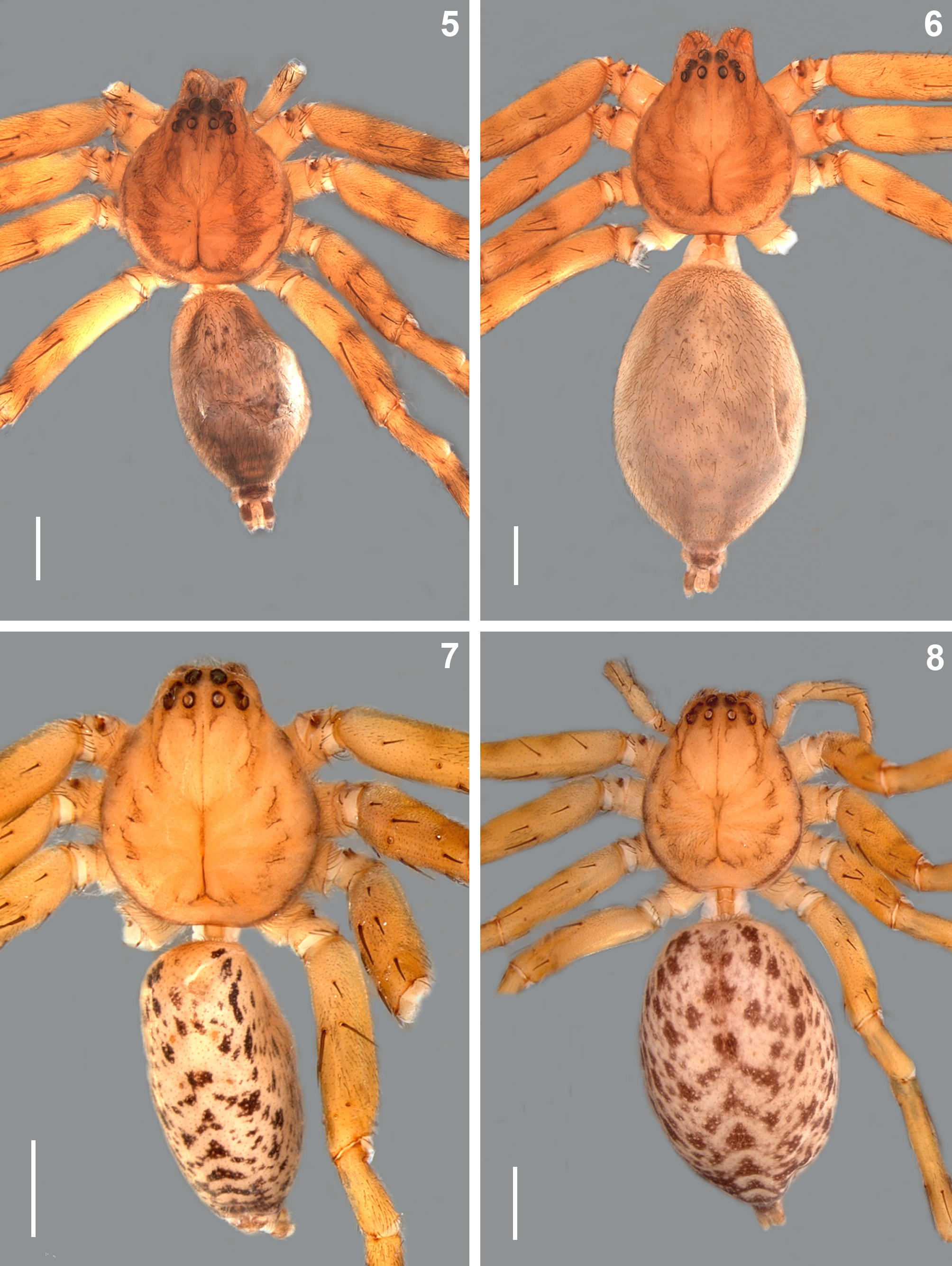
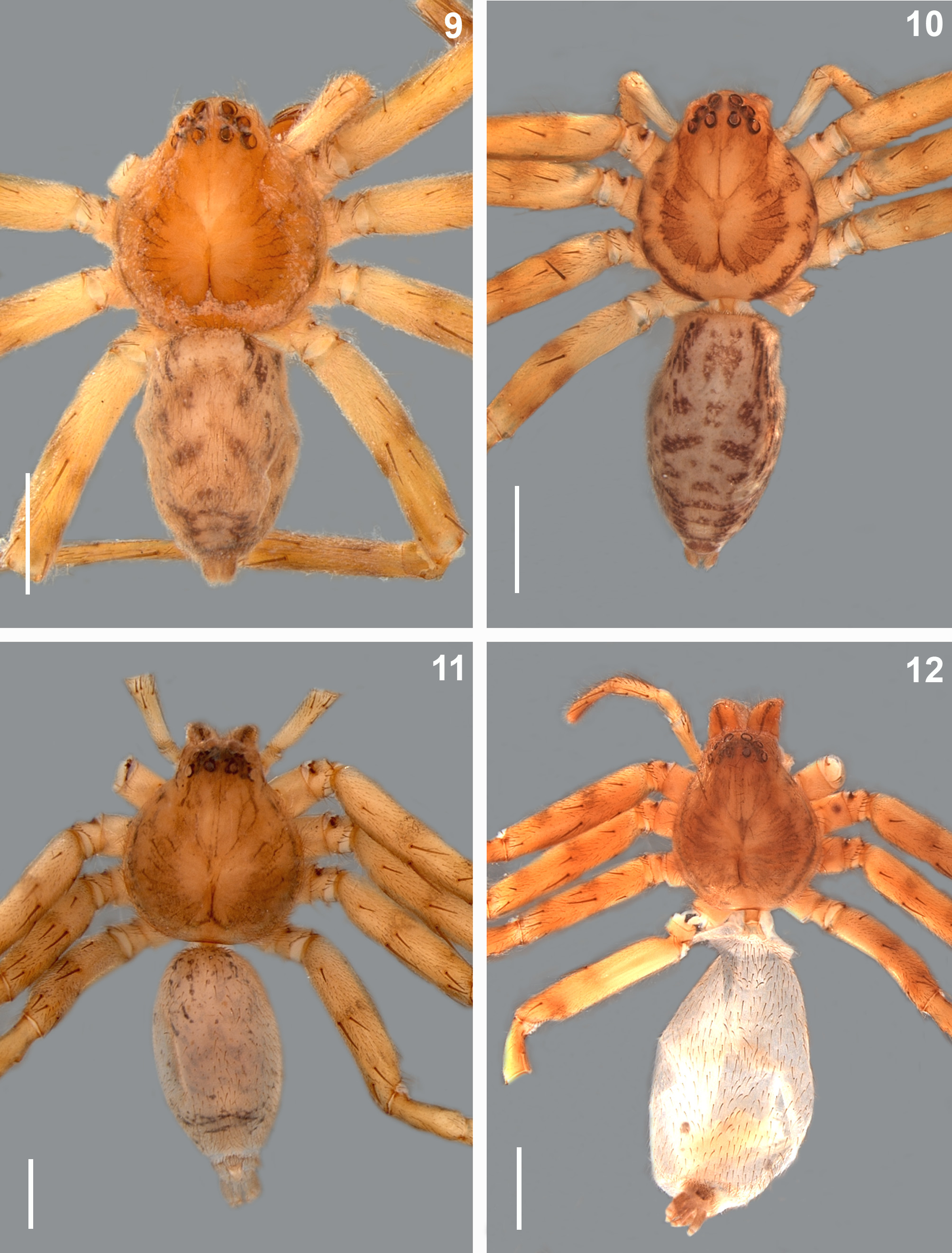
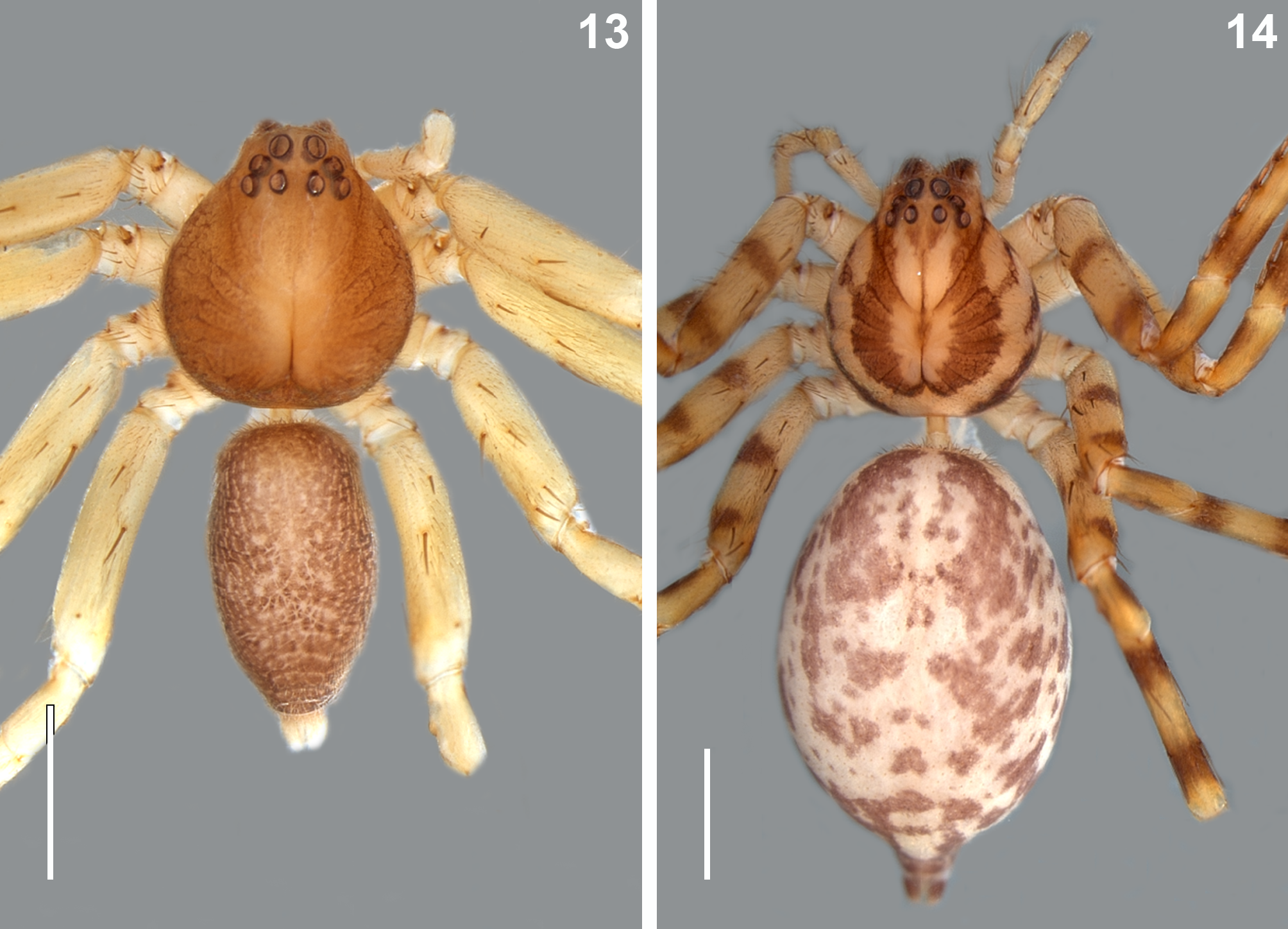
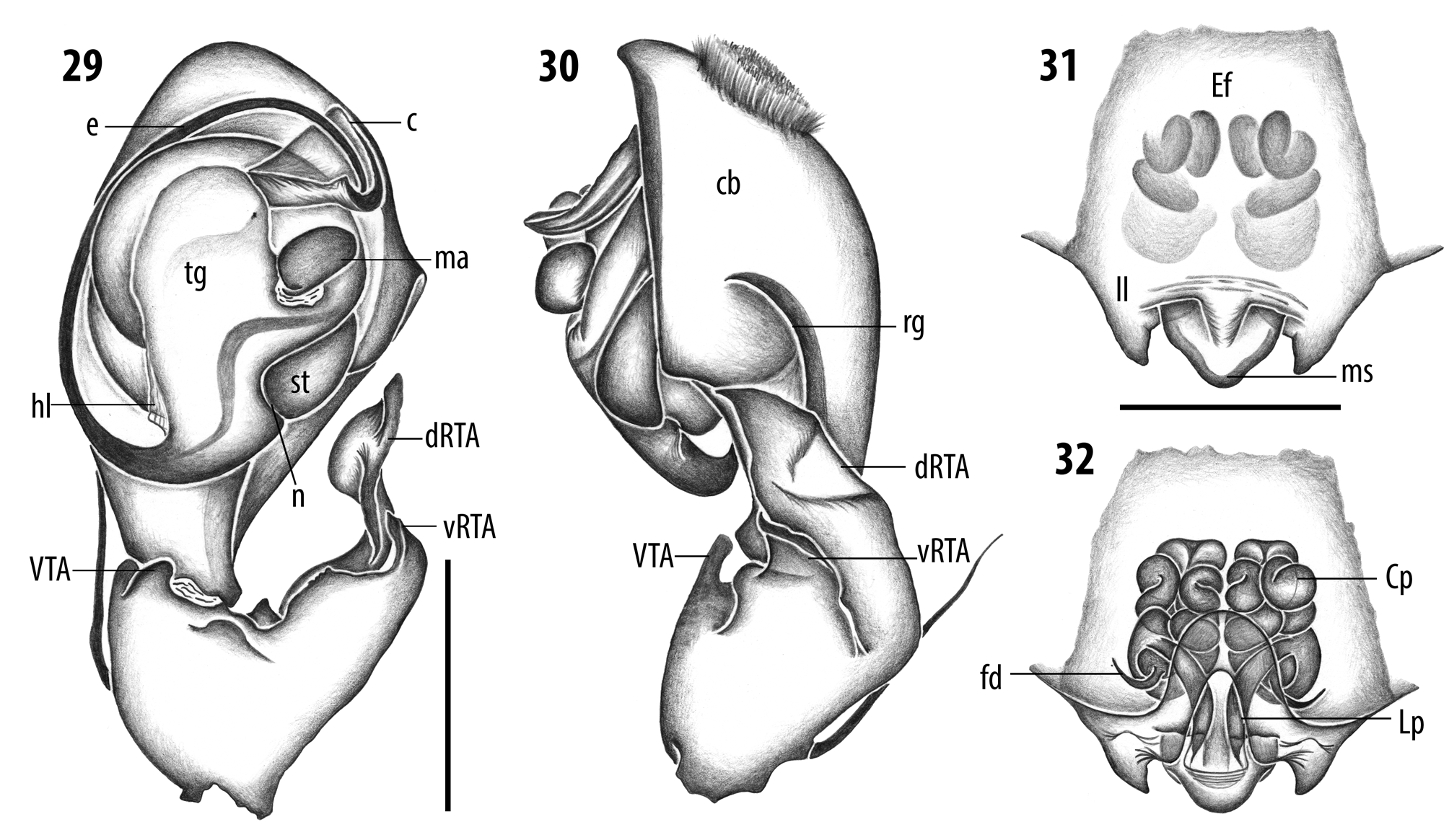
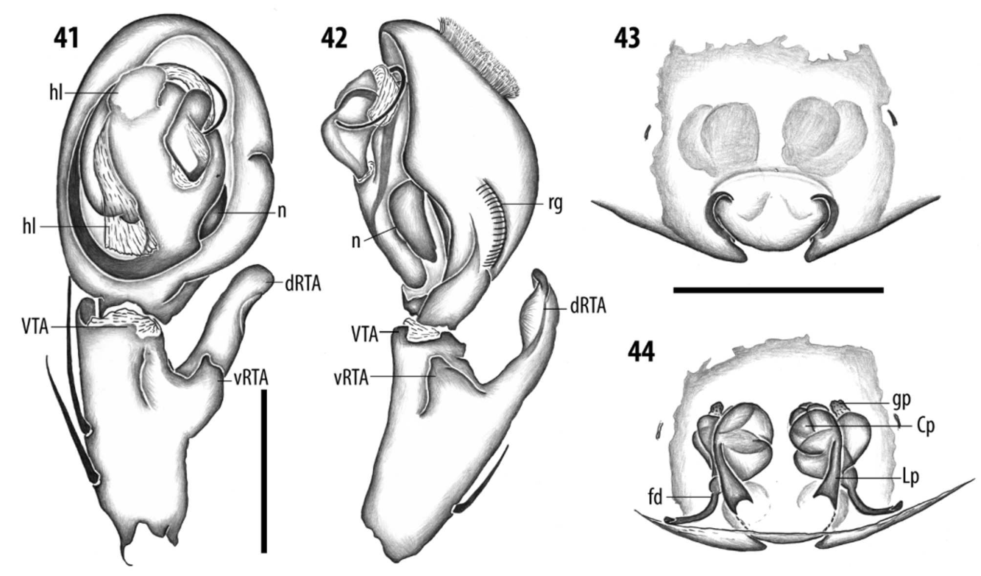
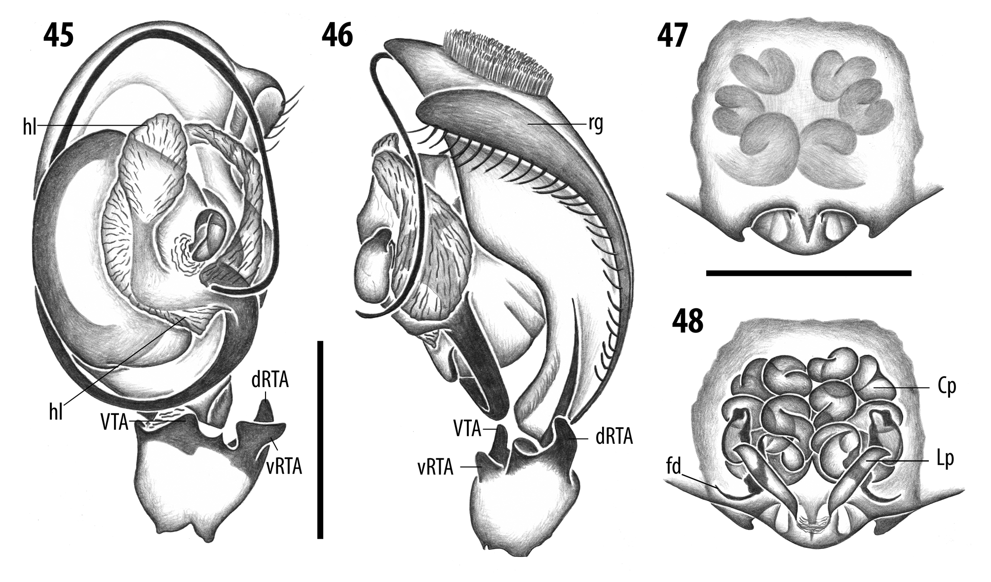
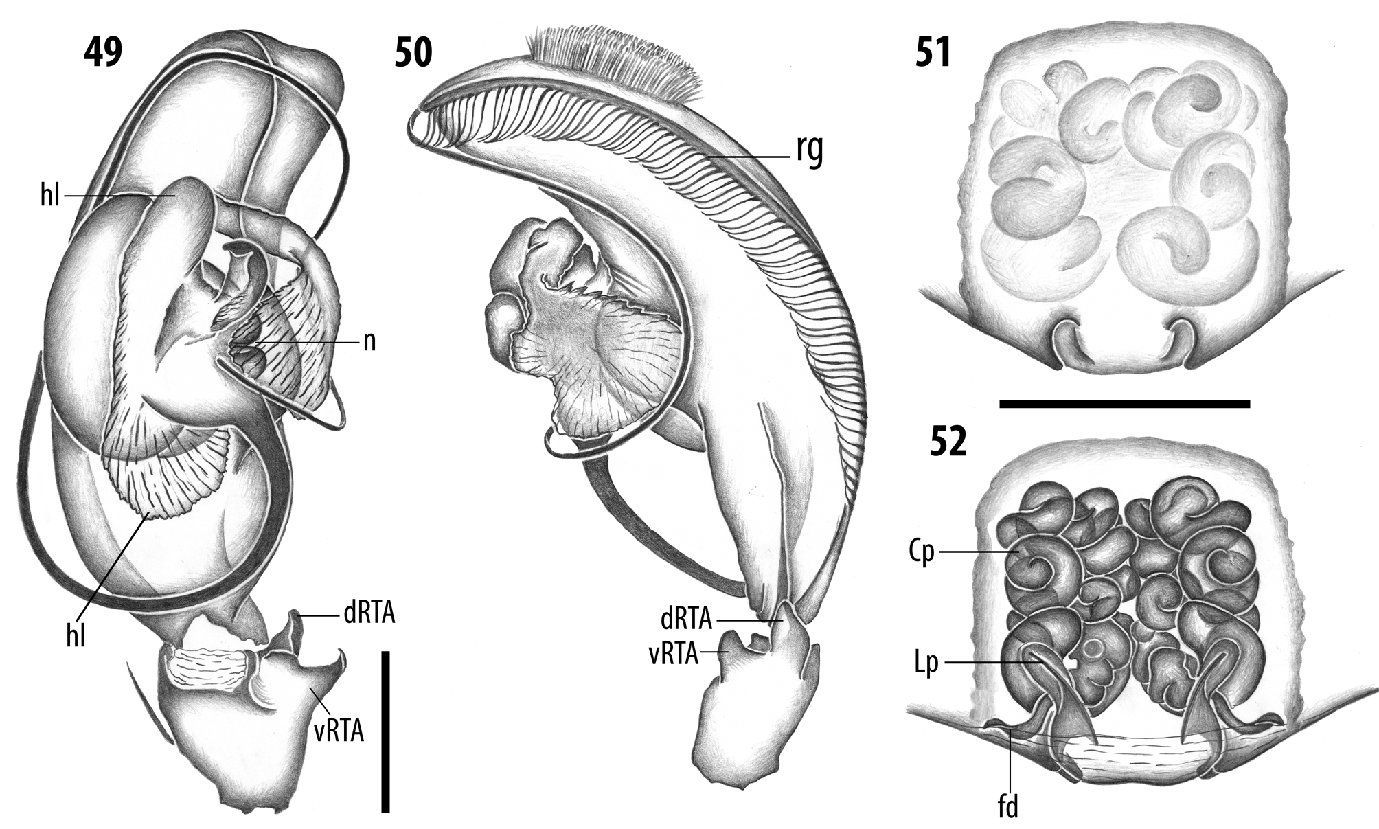
Diagnosis. Species of the genus Uaiuara gen. nov. are distinguished from the those of the remaining Neotropical Sparianthinae by the very recurved anterior eye row ( Figs 1–14 View FIGURES 1 – 4 View FIGURES 5 – 8 View FIGURES 9 – 12 View FIGURES 13 – 14 ) and the large number of ventral spines on leg tibiae I–II. Males are further distinguished by the retrolateral groove on the male palpal cymbium ( Figs 30 View FIGURES 29 – 32 , 34 View FIGURES 33 – 36 , 38 View FIGURES 37 – 40 , 42 View FIGURES 41 – 44 , 46 View FIGURES 45 – 48 , 50 View FIGURES 49 – 52 , 54 View FIGURES 53 – 56 ). Females resemble those of the Neotropical species of the genus Stasina Simon, 1877 described by Bryant (1940, 1948) in the general shape of the median septum and in the triangular projections of the lateral lobes ( Figs 31 View FIGURES 29 – 32 , 35 View FIGURES 33 – 36 , 39 View FIGURES 37 – 40 , 43 View FIGURES 41 – 44 , 47 View FIGURES 45 – 48 , 51 View FIGURES 49 – 52 ; Bryant 1940, figs 174, 182; 1948, fig. 99) of the epigynes. They are easily distinguished from the latter by the internal duct system of the vulva, which in Stasina comprises a short copulation duct and large spermathecae ( pers. comm.) and in Uaiuara a laminar and sclerotized part (Lp), arising from the copulation openings, followed by a longer, cylindrical and convoluted part (Cp), which ends at the fertilization ducts ( Figs 32 View FIGURES 29 – 32 , 36 View FIGURES 33 – 36 , 40 View FIGURES 37 – 40 , 44 View FIGURES 41 – 44 , 48 View FIGURES 45 – 48 , 52 View FIGURES 49 – 52 ).
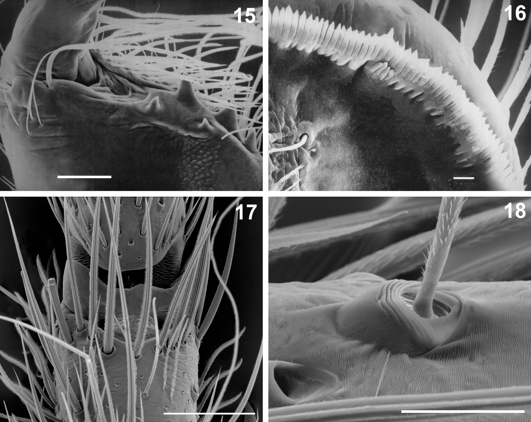
Description. Total length (males and females) 3.0–10. Prosoma slightly longer than wide, highest posteriorly. Thoracic fovea deep and line-like. Anterior eye row very strongly recurved; AME larger than ALE and more distant from each other than from laterals. Posterior eye row straight or very slightly recurved; eyes equal and equidistant ( Figs 1–14 View FIGURES 1 – 4 View FIGURES 5 – 8 View FIGURES 9 – 12 View FIGURES 13 – 14 ). Clypeus as high as diameter of AME. Chelicerae longer than wide. Cheliceral groove with three promarginal teeth, the most basal smallest, and two minute retromarginal teeth. One strong seta present at base of fang ( Fig. 15 View FIGURES 15 – 18 ). Intermarginal denticles absent. Labium rebordered, slightly longer than wide. Endites slightly convergent, longer than wide, with dense scopula on internal margin. Serrula with a single row of denticles ( Fig. 16 View FIGURES 15 – 18 ). Sternum as long as wide, slightly projected between coxae IV. Legs similar in size, usually 2143. Spination pattern in males: legs: femora I–III: p1-1-1, d0-1-1, r1-1-1; femur IV: p1-1-1, d0-1-1, r0-0-1; patellae I– IV: p0, r0; tibiae I–II: p1-0-1, d0-0-1, r1-0-1, v7–9 pairs; tibiae III–IV: p1-0-1, d0-0-1, r1-0-1, v2-2 -2; metatarsi I– II: p1-1-0, r1-1-0, v2 -0-0; metatarsi III–IV: p1-1-1, r1-1-1, v2-2 -0; palp: femur: p0-0-1, d0-1-2, r0-0-1; patella: p1, r1; tibia: p1-0-0, d1-0-0. Spination pattern in females: femora I–III: p1-1-1, d0-1-1, r1-1-1; femur IV: p1-1-1, d0-1- 1, r0-0-1; patellae I–IV: p0, r0; tibiae I-II: v7-9 pairs; tibia III: p1-0-1, r1-0-1, v2-2 -2; tibia IV: p1-0-1, d0-0-1, r1-0- 1, v2-2 -2; metatarsi I–II: p1-1-0, r1-1-0, v2 -0-0; metatarsi III–IV: p1-1-1, r1-1-1, v2-2 -0; palp: femur p0-0-1, d0-1- 2; patella p1, r1; tibia p2-1-0, d1-0-0; r1-1-0; metatarsus p2-1-0; r2-1-0. Trochanter smooth. Metatarsi I–IV with distal trilobate membrane with median lobe much smaller than laterals ( Fig. 17 View FIGURES 15 – 18 ). Trichobothria present on dorsal side of tibiae, metatarsi and tarsi, arranged in two or more rows on tarsi and one on metatarsi. Bothrium with dorsal plate with 6–7 distal grooves, projected over smooth basal plate ( Fig. 18 View FIGURES 15 – 18 ). Tarsal organ capsulate with slightly oval opening ( Fig. 19 View FIGURES 19 – 22 ), located dorsally on distal third of metatarsi. Metatarsi III–IV with distal, ventral preening comb ( Fig. 20 View FIGURES 19 – 22 ). Leg tarsi with pair of pectinate claws with 6–7 very slightly curved secondary teeth and claw tufts ( Fig. 21 View FIGURES 19 – 22 ). Female pedipalp with single, pectinate claw, with 4–5 short secondary teeth ( Fig. 22 View FIGURES 19 – 22 ). Opisthosoma oval, longer than wide. Six spinnerets on projected tegument ring: ALS contiguous, conical and bi-segmented, distal segment with several piriform gland spigots, one major ampullate gland spigot and one nubbin in male ( Fig. 23 View FIGURES 23 – 28 ) and two major ampullate gland spigots in female ( Fig. 26 View FIGURES 23 – 28 ); PMS short and truncated with one minor ampullate gland spigot and 5–7 aciniform gland spigots ( Figs 24, 27 View FIGURES 23 – 28 ); PLS conical and bi-segmented, distal segment with many aciniform gland spigots ( Figs 25, 28 View FIGURES 23 – 28 ). Male palp: femur two times patella length; tibia short, as long as or slightly longer than patella; RTA originating distally with dRTA slightly larger than vRTA ( Figs 30 View FIGURES 29 – 32 , 34 View FIGURES 33 – 36 , 38 View FIGURES 37 – 40 , 42 View FIGURES 41 – 44 , 46 View FIGURES 45 – 48 , 50 View FIGURES 49 – 52 , 54 View FIGURES 53 – 56 ); VTA present in U. amazonica (Simon) , U. dianae sp. nov., U. jirau sp. nov., U. ope sp. nov. and U. quyguaba sp. nov. ( Figs 29 View FIGURES 29 – 32 , 37 View FIGURES 37 – 40 , 41 View FIGURES 41 – 44 , 45 View FIGURES 45 – 48 , 53 View FIGURES 53 – 56 ). Cymbium slightly elongate with large oval alveolus and long retrolateral groove, smooth in U. amazonica (Simon) and U. dianae sp. nov. ( Figs 34 View FIGURES 33 – 36 , 38 View FIGURES 37 – 40 ) and flanked by short strong setae in U. barroana (Chamberlin) , U. jirau sp. nov., U. ope sp. nov., U. palenque sp. nov. and U. quyguaba sp. nov. ( Figs 42 View FIGURES 41 – 44 , 46 View FIGURES 45 – 48 , 50 View FIGURES 49 – 52 , 54 View FIGURES 53 – 56 ). Cymbial scopula small, round and located on distal third of cymbium. Bulb with subtegulum and tegulum notched retrolaterally at embolus base; tegulum flanked prolaterally by hyaline lamina, larger at base of embolus and at base of conductor, where it can be hyaline ( Figs 33 View FIGURES 33 – 36 , 45 View FIGURES 45 – 48 , 52 View FIGURES 49 – 52 ) or slightly membranous ( Figs 29 View FIGURES 29 – 32 , 37 View FIGURES 37 – 40 , 41 View FIGURES 41 – 44 , 49 View FIGURES 49 – 52 ); median apophysis small, arising at 3 o’clock; conductor hyaline ( Figs 33 View FIGURES 33 – 36 , 37 View FIGURES 37 – 40 , 41 View FIGURES 41 – 44 , 45 View FIGURES 45 – 48 , 49 View FIGURES 49 – 52 , 53 View FIGURES 53 – 56 ) or slightly sclerotized ( Fig. 29 View FIGURES 29 – 32 ) arising at 12 o’clock; embolus long, and slender, without projections, arising retrolaterally between 3 and 6 o’clock and curved prolaterally ( Figs 29 View FIGURES 29 – 32 , 33 View FIGURES 33 – 36 , 37 View FIGURES 37 – 40 , 41 View FIGURES 41 – 44 , 45 View FIGURES 45 – 48 , 49 View FIGURES 49 – 52 , 53 View FIGURES 53 – 56 ). Epigyne: Epigynal field as long as wide or slightly longer than wide; lateral lobes with triangular projections posteriorly; median septum triangular or slightly rectangular, extending beyond the epigastric furrow, with pair of anterior copulation openings ( Figs 31 View FIGURES 29 – 32 , 35 View FIGURES 33 – 36 , 39 View FIGURES 37 – 40 , 43 View FIGURES 41 – 44 , 47 View FIGURES 45 – 48 , 51 View FIGURES 49 – 52 , 55 View FIGURES 53 – 56 ). Vulva: internal duct system comprised of a laminar and sclerotized part (Lp), arising from the copulation openings, followed by a longer, cylindrical and convoluted part (Cp), which ends at short and slender fertilization ducts ( Figs 32 View FIGURES 29 – 32 , 36 View FIGURES 33 – 36 , 40 View FIGURES 37 – 40 , 44 View FIGURES 41 – 44 , 48 View FIGURES 45 – 48 , 52 View FIGURES 49 – 52 , 56 View FIGURES 53 – 56 ).
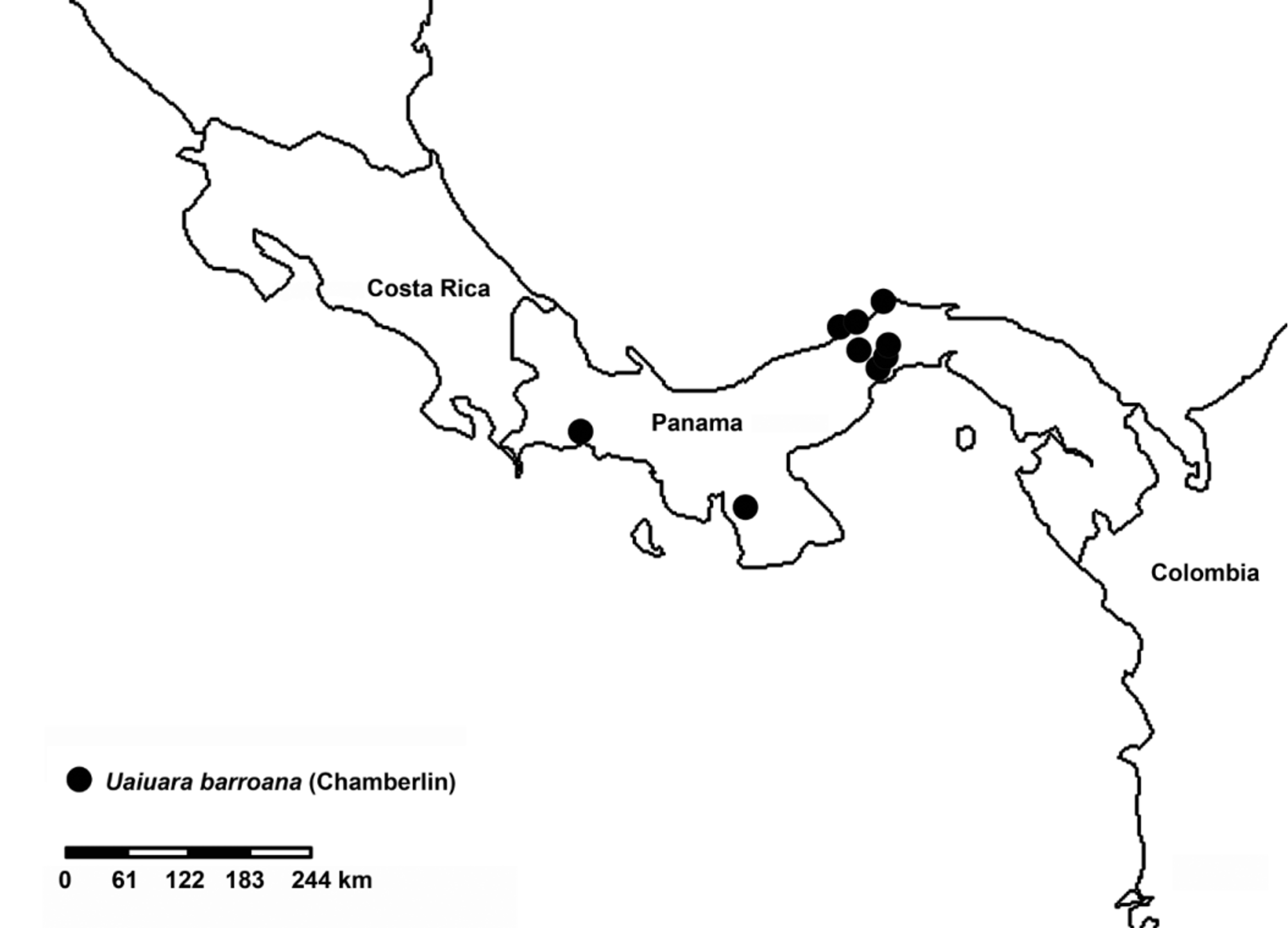
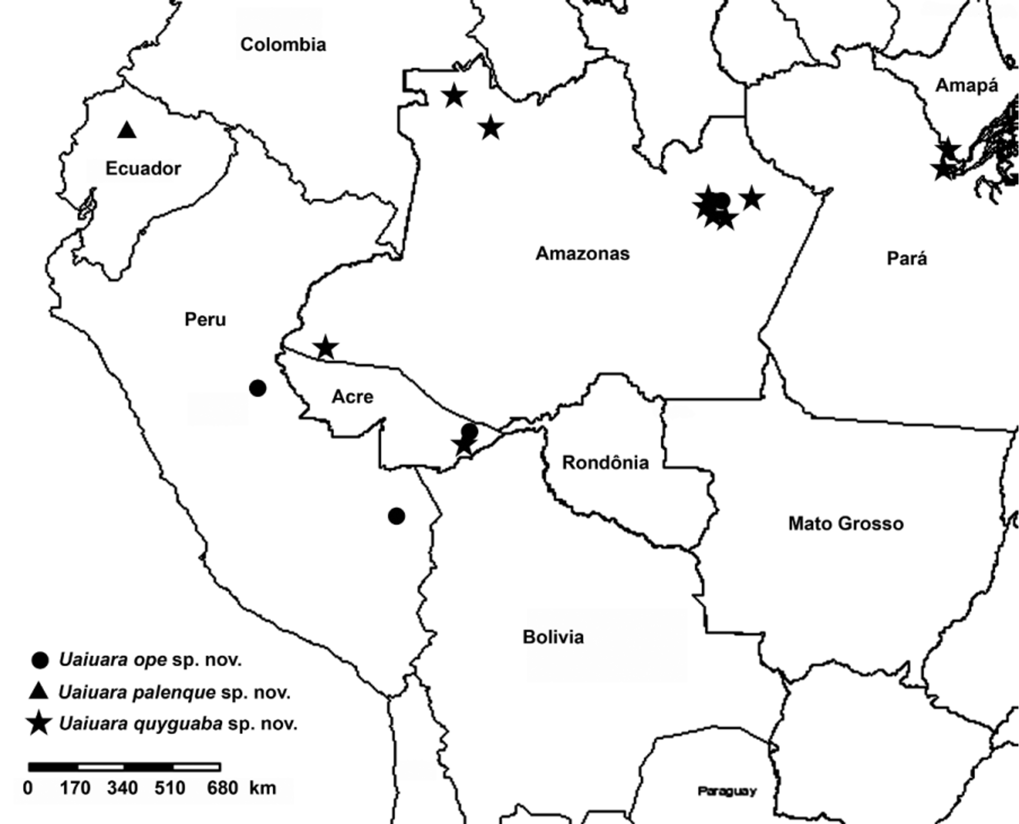
Distribution. Northern South America and southern Central America ( Figs 57–59 View FIGURE 57 View FIGURE 58 View FIGURE 59 ).
Composition. Seven species: Uaiuara amazonica (Simon) comb. nov.; U. barroana (Chamberlin) comb. nov.; U. dianae sp. nov., U. jirau sp. nov.; U. ope sp. nov.; U. palenque sp. nov.; and U. quyguaba sp. nov.


No known copyright restrictions apply. See Agosti, D., Egloff, W., 2009. Taxonomic information exchange and copyright: the Plazi approach. BMC Research Notes 2009, 2:53 for further explanation.