
Pinus
|
publication ID |
https://doi.org/10.2478/if-2018-0014 |
|
persistent identifier |
https://treatment.plazi.org/id/03C6D31A-3A09-FFAA-A4CB-F955FA52E8E3 |
|
treatment provided by |
Felipe |
|
scientific name |
Pinus |
| status |
|
Genus Pinus View in CoL View at ENA L., 1753
Pinus timleri KINKELIN in ENGELHARDT et KINKELIN, 1908 Pl. 1, Figs 1, 2, Pl. 2, Figs 1–3
Selected synonyms:
1887 Pinus cembra L. fossilis GEYLER et KINKELIN, p. 14, pl. I, fig. 8.
1900 Pinus aff. laricio POIR. fossilis KINKELIN, p. 127.
*1908 Pinus timleri KINKELIN in ENGELHARDT et KINKELIN, p. 205, pl. 25.
Pinus aff. laricio POIR. pliocaenica KINKELIN; Kinkelin in Engelhardt and Kinkelin, pro parte, p. 210, pl. 24, fig. 12.
1919 Pinus timleri KINKELIN ; Seward, p. 394, text-fig. 786A, 786B.
1939 Pinus timleri KINKELIN ; Mädler, p. 33.
1968 Pinus timleri KINKELIN in ENGELH. et KINKELIN; Kilpper, p. 161, pl. 44, figs 9–13.
1986 Pinus timleri KINKELIN ; Mai, p. 574, 578, pl. 45, fig. 1.
1989 Pinus timleri ; Wutzler, p. 10, pl. 8, fig. 2a, 2b.
1994 Pinus timleri ; Gregor, pl. 13, fig. 3.
1994 Pinus urani (UNGER) SCHIMPER ; Mai, p. 215 pro parte, pl. IV, figs 1–3.
2014a Pinus timleri KINKELIN in ENGELHARDT et KINKELIN; Kvaček et al., p. 21, pl. 1, pl. 2, figs 1–3, pl. 3, pl. 4, figs 1–2.
2014b Pinus timleri KINKELIN ; Kvaček et al., p. 137, pl. 1, figs 3–10, pl. 2, pl. 3, figs 1–5.
N o t e. Kvaček et al. (2014a) and Kvaček et al. (2014b) gave a description of the overall taxon. The present work is focused on the specific external morphology of the seed cones.
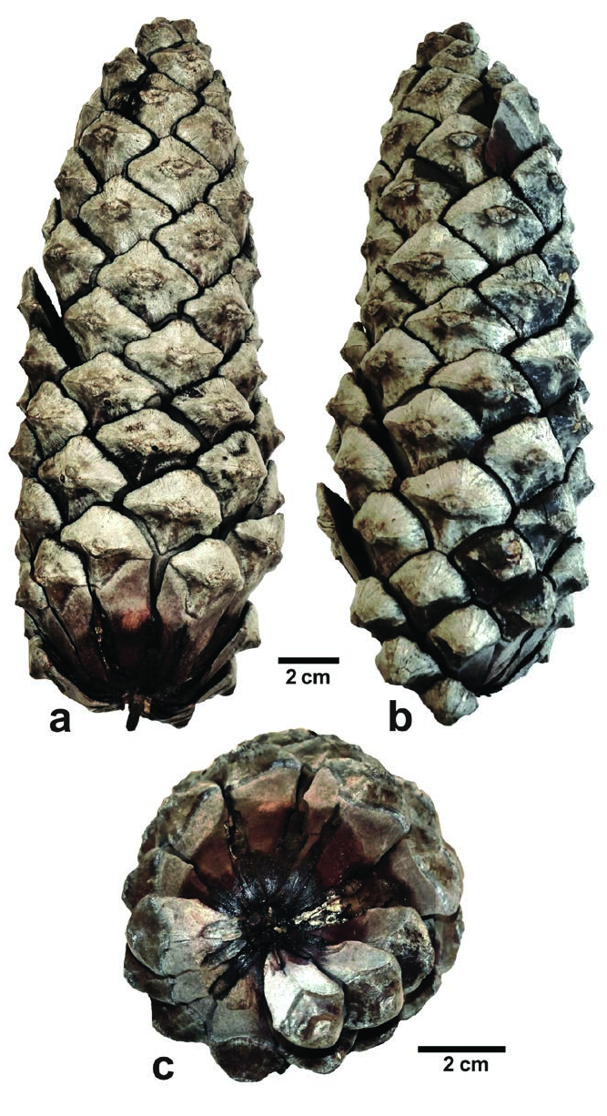
D e s c r i p t i o n o f s e e d c o n e s. Based on well preserved female cones and cone fragments of P. timleri without transport damage, the articulated appearance of the cones is asymmetric and features a highly variable morphology of the apophyses. The whole P. timleri seed cones have a total length of 15.2 to 17.2 cm and a maximum width of 7.2 to 9.5 cm (see Pl. 1, Figs 1a, b, 2b–d, Pl. 2, Fig. 1a, b) (other seed cones show a length up to nearly 28 cm). The cone axis is bent concavely towards the smooth side (Pl. 1, Fig. 1a), while the opposite side shows a convex shape. Incomplete seed cones tend to disarticulate towards the base so that some cone scales must be picked up from the sediment while the moist cone apex resists disarticulation until desiccation (Pl. 2, Figs 2a, b, 3). Only rarely are cones preserved with the short peduncle (Pl. 1, Fig. 2f), most specimens are broken off at the base (Pl. 1, Fig. 1d). The apex has a rounded shape and compact structure (Pl. 1, Fig. 2e, Pl. 2, Fig. 2c).
Most cones show broad-rhombic shaped apophyses with a mucro on the smooth side, sometimes with slight horizontal ridges (Pl. 1, Fig. 1a, 2c, Pl. 2, Fig. 2a). The small umbo of the smooth side is not concave (denticulato-mucronate; Pl. 1, Fig. 2c). On the opposite side, the (excentrodenticulate to mucronate) umbo is strongly conical and sometimes hookshaped (Pl.1, Fig. 2d) or nearly hemispherical (Pl. 1, Fig. 2f) and can feature radial grooves (Pl. 1, Fig. 1f). The raised umbo may also bear a roundish erect navel (Pl. 2, Fig. 1c), which can feature a recurved mucro (prickle) (Pl. 2, Fig. 1b). Occasionally, the mucros are eroded on the side with the thinner umbos. In this case, the apophyses appear slightly concave. This feature is therefore of taphonomic origin and not a morphological character. It can be observed in the scales of the Frechen cone (Pl. 2, Fig. 3).
E x t i n c t a n d e x t a n t s e e d c o n e s. Seed cones of P. timleri apparently most often broke off the tree in an incomplete state since their bases remained with scales on the branch or even initially on the trunk. Only later did the base separate or decompose in place on the dead tree. Extant large heavy seed cones, e.g. Pinus canariensis SWEET ex K. SPRENGEL , P. pinea L. or P. radiata D.DON (see Page 1974, cf. Klaus 1980, Krüssmann 1983, Klaus 1989, Schütt et al. 2004, Farjon 2005) show the same pattern.
In addition to the usual variation, the morphology of a seed scale depends on the position of the scale on the cone. In many extant Pinus species , the shadow-exposed sides of the cone feature flat apophyses. On the opposite side that is exposed to direct light the apophyses of cone scales are thickened, presumably as protection against seed predation. This is particularly true of P. canariensis SWEET ex K.SPRENGEL ( Text-fig. 3a–c View Text-fig ) and P. roxburghii SARG , the species morphologically most similar to P. timleri (see Page 1974, Klaus 1980, Krüssmann 1983, Schütt et al. 2004, Kvaček et al. 2014a, b). We hypothesise that the same relationship of direct light exposure and asymmetric growth was present in P. timleri and similar fossil seed cone taxa.
No known copyright restrictions apply. See Agosti, D., Egloff, W., 2009. Taxonomic information exchange and copyright: the Plazi approach. BMC Research Notes 2009, 2:53 for further explanation.