
VIPERINAE, Oppel, 1811
|
publication ID |
https://doi.org/ 10.5252/geodiversitas2020v42a20 |
|
publication LSID |
urn:lsid:zoobank.org:pub:8FF2A078-CE45-4BF1-A681-00136F57375E |
|
DOI |
https://doi.org/10.5281/zenodo.4447734 |
|
persistent identifier |
https://treatment.plazi.org/id/03C587C7-431C-FFF8-FC13-F92F4990F95E |
|
treatment provided by |
Felipe |
|
scientific name |
VIPERINAE |
| status |
|
VIPERINAE (‘ Oriental vipers’ group)
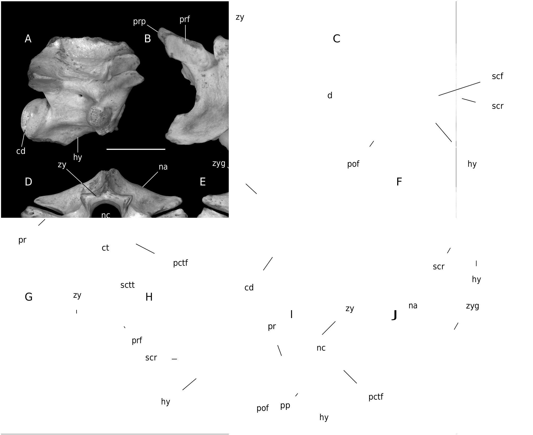
( Fig. 14 View FIG A-N; Fig. 15 View FIG A-J)
Vipera sp. 1 (‘ Oriental vipers’) – Ivanov & Musil 2004: 230.
Vipera sp. (‘ Oriental vipers’ group) – Ivanov et al. 2006: 229, table 2.
MATERIAL. — MWQ, early Miocene, Burdigalian, Orleanian, MN 4: 1/2001 Turtle Joint: one left maxilla (Pal. 1501), isolated fang (Pal. 1502), 30 trunk vertebrae (Pal. 1503-1532). 2/2003 Reptile Joint: 28 trunk vertebrae (Pal. 1994-2021).
DESCRIPTION
Maxilla
Only a left maxilla is preserved ( Fig. 14A, B View FIG ). In rostral view, the body of the bone is high, especially along its medial margin. The ascending process is long, and it is inclined medially. A small process for the prefrontal connection occurs medially at its distal termination. A ridge extends from the base of the ascending process as far as its distal termination. A large, slightly oval, foramen occurs opposite to the medially directed maxillary-prefrontal process. In caudal view, a wide groove for connection with the ectopterygoid occurs above the base of the fangs. This groove is deep, and it is restricted dorsally by a transverse ridge. This ridge extends from the medial margin of the bone (where it forms two small processes) as far as the middle of the ascending process width. In medial view an oval foramen occurs within the deep, wide orifice. This orifice is restricted dorsally by a ridge.
Dentition
A single fang of about 1 cm length is preserved ( Fig. 14C View FIG ). It is long and slender, and is curved slightly caudally. A canal extends within the tooth and has a strongly elongated/slit-like orifice situated mesially, close to the pointed tip of the fang. The slightly widened basal portion of the fang is damaged and does not preserve the orifice of the venom canal.
Trunk vertebrae
Numerous fragmentary trunk vertebrae ( Fig. 14 View FIG D-N; Fig. 15 View FIG A-J) are preserved, mostly with broken-off neural spines and hypapophyses. In lateral view, the neural spine was about as high as long in anterior precaudal vertebrae.
The most complete neural spine lacks its cranial margin, but it seems possible that it was vertical or slightly anteriorly inclined. The interzygapophyseal ridges are welldeveloped and sometimes rather sharp. Lateral foramina are large and are situated in shallow depressions. The parapophyses are well-separated from the diapophyses and the parapophyseal processes are strongly built and directed antero-ventrally. The subcentral ridges are conspicuous. The condyle is developed on a very short neck. The rarely preserved hypapophyses of anterior trunk vertebrae are long, straight, and directed posteroventrally. The pointed distal termination of the hypapophysis of middle and posterior trunk vertebrae is directed caudally. The hypapophysis of the posteriormost trunk vertebrae have indication of bifurcation on its distal tip. In dorsal view, the vertebrae are markedly short and wide. The cranial margin of the zygosphenal lip is concave, straight, or with a small medial lobe. The medial lobe is better developed in posterior trunk vertebrae. The prezygapophyseal articular facets are widely oval to subtriangular (although partial damage cannot be excluded). The prezygapophyseal processes are broken-off close to their bases. Epizygapophyseal spines are absent. In ventral view, the subcentral grooves are shallow, and the large subcentral foramina are situated on both sides of the base of the wide hypapophysis. The ventromedial margin of parapophyseal processes is always medially enlarged and it is usually fused with the ventrolateral extensions of the cotylar rim (subcotylar tubercles). The blood vessels of the circulatory system passed through the canals on both sides of the hypapophyseal base. The postzygapophyseal articular facets have an irregularly triangular outline. In cranial view, the neural arch is strongly flattened dorsoventrally. The cranial margin of the zygosphene is straight. The prezygapophyses are tilted up dorsally. The paracotylar foramina are situated on both sides of the rounded cotyle. The subcotylar tubercles are usually fused with bases of parapophyseal processes. The vertebral dimensions of the largest vertebrae from 1/2001 Turtle Joint are as follows (n = 7): cl: or = 6.98-7.76 mm; naw: or = 6.31-7.03 mm; cl/ naw: or = 1.02-1.15, mean 1.09 ± 0.05.
REMARKS
The maxilla partially resembles that of the ‘ xanthina ’ clade of Montivipera in the shape of the medial margin of the body of maxilla. The maxilla of Viperinae (‘Oriental vipers’ group) differs from that of extinct Macrovipera gedulyi ( Bolkay, 1913) (for current generic allocation see Cordea et al. 2017) in the medially strongly inclined ascending process. However, the presence of a sharp ridge situated on the rostromedial margin of the process, as well as the single orifice of the dental canal being located close to the distal termination of the process typically occur in this extinct species ( Szyndlar & Rage 2002; Cordea et al. 2017). A study at the late Miocene (MN 13) Polgárdi site in Hungary shows that maxilla morphology is highly variable in M. gedulyi ( Szyndlar & Rage 2002: 421, fig. 4). Findings of venomous fangs are rarely discussed in palaeoherpetological literature because isolated teeth do not allow identification even at the subfamily level ( Szyndlar & Rage 2002). However, the venom fang is typified by its large dimensions, and the maxilla as well as almost all the viperid vertebrae at MWQ belonged to ‘Oriental vipers’. Therefore, it is almost certain that the isolated venom fang belonged to a large ‘Oriental viper’.
The large massive vertebrae with a low cl/naw ratio as well as long,straight hypapophyses and strongly dorsoventrally depressed neural arches enable identification of vertebrae as belonging to ‘ Oriental vipers’. Distinct subcotylar tubercles are not usually observed in recent ‘ Oriental vipers’ ( Szyndlar & Rage 1999; pers. observation) but in fossil representatives, at least small subcotylar tubercles frequently occur ( Szyndlar 1988; Zerova 1992; Szyndlar & Rage 1999). However, the fusion of the ventromedial margin of the parapophyseal processes with strongly developed subcotylar tubercles has not been observed in either extant or extinct ‘ Oriental vipers’. A medial elongation of the medial margin of the parapophyseal processes was reported in posteriormost trunk vertebrae just anterior to the cloacal region, e.g. in Macrovipera ukrainica ( Zerova, 1992) ( Zerova 1992: fig. 10). The strange development of the parapophyseal region in trunk vertebrae of ‘ Oriental vipers’ from MWQ is probably not due to intraspecific variation or abnormal (pathological) development because the same vertebrae have been reported from the coeval (MN 4), still unpublished, locality 3/ 2005 in Mokrá-Central Quarry. Although ‘ Oriental viper’ from MWQ is not identified below the subfamily level, it is probable that this true viper could represent the genus Macrovipera . If it is true, the first occurrence of the genus Macrovipera could be placed to the late early Miocene.

| MN |
Museu Nacional, Universidade Federal do Rio de Janeiro |
No known copyright restrictions apply. See Agosti, D., Egloff, W., 2009. Taxonomic information exchange and copyright: the Plazi approach. BMC Research Notes 2009, 2:53 for further explanation.
|
Kingdom |
|
|
Phylum |
|
|
Class |
|
|
Order |
|
|
SuperFamily |
Scincoidea |
|
Family |
|
|
SubFamily |
Viperinae |
|
Genus |
VIPERINAE
| Ivanov, Martin, Čerňanský, Andrej, Bonilla-Salomón, Isaac & Luján, Àngel Hernández 2020 |
Vipera
| IVANOV M. & MUSIL R. & BRZOBOHATY R. 2006: 229 |
Vipera
| IVANOV M. & MUSIL R. 2004: 230 |