
Adinoplana, 2023
|
publication ID |
https://doi.org/10.1093/zoolinnean/zlac072 |
|
publication LSID |
lsid:zoobank.org:pub:010109AB-79F5-4E6D-909B-08BB1803E589 |
|
DOI |
https://doi.org/10.5281/zenodo.7797635 |
|
persistent identifier |
https://treatment.plazi.org/id/03C49B73-6966-FFAB-FEA6-712EB5A37044 |
|
treatment provided by |
Plazi |
|
scientific name |
Adinoplana |
| status |
sp. nov. |
ADINOPLANA ALERNA ALMEIDA & CARBAYO SP. NOV.
( FIGS 3–7 View Figure 3 View Figure 4 View Figure 5 View Figure 6 View Figure 7 )
Zoobank registration: urn: lsid: zoobank. org:act: EE822DBC-D6C1-426C-B11A-D36B474B347D
Holotype: MNHNCL PLAT-15048 (Field code, F4925). Parque Nacional Alerce Andino , Región de Los Lagos, Chile ( 41°34′48.0′′S, 072°36′36.0′′W), coll. F. Carbayo et al., 15 December 2010 GoogleMaps . Cephalic region: transverse sections on 20 slides; body portion immediately behind the head: horizontal sections on 27 slides; pre-pharyngeal region: transverse sections on 28 slides; pharynx and male component of the copulatory apparatus: sagittal sections on 113 slides; female component of the copulatory apparatus: sagittal sections on 20 slides.
Paratype: MZUSP PL 2285 (Field code, F4921): Monumento Nacional Nahuel Ñadi, Puerto Montt, Provincia Llanquihue, Región de Los Lagos, Chile, Coord. –41.406251, –73.033312, coll. F. Carbayo et al., 14 December 2010. Preserved in 80% ethanol.
Type locality: Parque Nacional Alerce Andino, Región de Los Lagos, Chile. Species only known from this locality.
Diagnosis: Adinoplana species with dorsal colour black blue, with yellowish marks in the lateral region; anterior and posterior extremities of the ventral side covered with large black-grey spots; prostatic vesicle enters the anterior aspect of the penis bulb; copulatory apparatus with two types of musculoglandular organs; proximal portion of the ejaculatory duct runs ventrally; ejaculatory duct widened distally.
Etymology: The specific epithet is an abbreviated anagram composed of the first and last letters of Alerce Andino.
Description
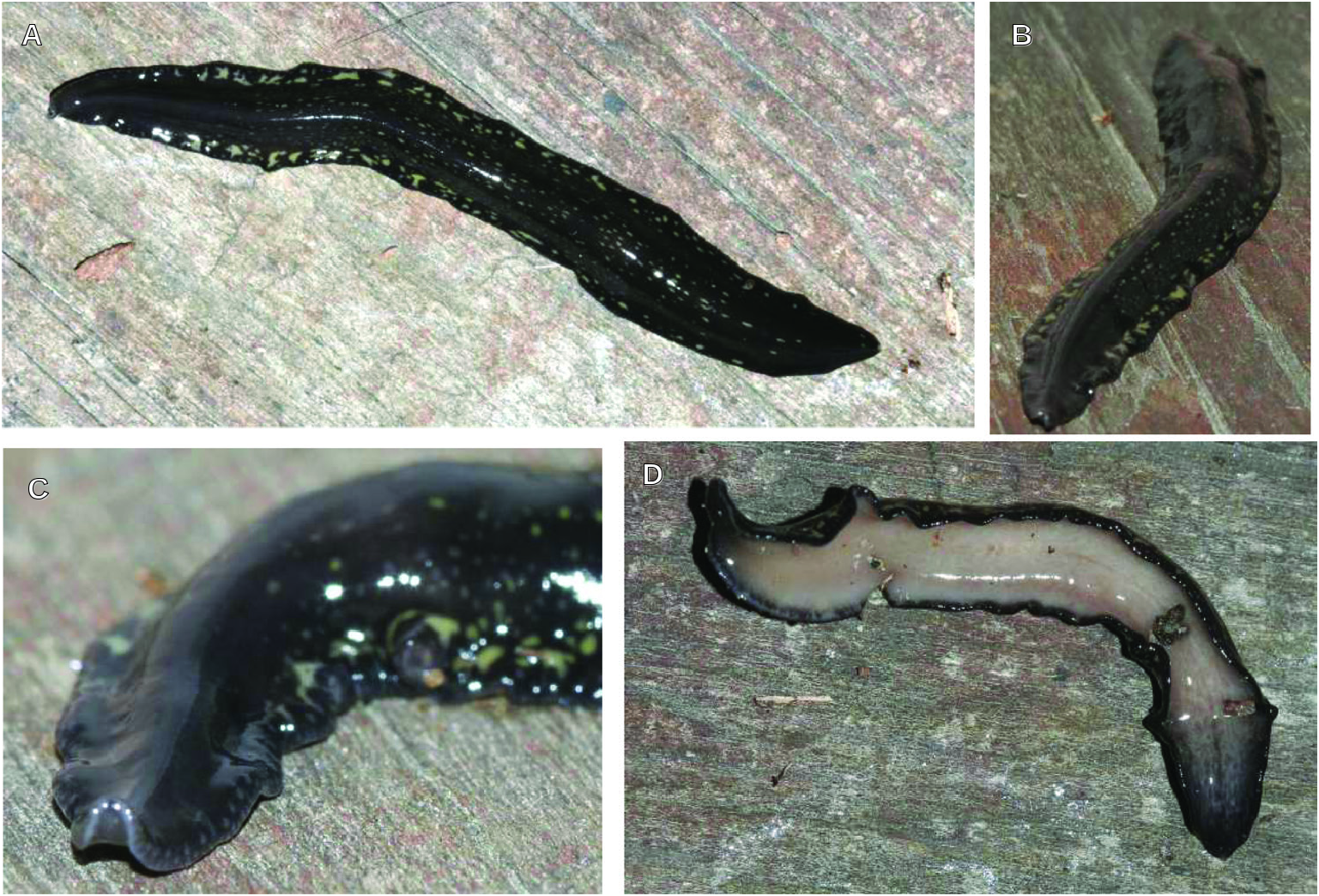
External aspect: The live holotype was approximately 50 mm long and 6 mm wide. The preserved specimen measured 45 mm long, 8 mm wide and 2.5 mm high. Live animals display nearly parallel body margins throughout most body length ( Fig. 3A, B View Figure 3 ). The body tapers gradually toward the anterior, rounded tip and less gradually toward the posterior, obtuse tip. The dorsum is flattened except for a carina that occupies the median region ( Fig. 3C, D View Figure 3 ). The ventral side is flat. The body exhibits undulating body margins when the animal is contracted ( Fig. 3B View Figure 3 ). The colour of the dorsum is black-blue (RAL 5004) with small green-beige (RAL 1000) dots on the median region and larger, irregular green beige patches in the submarginal region ( Fig. 3A, C, D View Figure 3 ). The ventral side is a cream colour (RAL 9001), covered with an increasing number of blackgrey (RAL 7021) pigment toward the extremities of the body. The eyes are of a single-cup measuring 50–70 µm in diameter. Each eye is in the middle of a round, light-grey halo ( Fig. 3C View Figure 3 ). The eyes are organized in a uniserial row around the anterior tip and 10 mm posteriorly they spread onto the dorsal side to the extent of 27% of the body width on either side. The sensory pits are simple invaginations, 50–57 µm deep, located ventromarginally and distributed in a single row from the anterior body tip up to the prepharyngeal region (42% of body length). These pits occur in the middle of light-grey haloes at the anterior tip ( Fig. 3C View Figure 3 ). The mouth is positioned at a distance from the anterior extremity equal to 47% of the body length; the gonopore at 68%.
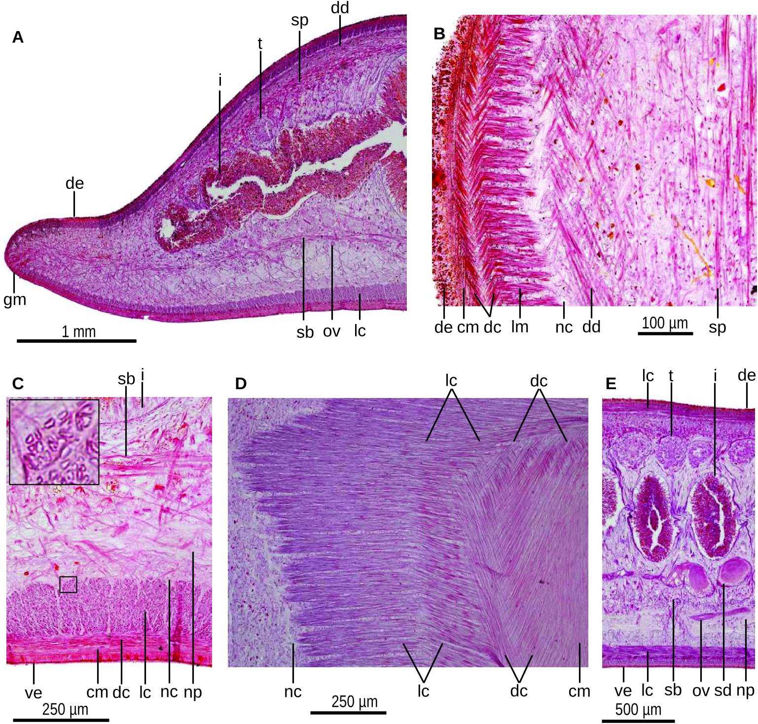
Internal morphology: The epidermis in the cephalic extremity is ciliated dorsally and ventrally. Behind the cephalic extremity, it is only ciliated on the creeping sole, with this sole being 95% of the body width. Erythrophil granules and rhabdites are discharged through the dorsal and ventral epidermis, both being more abundant dorsally. Gland cells discharge erythrophil granules through the ventral epidermis. A glandular margin is moderately developed ( Fig. 4A View Figure 4 ). This margin consists of two types of gland cells, producing xanthophil and erythrophil granules, respectively.
The cutaneous musculature comprises three layers: a subepithelial, 5 µm thick circular layer; a dense, double layer with decussate fibres (15 µm thick dorsally, 45 µm ventrally); and an innermost, welldeveloped longitudinal muscle layer ( Fig. 4A–D View Figure 4 ). The longitudinal muscle is 40 µm thick dorsally and 145 µm ventrally. The ventral portion of the longitudinal muscle becomes gradually thinner toward the body margins ( Fig. 4A View Figure 4 ). The longitudinal muscle fibres lying immediately under the decussate ones are orientated at a slight angle to the longitudinal axis ( Fig. 4D View Figure 4 ). The longitudinal muscle fibres are hollow ( Fig. 4C View Figure 4 , inset). The cutaneous musculature thickness relative to body height at the pre-pharyngeal region is 10%. A cephalic retractor muscle is absent.
Three parenchymal muscle layers are present throughout the body: a dorsal layer of decussate fibres (60 µm thick, or 2.4% of the body height) located to the inside of the peripheral nervous plexus ( Fig. 4B View Figure 4 ); a dense supraintestinal layer of transverse fibres (60 µm thick; 2.4%); and a dense subintestinal layer of transverse fibres (70 µm thick; 2.8%) ( Fig. 4A View Figure 4 ).
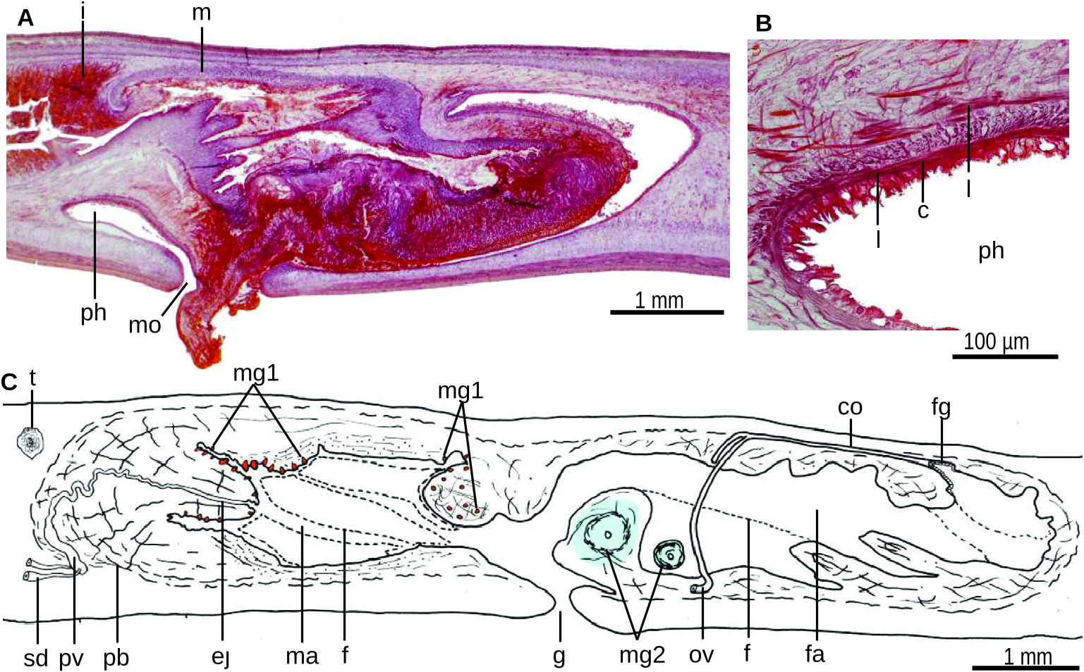
The mouth is situated at a distance from the root of the pharynx, equivalent to 27% of the length of the pharyngeal pouch length. The pharynx is collarshaped, with the dorsal insertion shifted backward by a distance equivalent to 70% of its length ( Fig. 5A View Figure 5 ). An oesophagus is absent. The outer pharyngeal musculature consists of a subepithelial layer of longitudinal muscle (10 µm thick), followed by a layer of circular muscle (20 µm) and a third layer of longitudinal fibres (30 µm thick). The inner pharyngeal musculature consists of a 100 µm thick layer of circular muscle, followed by a 10 µm thick layer of longitudinal muscle ( Fig. 5B View Figure 5 ).
The copulatory apparatus occupies 85% of the body height and is six times longer than its height. The testes are rounded, measuring about 180 µm in diameter. They are dorsally located beneath the transverse supraintestinal parenchymal muscle and between the intestinal branches ( Fig. 4E View Figure 4 ). The anteriormost testes are placed at a distance from the anterior body tip equivalent to 38% of the body length (i.e. considerably behind ovaries; see below); the posteriormost testes are located at 59% of body length (i.e. at the level of the penis bulb, see below).
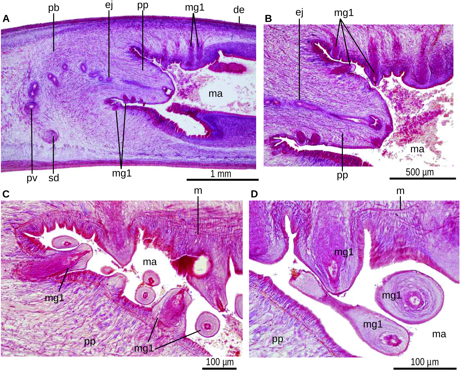
The sperm ducts run above the subintestinal parenchymal muscle and dorsally to the ovovitelline ducts ( Fig. 4E View Figure 4 ). These ducts contain sperm except their most distal portion, which is narrow and separately open into the proximolateral region of the prostatic vesicle ( Fig. 5C View Figure 5 ). The prostatic vesicle is tubular, sinuous and more or less C-shaped in lateral view. This vesicle is extrabulbar, but some fibres of the common muscle coat enclose it dorsally. The vesicle penetrates the anterior region of the well-developed penis bulb. The prostatic vesicle is lined with a cuboidal, ciliated epithelium and is surrounded by 30 µm thick circular muscle. Gland cells discharge erythrophil granules into the lumen of the prostatic vesicle. The ejaculatory duct traverses the centre of the penis papilla and its distal region widens to form a small, irregular cavity at the tip of the penis papilla. The ejaculatory duct is lined with a cuboidal, ciliated epithelium and is underlain by a 5 µm thick circular muscle. The small cavity is lined with a cuboidal, non-ciliated epithelium.
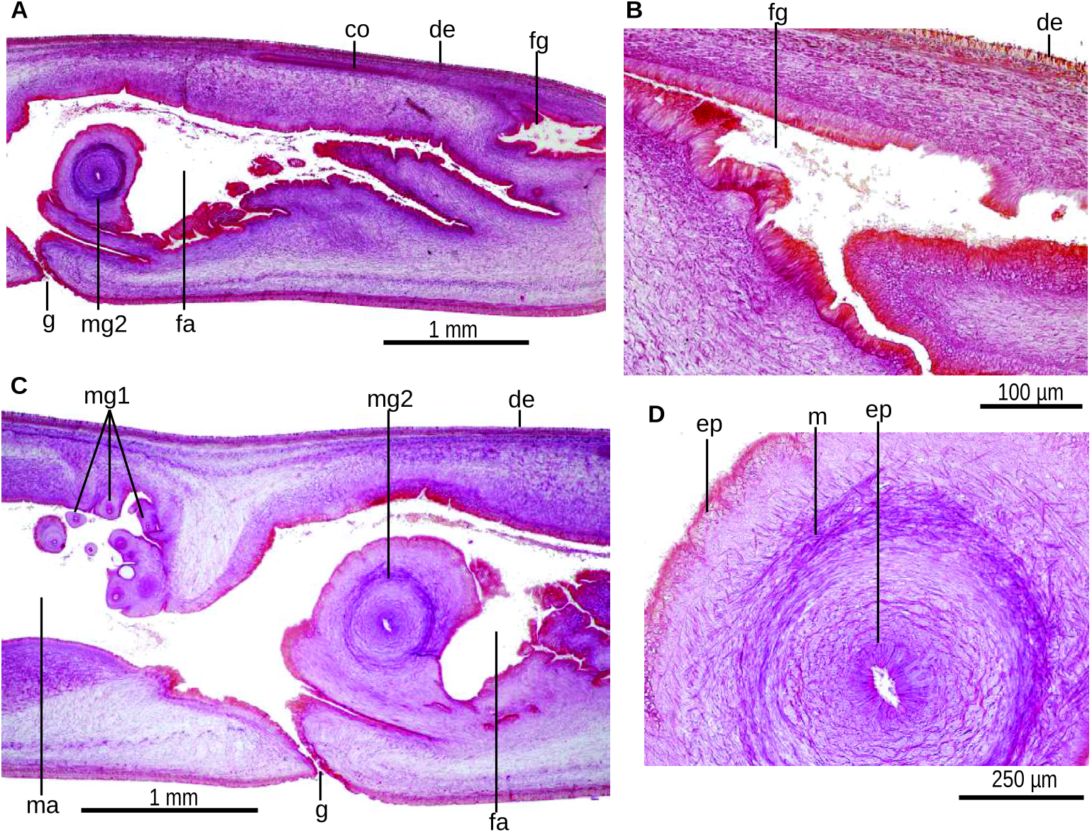
The penis papilla is conical-to-cylindrical and slightly inclined posteroventrally. This papilla occupies the anterior 20% of the male atrium ( Fig. 5C View Figure 5 ). The surface of the papilla has a spiny aspect due to the presence of protruding small musculoglandular organs (abbreviated mg 1 in the figures). Up to ten of these musculoglandular organs are visible in a single sagittal section. These organs are 65–70 µm wide and 30–130 µm long, with half of this length constituted by a sort of cone protruding beyond the surface of the penis papilla ( Fig. 6A–D View Figure 6 ). The musculoglandular organs contain a blind cavity opening at the tip of the organ. The cavity receives fine erythrophil granules produced by gland cells located outside the penis bulb. A finely granular cyanophil mass is placed beneath the squamous epithelium lining the organ ( Fig. 6B–D View Figure 6 ). The basis of the musculoglandular organ is delimited by a muscle net ( Fig. 6B–D View Figure 6 ).
The penis papilla is lined with a squamous epithelium and is underlain by a layer of circular muscle (8 µm thick), followed by a 10 µm thick layer of longitudinal muscle. Gland cells with 10 µm thick necks producing cyanophil granules also pierce the penial epithelium, especially in its basal half.
The male atrium is long and presents two to thtee large, longitudinal, lateral folds ( Fig. 5C View Figure 5 ). Anterior and posterior regions of the roof of the male atrium also possess musculoglandular organs, being different from those of the penis papilla and being larger in size than the latter, namely, 100–150 µm long ( Fig. 6A–D View Figure 6 ). The male atrium is lined with a columnar epithelium, which is underlain by an 80–100 µm thick coat of intermingled circular and longitudinal muscle fibres. The epithelium of the anterior quarter of the male atrium is traversed by the necks of gland cells secreting cyanophil granules. Additionally, gland cells producing erythrophil granules apparently pierce the entire epithelium of the male atrium.
The incompletely developed ovaries are rounded-toovoid and have a diameter of about 300 µm. They lie immediately above the ventral nerve plate and are located 6.3 mm anterior to the anteriormost testes (or the equivalent to 14% of body length), and at a distance from the anterior extremity of the body equivalent to 24% of the body length. The ovovitelline ducts emerge from the dorsolateral side of the ovaries. They run between the subintestinal parenchymal muscle layer and the ventral nerve plate ( Fig. 4E View Figure 4 ). These ducts bend abruptly dorsad laterally to the female atrium and then join the female atrium to form a 2 mm long, horizontal common ovovitelline duct ( Fig. 7C View Figure 7 ). Shell glands are absent. The common ovovitelline duct communicates with the female genital canal ( Fig. 7B View Figure 7 ), which is funnelshaped and projects anteriorly from the dorsoposterior portion of the female atrium ( Fig. 7B View Figure 7 ). The female genital canal and female atrium are lined with a columnar epithelium with conspicuous cilia. The female atrium is long, 1.2 times longer than the male atrium, and its lumen is narrowed by two to three lateral, longitudinal folds ( Figs 5C View Figure 5 , 7A, C View Figure 7 ). Three types of gland cells discharge xanthophil, erythrophil and cyanophil granules, respectively, into the female atrium. The latter type is more abundant in mid-atrium. The epithelium of the female atrium is underlain by a 100–200 µm thick muscle layer of circular and longitudinal fibres. Two large musculoglandular organs (mg 2 in the figures) are embedded in the lateral wall of the anterior section of the female atrium, each one on either side of the atrium ( Fig. 7A, C, D View Figure 7 ). These large musculoglandular organs project toward the midsagittal body plane. The conical, protruded portion of the musculoglandular organs is approximately 300 µm high and 500–700 µm in diameter. The embedded, semi-spherical portion is about 200 µm deep, while these musculoglandular organs present a blind canal that opens at the tip of the organ. The outer epithelium of the large musculoglandular organs is similar to that of the female atrium. At the same time, the inner one is columnar and ciliated, and is pierced by the necks of two types of gland cells producing erythrophil and cyanophil granules, respectively. A small quantity of cyanophil mass is located in the canal ( Fig. 7D View Figure 7 ). The semi-spherical, embedded portion consists of a well-developed muscularized ring, followed by oblique muscle fibres.
The common muscle coat is mainly comprised of longitudinal fibres, and envelops the male and female atria, the female genital canal, and also dorsally covers part of the prostatic vesicle.
| MZUSP |
Museu de Zoologia da Universidade de Sao Paulo |
| PL |
Západoceské muzeum v Plzni |
No known copyright restrictions apply. See Agosti, D., Egloff, W., 2009. Taxonomic information exchange and copyright: the Plazi approach. BMC Research Notes 2009, 2:53 for further explanation.
|
Kingdom |
|
|
Phylum |
|
|
Order |
|
|
Family |
|
|
Tribe |
Adinoplanini |