
Circamustela peignei, Valenciano & Pérez-Ramos & Abella & Morales, 2020
|
publication ID |
https://doi.org/10.5252/geodiversitas2020v42a8 |
|
publication LSID |
urn:lsid:zoobank.org:pub:CB141709-74F3-49FC-A689-059A907908FF |
|
DOI |
https://doi.org/10.5281/zenodo.3854806 |
|
persistent identifier |
https://treatment.plazi.org/id/03C287B8-FF97-5A40-FED1-F8AB6FA1F8AA |
|
treatment provided by |
Valdenar |
|
scientific name |
Circamustela peignei |
| status |
sp. nov. |
Circamustela peignei n. sp.
( Figs 2-6 View FIG View FIG View FIG View FIG View FIG ; 7 View FIG A-D)
Mustelidae gen. et sp. indet. aff. Circamustela dechaseauxi – Valenciano 2017: 331.
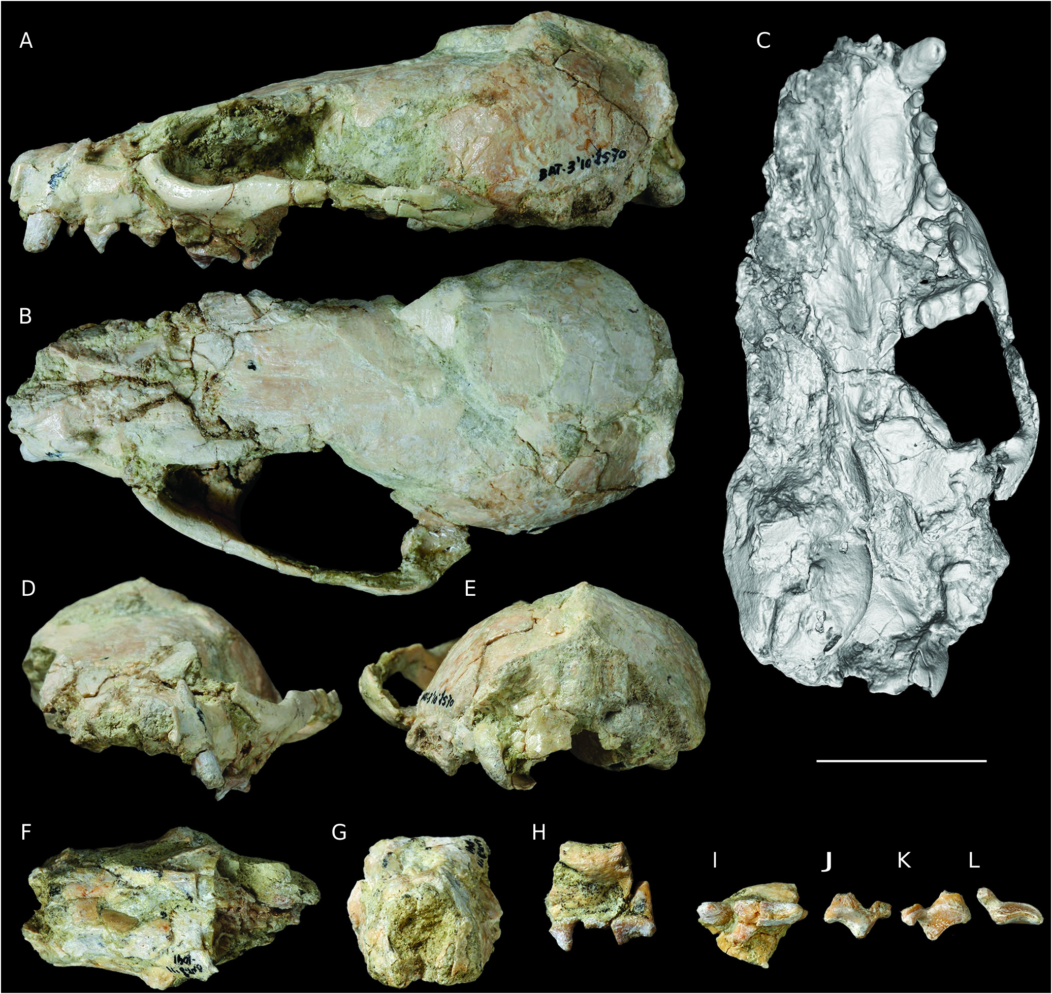
HOLOTYPE. — BAT-3 ’10.1570, complete cranium with C, P1-4 and M1 ( Fig. 2 View FIG A-E).
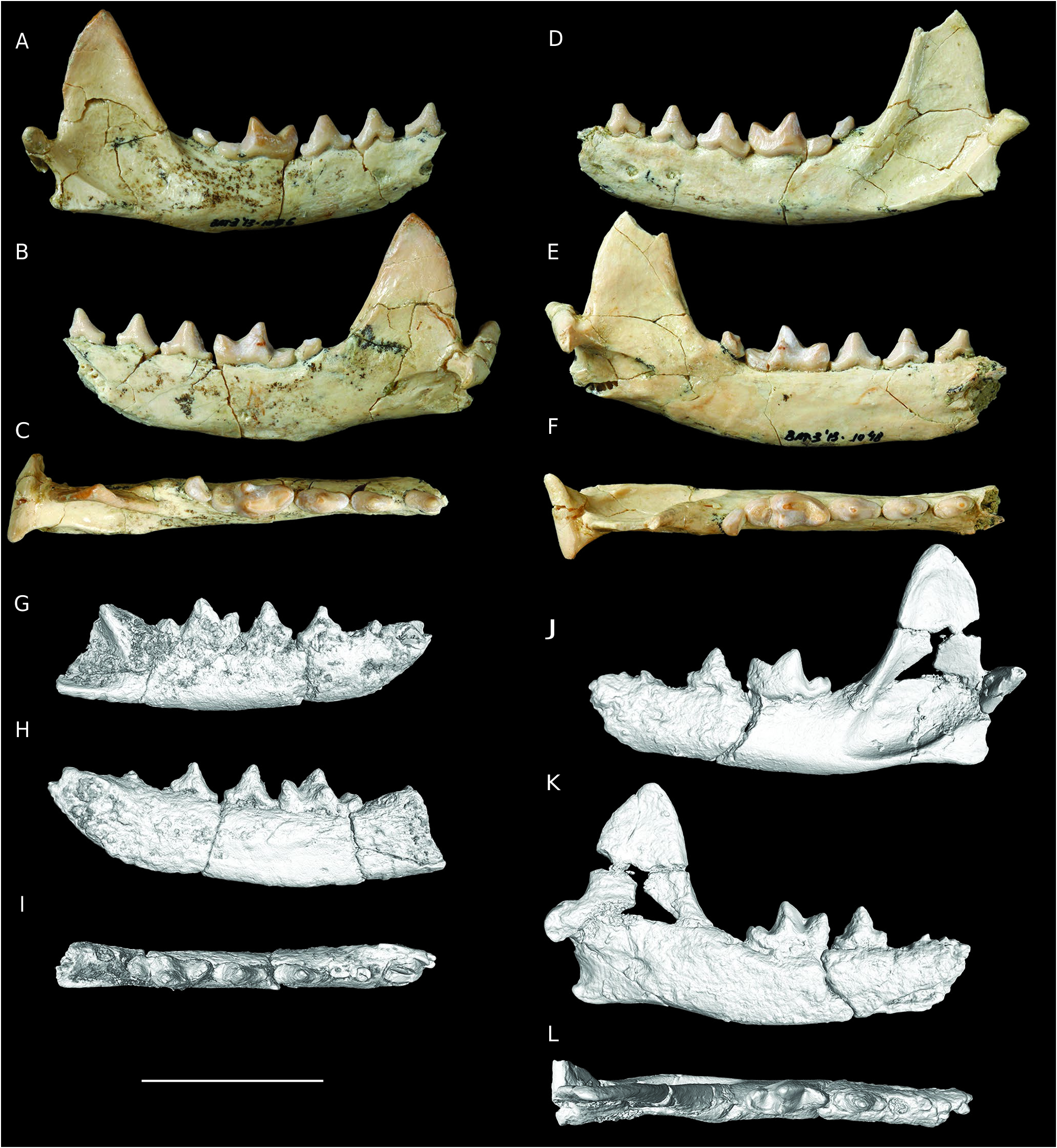
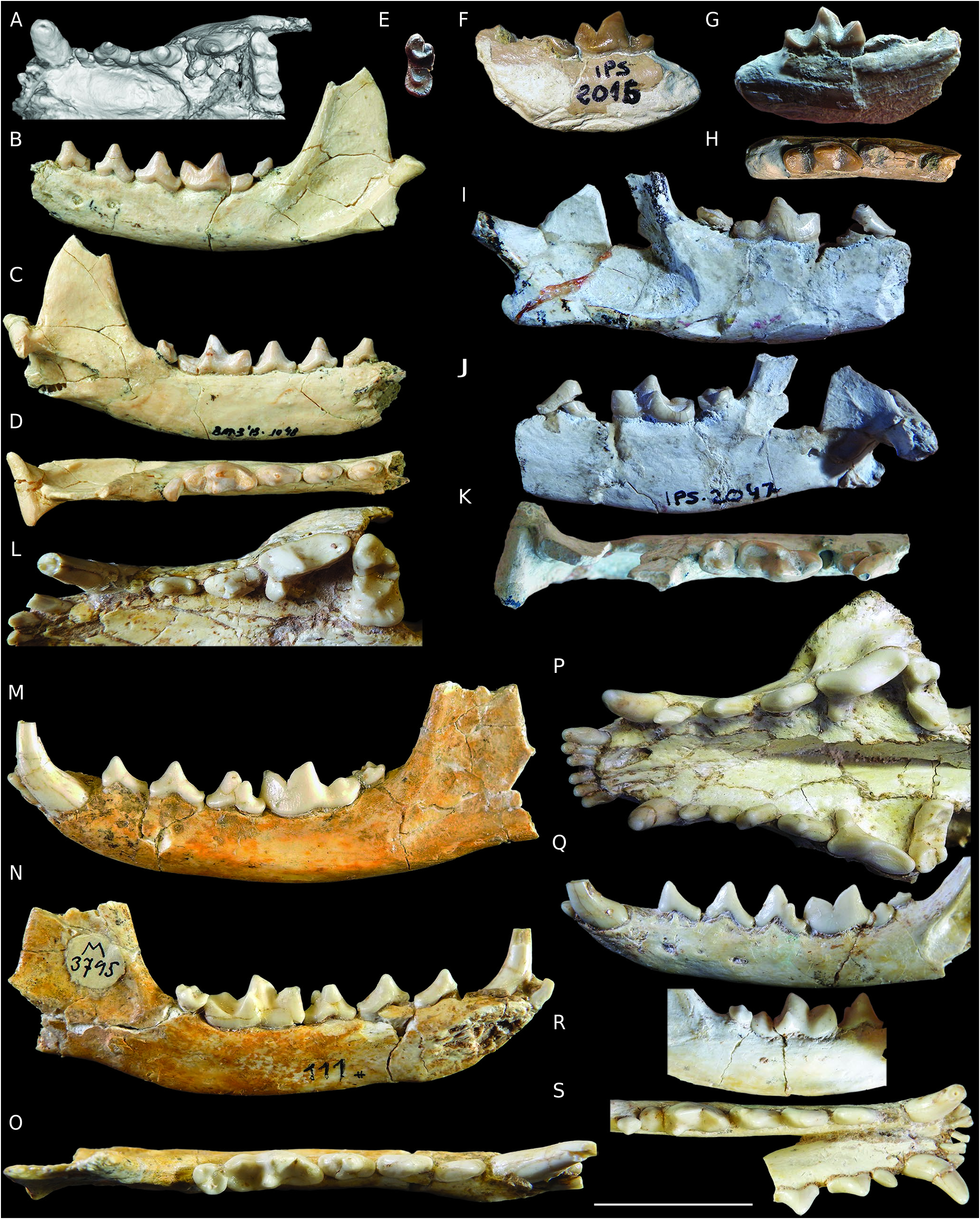
PARATYPE. — BAT- 3’13.1048, nearly complete left hemimandible with p2-m2 ( Fig. 5 View FIG D-F).
ETYMOLOGY. — In memory of Dr Stéphane Peigné, expert on Neogene carnivorans from Eurasia and Africa.
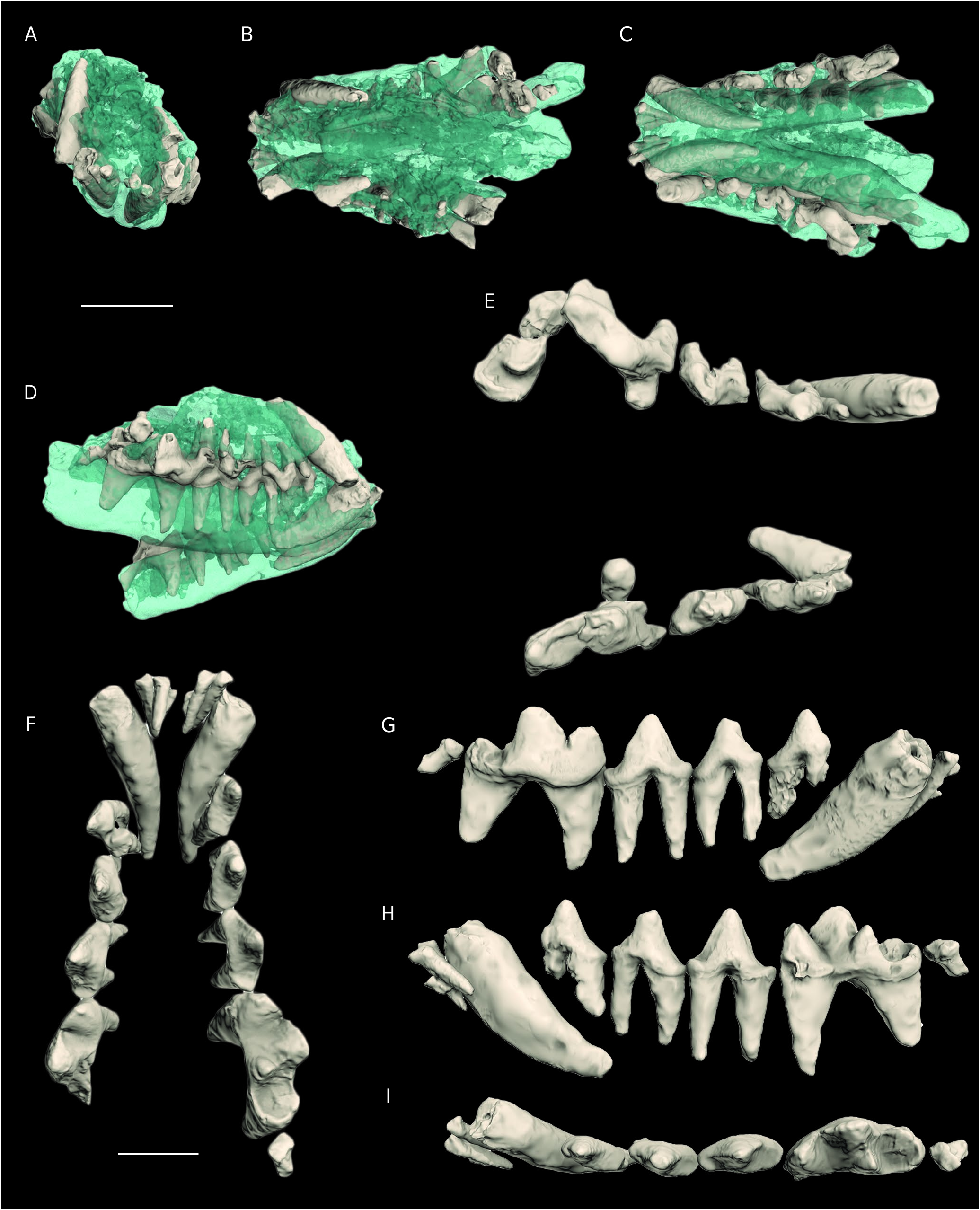
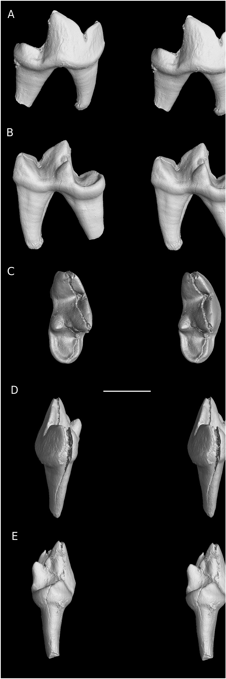
HYPODIGM. — BAT-3’11.1041 ( Fig. 2 View FIG F-L): fragmentary cranium, comprising the muzzle, a left fragment of the maxillary with a fragmented P3 and a complete P4 and isolated right P4; BAT-3 ’10.1246: fragmentary cranium, comprising the muzzle and attached mandible, including C, P1-4, M1 and i1-3, c, p2-4, m1-2 ( Fig. 3 View FIG ) ; BAT-3 ’13.1086: nearly complete right hemimandible with p2-m2 ( Fig. 5 View FIG A-C) (same individual as the paratype) ; BAT-3 ’10.1570A ( Fig. 5 View FIG J-L): fragmentary left hemimandible with p4 and m1 (same individual as the holotype) ; BAT-3 ’10.1570B ( Fig. 5 View FIG G-I): fragmentary right hemimandible with a broken p2 and complete p3-m1 (same individual as the holotype) ; Bat5-’10.G14.129: right m1 ( Fig. 6 View FIG ).
TYPE LOCALITY. — Batallones-3 (late Miocene, Vallesian, MN 10).
OTHER LOCALITY. — Batallones-5 (late Miocene, Vallesian, MN 10).
AGE. — Late Miocene, Vallesian, MN10.
DIAGNOSIS. — Mustelid of a size comparable to Circamustela dechaseauxi. Relatively long muzzle; P1 present; P2-3 unicuspid and elongated; P3 distally widened; P4 long with conical and slender protocone mesially located, low parastyle and lingual cingulum; M1 buccolingually elongated and mesiodistally reduced, with a large parastylar area, paracone larger than metacone, the latter being distinctive, high mesially located protocone; long and low mandibular corpus; high coronoid process and shallow masseteric fossa; p2-4 elongated; p2-3 unicuspid; p4 with low distal accessory cuspid; m1 metaconid lingually expanded; oval and short m2 with small protoconid and metaconid.
DIFFERENTIAL DIAGNOSIS. — Differs from Circamustela dechaseauxi in more developed M1 metacone, lesser developed metastylar area, higher and mesially located protocone, lesser development of the cingulum on the lingual platform; more developed m1 metaconid, and a more conical hypoconid; Differs from Martes melibulla in the shorter mandibular corpus, reduced m1 talonid with a much shallower talonid basin, with reduced m1 entocristid and reduced m2; Differs from “ Martes ” sansaniensis, “ Martes ” filholi, Martes woodwardi, Martes ginsburgi, Pekania palaeosinensis, Pekania occulta, and Paramartes pococki, in smaller size, reduced lingual platform of the M1 and more reduced m1 talonid.
DESCRIPTION
Cranium and upper dentition
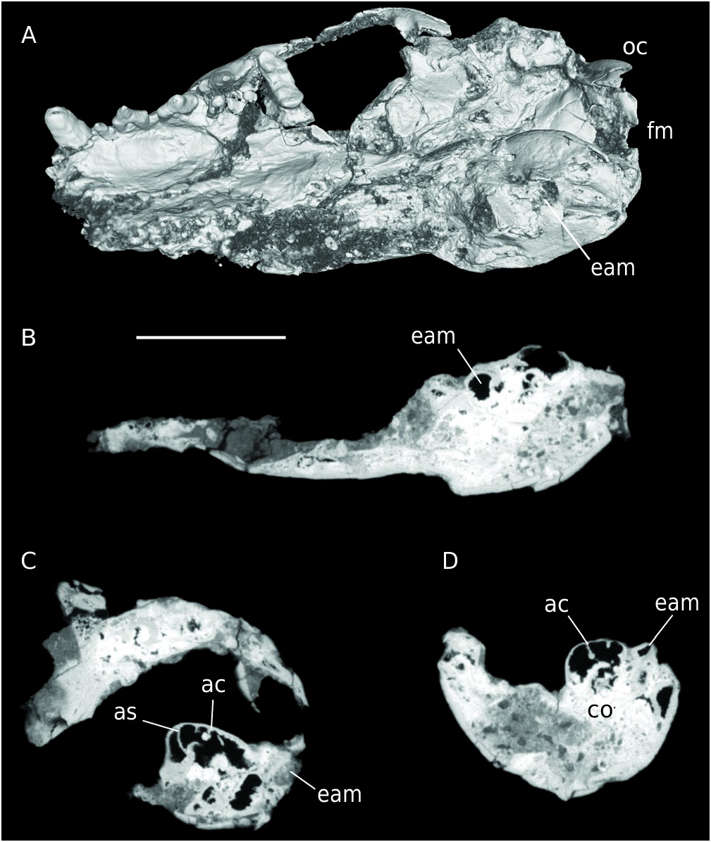
Circamustela peignei n. sp., has a medium-sized cranium about as large as that of the American marten ( Martes americana ) and a long rostrum similar to that of the North American fisher ( Pekania pennanti). Three fragmentary skulls have been found at Batallones-3. Specimen BAT-3’10.1570 ( Fig. 2 View FIG A-E) is the most complete, and with a basal cranial length of 80.7 mm. It is dorso-ventrally compressed and the left frontal bone is collapsed. The cranium is partially crystallized in its interior, affecting the internal cavities and roots, and some superficial bone, such as of the right maxilla and pre-maxilla is dissolved. The nuchal and ventral regions are also damaged. The right zygomatic arch, left bulla, and both right mastoid process and occipital condyle are missing. The nasal aperture is broken. The orbit is large. The rostral margin of the orbit ends at the level of the mesial margin of the P4 parastyle. The postorbital processes are not preserved. The infraorbital foramen is located above the P3 and P4. It has a well-developed sagittal crest ( Fig. 2A View FIG ), which suggests that it was a male individual ( Larivière & Jennings 2009). In caudal view ( Fig. 2E View FIG ), the nuchal area is triangular, rather flat, and no muscular attachments are preserved on the supraoccipital bone. The left zygomatic arch is similar to that of living martens, so it is not especially robust. The frontal process of the zygomatic arch is triangular and of moderate size. On the palate, the incisive foramina are not preserved. There is an oval concavity in the left maxilla from the canine at the P3 level. The right bulla is preserved. It is swollen, oval and rostrocaudally larger. It has three small perforations on its surface due to erosion. The scan images show a highly septate tympanic bulla, with a more developed anterior septum that partially divide the bulla ( Fig. 3 View FIG B-D), as it happens in the late Miocene African mustelid Howellictis valentini (de Bonis et al. 2009). No additional features or foramina from the basicranial area are preserved.
urn:lsid:zoobank.org:act:
Specimens BAT-3’11.1041 ( Fig. 2 View FIG F-L) and BAT-3’10.1246 ( Fig. 4 View FIG ) preserve the anterior part of the rostrum. Both show signals of dissolution by soil acids. BAT-3’11.1041 is not distorted. It shows a higher than broad nasal aperture and a high and relatively robust muzzle ( Fig. 2 View FIG F-G). The rostral part of the cranium BAT-3’10.1246 has both hemimandibles attached, as seen in the virtual reconstruction ( Fig. 4 View FIG ).
The preserved upper dentition comprises the C, P1-4 and M1 ( Figs 2-4 View FIG View FIG View FIG ; Table 1 View TABLE ). Most teeth show signs of dissolution. The C is oval and long. Both BAT-3’10.1570 and BAT-3’10. 1246 have a large wear facet on the tip. There are diastemata between all the upper premolars. The P1 is robust and unicuspid. Both P2 and P3 are elongated, unicuspid and show a concave buccal wall. The P3 is distally widened. The P4 is relatively long. It possesses an eroded parastyle in BAT-3’10.1570 ( Fig. 2C View FIG ). There is an inflection between the parastyle and the protocone. The protocone is very isolated and mesially located. Additional P4s associated with the crania BAT-3’11.1041 ( Fig. 2 View FIG H-L) and BAT-3’10.1246 ( Fig. 4E View FIG ) indicate that the parastyle is of moderate size. The protocone is conical and slender with moderate height. A lingual cingulum is present on the whole length of the lingual wall ( Fig. 2 View FIG K-L).Two M1 are preserved in the crania BAT-3’10.1570 and BAT-3’10.1246. The M1 BAT-3’10.1570 is buccolingually elongated and mesiodistally reduced ( Fig. 2C View FIG ). It has a large parastylar area. The paracone is larger than the metacone. Both mesial and distal walls are very straight. The protocone is high and mesially located. The lingual platform is reduced in comparison with living gulonines (e.g., Martes martes , Martes foina, Pekania pennanti). The CT-scan allowed us to examine the M1 BAT-3’10.1246 ( Fig. 4E View FIG ). It is broken at the paracone-metacone level and the metacone is dissolved.Its overall morphology is close to BAT-3’10.1570, showing a short lingual platform and a high and distally placed protocone ( Figs 2C View FIG ; 4E View FIG ).
Mandibles and lower dentition
Three incomplete mandibles have been found in Batallones-3 comprising i1-3, c, p2-4 and m1-2 ( Figs 4 View FIG , 5 View FIG ; Table 2 View TABLE ). Both BAT-3’10.1570A and B are poorly preserved and the mandibular corpus and teeth are dissolved and carbonated, having several small concretions on the surface ( Fig. 5 View FIG G-K).
The mandible of C. peignei n. sp. is slender with a low mandibular corpus.Laterally, there are two rounded mental foramina, one under the distal part of the p2 and the other beneath the middle cuspid of the p3. The mandibular symphysis is elongated and curved ( Fig. 5 View FIG G-H). Its medial surface is rough, and the attachment of the fibrocartilage pad reaches the mesial part of the p3. The coronoid process is high, sharp and vertically oriented. The masseteric fossa is shallow, although in BAT-3’10.1570A it is deeper. Its rostral margin lies at the level of the m2. The tooth row is straight and is aligned with the articular process. The articular process is large and close to the angular process. The angular process is robust and caudally directed. It is more developed in BAT-3’10.1570A.
BAT-3’10.1246 preserves all the lower incisors on both sides ( Fig. 4 View FIG ). The i1 is very small and peg-like. The right i1 only preserves part of the root. The i2 and i3 are bilobed, with i3 mesiodistally wider. The root of the i2 is imbricated between the i1 and the 13. The canine is large and has a lingual cingulid ( Fig. 4 View FIG F-I). BAT-3’10.1570A also shows an alveolus for p1. The p2-3 are elongated and unicuspid. The cuspid of p2 is placed mesially, but in the p3 it is located in the middle portion of the tooth. Both p3-4 have high mesial and distal cingulids. The p4 has a tiny distal accessory cuspid ( Fig. 4 View FIG F-I, 4A-F), which is more developed in BAT-3’10.1570A ( Fig. 5 View FIG J-L). The m1 is long; its trigonid forms more than two thirds of the total length of the tooth; the protoconid is higher than the paraconid. The metaconid is distinct, attached to the protoconid but not exceeding its posterior edge. It is lingually extended ( Figs 4F View FIG , H-I; 5Q; 6), and is more developed in BAT-3’13.1086 ( Fig. 5 View FIG A-C), and in the specimen from Batallones-5 ( Fig. 6 View FIG ). The maximum width is located at the protoconid-metaconid level. The talonid is reduced in length. The hypoconid is low and lingually bevelled ( Figs 4 View FIG F-I; 5C, E). There is no entocristid; instead a posterior cristid of the metaconid extends along the lingual edge of the talonid, reaching the hypoconulid. Between the hypoconid and the aforementioned edge there is a shallow basin. The hypoconulid is more developed in BAT-3’10.1570A ( Fig. 5 View FIG J-L) and BAT-5’10.G14.129 ( Fig. 6 View FIG ). The m2 is reduced and oval. Its protoconid and metaconid are of similar height ( Fig. 5 View FIG A-C).
| MN |
Museu Nacional, Universidade Federal do Rio de Janeiro |
No known copyright restrictions apply. See Agosti, D., Egloff, W., 2009. Taxonomic information exchange and copyright: the Plazi approach. BMC Research Notes 2009, 2:53 for further explanation.
|
Kingdom |
|
|
Phylum |
|
|
Class |
|
|
Order |
|
|
Family |
|
|
Genus |