
Indarctos punjabiensis ( Lydekker, 1884 )
|
publication ID |
https://doi.org/ 10.5252/geodiversitas2019v41a23 |
|
publication LSID |
urn:lsid:zoobank.org:pub:4174E526-4A7D-4D2E-9233-6FD43F145F07 |
|
DOI |
https://doi.org/10.5281/zenodo.3704196 |
|
persistent identifier |
https://treatment.plazi.org/id/03C1E33A-8024-FF8B-FF5C-33AE8E11F82C |
|
treatment provided by |
Valdenar |
|
scientific name |
Indarctos punjabiensis ( Lydekker, 1884 ) |
| status |
|
Indarctos punjabiensis ( Lydekker, 1884)
Hyaenarctos punjabiensis Lydekker, 1884: 226 .
Agriotherium cf. A. roblesi – Alcalá 1994: 103, 104, pl. 3, fig. o.
SYNTYPE. — Fragments of the left and right maxillae with P4-M1, figured by Lydekker (1884: pl. XXX, fig. 2).
TYPE LOCALITY. — Hasnot Siwaliks.Salt Range, Jhelum District. Punjab.
AGE. — Late Miocene. MN 10-13.
EMENDED DIAGNOSIS. — Medium to large sized Ursidae , with marked sexual dimorphism. Plantigrade. Dolichocephalic skull, base of the zygomatic arch coincides with the M1 and the mesial part of M2. The premolars are smaller in relative size and number compared to the other members of the genus, being crowded to some degree. Both the P2 and the P3 are double rooted, with the tendency to be fused in some individuals. The mesio-distal length of the P4 is equal to that of M1, and it has a quite developed parastyle, with two crists, une lingual and one labial. Wide upper molars, almost as wide as they are long; from sub-rectangular to trapezoid in shape. The talon in M2 is large, but not as long as in the derived Ursinae . Generally low, but robust jaw. Relatively long m1, with equally long talonid and trigonid; tall entoconid, taller than the hypoconid; m1 talonid similar to that of m2. The m3 morphology varies from rounded to elongated.
DESCRIPTION
Upper dentition
The description has been carried out on the possibly associated upper dentition, using both sides when possible.
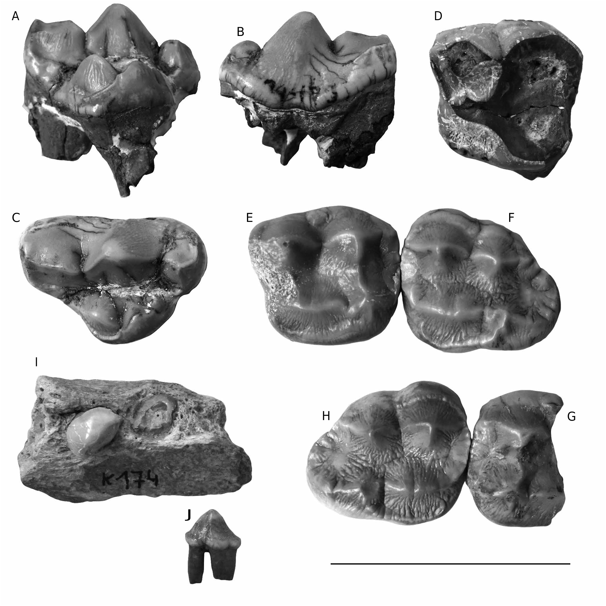
P1 ( Fig. 2I View FIG ). Only a single root is preserved. Smallest of the upper premolars.
P2 ( Fig. 2I View FIG ). Reduced, ovoid premolar, with only one root. Single cusp with two ridges, one mesial and one distal, that curve towards the lingual side; the distal cusp is blunter than the mesial one. This tooth is rotated relative to the main axis of the maxilla and somehow crowded, due to the shortening of the muzzle.
P3 ( Fig. 2I View FIG ). Only a single root is preserved. Larger than the preceding upper premolars.
4
P
W
1
M
W
L M1
P4 ( Fig. 2 View FIG A-C). Complete upper carnassial. Deeply wrinkled enamel. Well-developed parastyle with two ridges, one labial and one lingual. High paracone, almost flat in its lingual part but convex in the labial one. Metastyle lower than paracone, but more sectorial; wide at its base, with two cingula, a weak one in the labial part and a slightly more developed one in the lingual part. Where the two cingula join they create a distal pointed edge. The protocone is complex. It has three cusps, which grow taller towards its distal part, with the latter being the tallest, practically at the level of the notch between paracone and metastyle, and the only one partly surrounded by the labial cingulum of the cusp. The protocone occupies an intermediate position with respect to the paracone, and is projected lingually.
M1 ( Fig. 2D View FIG ). Very worn first upper molar, much more in the distal than in the mesial part. This kind of asymmetric wear is quite common in ursoids, and in this case prevents the description of many of the dental features. It is nearly square in shape. The contact facet for P4 is relatively large and concave instead of flat, while that for the M2 is located almost at the labio-distal corner of the tooth. Towards the labial side, between the paracone and the metacone, there is an expansion zone that widens towards the metacone; this structure has a slight vertical wear facet.
M1 ( Fig. 2 View FIG E-G). Molars with a clear quadrangular shape, almost as wide as long. The enamel has the typical young ursid roughness. It is barely worn and the roots were not completely formed before death and are not recovered. As in the worn M1, these two also have a medial-lingual thickening of the labial cingulum between the paracone and metacone. The paracone is larger (in both width and height) than the metacone. Both parastyle and metastyle are present, although these are very weak compared to the main cusps. There is a well-developed cingulum occupying almost all of the lingual face. The contact with P4 is vertical and straight. The protocone is large and cristiform. Distal to the protocone there is a robust hypocone, which is taller than the protocone, and although being the small- est of the four main cusps. Hypocone is more complex, having a ridge on each side. The mesial ridge is close to the protocone, whereas the lingual one joins the lingual cingulum. The medial one meets the ridge coming down the central valley of the tooth from the metacone and the distal ridge ends at the most distal edge of the tooth. In the labial side there is other cingulum, which is considerably thickened in the distal region.
M2 ( Fig. 2F, H View FIG ). A wide tooth, with large cingula on both in its labial and its lingual side, with the latter the most developed. In occlusal view the talon is slightly rotated labially while the labial cingulum is labially tilted, giving the teeth two different crushing surfaces. The tooth enamel is clearly rough, which is most evident on the lingual surface. The protocone is continued by the ridge that originates at the front of the paracone, continuing throughout the mesial part of the molar towards the lingual side, but not forming a clear parastyle. Unlike M1, the M2 protocone is slightly bilobed in its central part. The paracone is taller than the metacone although the difference is slightly smaller than in the M1. The talon is very rounded and relatively short, and although it joins the metacone, it has a more labial position. It has a ridge that runs from the hypocone to the metacone, forming a distal semicircle divided into at least six small cusps.
Lower Dentition
px ( Fig. 2J View FIG ). Biradicular premolar. Longer than wide. It has only one cusp in the center of the crown, with two cristids, one slightly mesio-lingual, which approaches the mesial edge, and other one distal.
Postcranial skeleton
Right Mc IV ( Fig. 3 View FIG ). This metapodial has a robust general morphology. Epiphyses are wider than the diaphysis, the latter having a homogeneous width in dorsal view throughout its length. However, in lateral view, there is a difference in height because the distal part of the diaphysis is clearly flatter. The proximal epiphysis is robust, convex in the most proximal zone, the surface that serves as articulation for the unciform. Its contact facet for the Mc III has two lobes, a convex dorso-proximal one and a flat palmar-proximal one. The articulation for the Mc V has also two facets, a slightly convex dorsal one and a noticeably concave palmar one. The distal epiphysis is slightly wider and somewhat rounded in its dorsal view, thus being more flattened in lateral view.

| MN |
Museu Nacional, Universidade Federal do Rio de Janeiro |
No known copyright restrictions apply. See Agosti, D., Egloff, W., 2009. Taxonomic information exchange and copyright: the Plazi approach. BMC Research Notes 2009, 2:53 for further explanation.
|
Kingdom |
|
|
Phylum |
|
|
Class |
|
|
Order |
|
|
Family |
|
|
Genus |
Indarctos punjabiensis ( Lydekker, 1884 )
| Abella, Juan, Hontecillas, Daniel, Valenciano, Alberto, Montoya, Plinio, Morales, Jorge, Pesquero, María Dolores & Alcalá, Luis 2019 |
punjabiensis
| LYDEKKER R. 1884: 226 |