
Sclerosococcus williamsi Granara de Willink, 2023
|
publication ID |
https://doi.org/10.5281/zenodo.10834645 |
|
publication LSID |
lsid:zoobank.org:pub:2A1FB016-1B67-4861-BB8B-2011B26679F1 |
|
DOI |
https://doi.org/10.5281/zenodo.8222330 |
|
persistent identifier |
https://treatment.plazi.org/id/03C0696F-DF4F-FFF3-FF49-77B4AF894AA9 |
|
treatment provided by |
Felipe |
|
scientific name |
Sclerosococcus williamsi Granara de Willink |
| status |
sp. nov. |
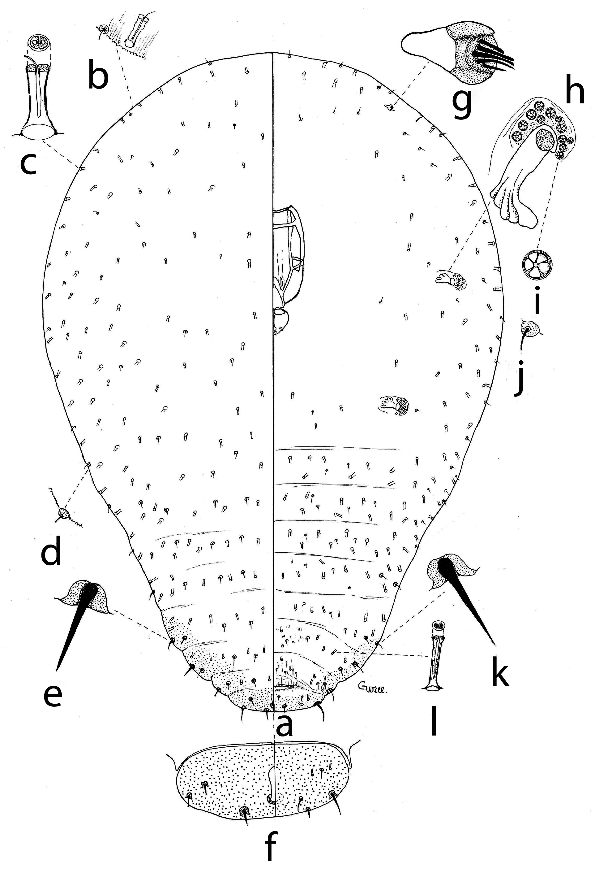
Sclerosococcus williamsi Granara de Willink , new species
( Fig. 7 View Figure 7 , 8 View Figure 8 )
Type material. Holotype. Female marked by a black circle. Argentina, Salta, Alemanía, v/1991, on Bromeliaceae , possibly Deuterocohnia longipetala (Baker) Mez in Mart, Granara de Willink col. 1(2). Paratypes. 6 ( 13, 2 N 2); same data as holotype. IFML.
Material examined. Argentina, Salta, Alemanía, iii/1993, 4 ( 16, 4 N 1, 1 N 2); vii/1990, 1(3); Salta, Cafayate, iv/1997, 5 ( 4, 7 N 2, 1 male); Salta, El Anfiteatro, near Cafayate, 22/v/2009, 10 ( 31, 17 N 1, 4 N 2), all collected on Deuterocohnia Mez , possibly D. longipetala (Baker) Mez (Bromeliaceae) by Granara de Willink. IFML.
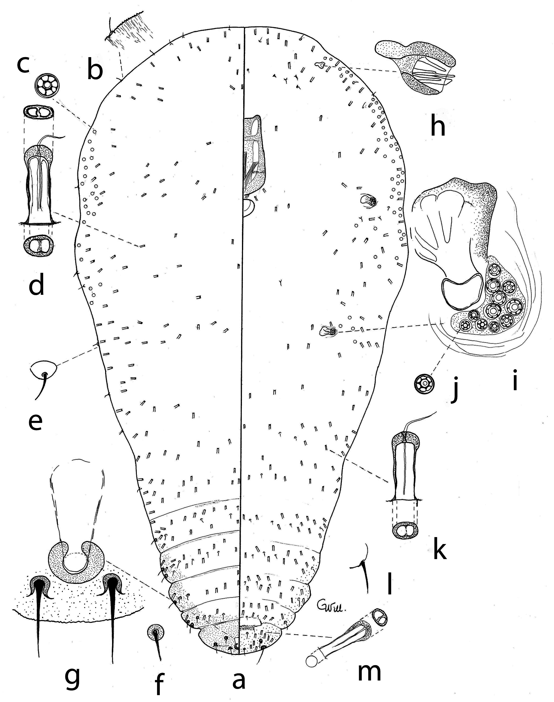
Diagnosis. Multilocular pores with 5–9 locules present in spiracular atrium. Multilocular pores absent or present on marginal and submarginal cephalothorax. Multilocular pores absent on dorsal and ventral abdomen. Numerous eight-shaped pore ducts of varying sizes on dorsum and venter. Marginal setae of the last four abdominal segments and of the anal lobe strong, with a bulbous base, inserted into a sclerotized depression.
Habitus. This species was found in flower calices. Apparently, it does not make pits. Each female forms a network of thin and long wax filaments that surround and protect it, leaving the posterior part of the body free. Without filaments, it shows little powdery wax and a yellowish color.
Microscopic description. Body elongated oval or pyriform; length 1.62 (0.94–1.86) mm, width 0.66 (0.51–1.22) mm. Without legs. Dermis membranous, striated (easily seen at body margin); last abdominal segment sclerotized, segments V-VIII submarginally sclerotized.
Dorsum. With thick setae with a wide and tall sclerotized base; setae with different sizes; with one or two marginal setae on abdominal segments IV-VII, 19–29 (24–33) μm long and 2.5–5 μm at the base. Setae rarely present on thorax and anterior abdominal segments; if present, then shorter and thinner than other setae, short and stout, 2.5 µm wide at the base and 2.5 (2.5–7) µm long; six setae on abdominal segment V, 10 setae on VI, eight setae on VII; four setae in anterior region, 10 µm long. Marginal and submarginal multilocular pores occasionally occur on the cephalothorax. Numerous eight-shaped pore ducts on the internal end, with a simple wide aperture, and a short and thin filament, 10–15 µm long, scattered; ducts arranged in transverse rows on abdominal segments, anterior segments with a double row of ducts, segment VI with 11 ducts, VII with four ducts, and VIII with six ducts. Horseshoe-shaped anal ring, 15 (14–17) µm wide, without pores or setae, anal opening 10 (7–12) µm wide. With sclerotized anal tube.
Venter. Antennae 22 (17–26) µm long and 12 (12–17) µm wide, with two segments and fleshy setae. Anterior and posterior spiracles of same size, 32 (46–57) µm long and 11 (21–33) mm wide, peritreme diameter 11 (24–27) µm. Clypeolabral shield 290 (216–288) µm long and 165 (127–149) µm wide. Labium one-segmented, 75 (45–96) µm long, 85 (55–108) µm wide, with four setae. With few short, slender, and tack-shaped marginal and submarginal setae, 5–7 µm long and 5–6 µm wide at the base, up to segment III; setae absent in middle of meso- and metathorax; 4–5 setae in transverse rows in the middle of segments IV-VIII. With one broad-based seta, similar to dorsal ones, 24 µm long, on the margin of each of the segments IV-VIII. With microspicules in the middle of segment VI. With multilocular pores with 5–9 locules, approximately 5 µm in diameter, in spiracular atria; with 5–12 pores in anterior and 5–16 pores in posterior atria, respectively. Multilocular pores with 7–9 locules found on margin and submargin of thorax. These are either absent or very numerous (up to 400) and are distributed either between the anterior and posterior spiracles, between the posterior spiracles and antennae, or, forming a continuous row including the cephalic area, from the posterior spiracle of one side to the posterior spiracle of the opposite side. Multilocular pores are absent on abdominal segments. With eight-shaped pore ducts, similar to the dorsal ones, 17–21 (15–24) μm long and 5 μm wide, numerous in marginal and submarginal areas on cephalothorax, but also in the medial area, and arranged in transverse rows on abdomen, smaller in posterior than in anterior segments. Vulva between segments VII-VIII, apparently covered by a plate-like structure, with 4 (4–6) ducts and 4 (4) setae. With a pair of setae posterior to the vulva on segment VIII, 6 μm in diameter at the base and 10–14 μm long; laterally a seta and seven ducts on each side; marginal seta 48 (30–61) μm long, with a deep and broad base, 20 μm in diameter and 8 μm deep.
Observations. Apparently, there are two types of females; one with multilocular pores on the cephalothorax and the second one without pores in that position. Unborn nymphs were observed in both types of females, indicating viviparity.
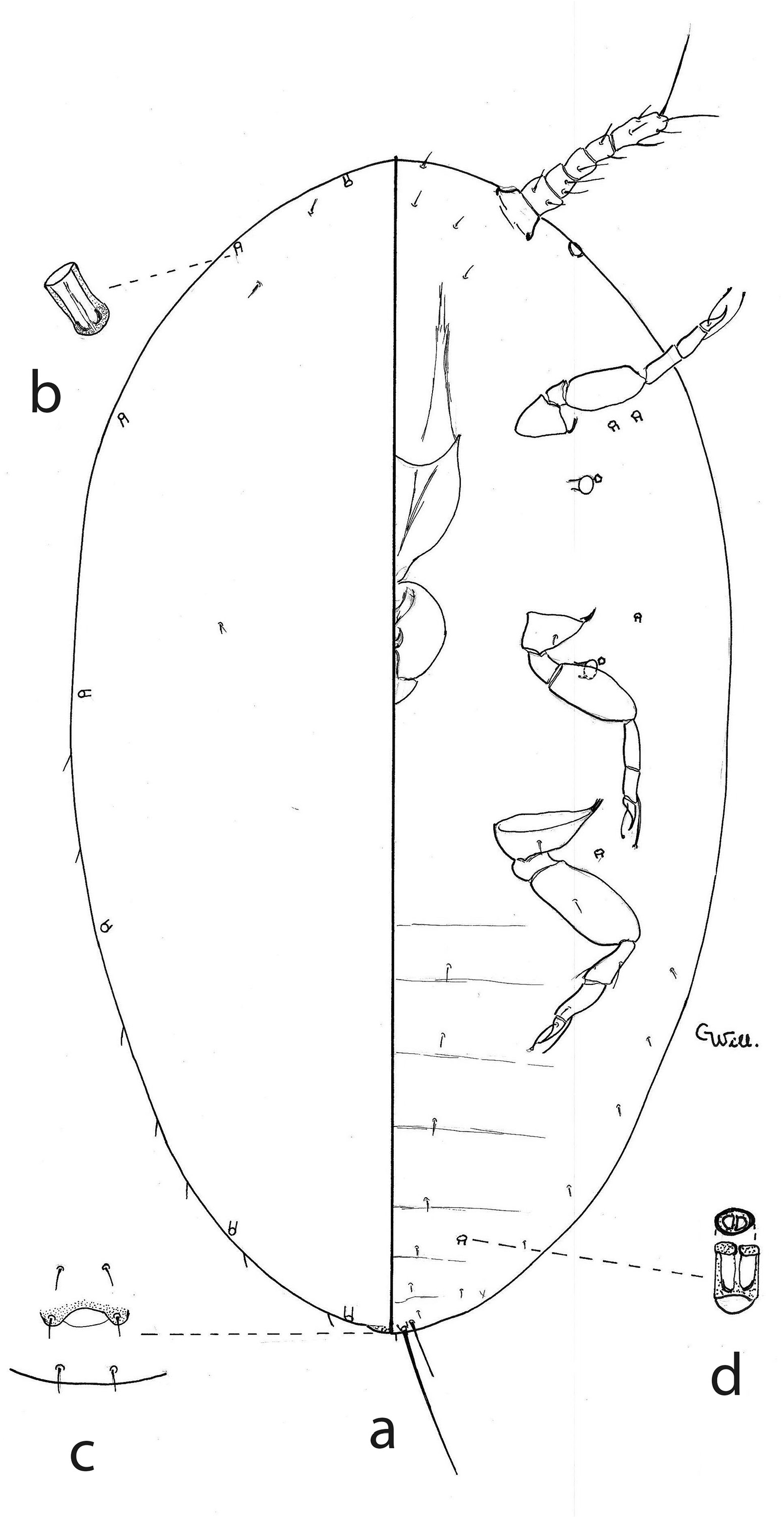
First instar nymph ( Fig. 9 View Figure 9 ). Oval body shape, 366 µm long and 148 µm wide. Dorsum: Eight-shaped pore ducts, short and wide, 7–12 μm long and 2.5–5 μm wide, marginal, one in the cephalic region, two on the thorax, and three on the abdomen. Anal lobes not developed, represented by a ventral seta, 36 µm long, an apical seta, 60 µm long, and three dorsal setae on each side of the anal ring. Venter: Antennae 60–67 μm long and 10 μm wide, with six segments. Eyes poorly developed. Labrum 67–79 µm long and 42–45 µm wide. Labium 31–36 µm long and 24–26 µm wide. Legs developed, metathoracic coxa 12–17 µm long, trochanter plus femur 33–56 µm long, tibia 12–14 µm long, tarsus 10–17 µm long, and claw 7 µm long. With thin setae with circular bases, submarginal and medial on abdominal segments, and four pairs between the antennae. With a quinquelocular pore close to each spiracle.
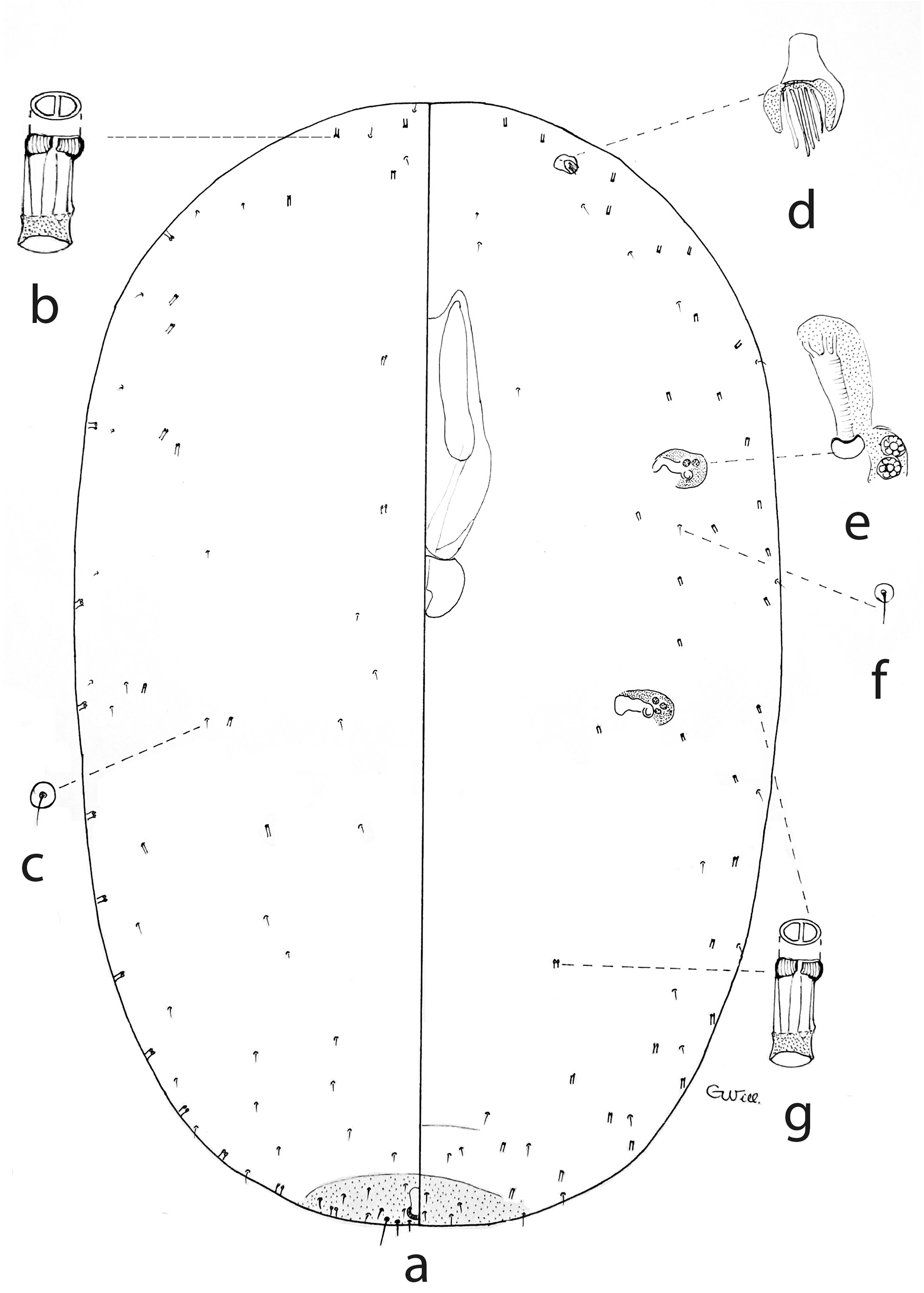
Second instar female nymph ( Fig. 10 View Figure 10 ). Oval body shape, 510–660 µm long and 346–420 µm wide, without legs. Dorsum: Small dorsal setae with a conical base, 2.5 µm wide at base and 7–10 µm long, on thorax, scattered between ducts. Marginal setae 12 µm long. With eight-shaped pore ducts, similar to those of the adult, 9–12 µm long and 5 µm wide, submarginal and lateral along body. Horseshoe-shaped anal ring, without setae. Undifferentiated anal lobes, represented by two setae, 5 µm and 10 µm long, respectively. Last abdominal segment slightly sclerotized. Anal opening 9.6–12 µm long. Venter: Antennae one-segmented, 12–16 µm long and 10–12 µm wide, with five fleshy setae. Spiracles 26–31 µm long and 9–12 µm wide. Clypeolabral shield 160–170 µm long and 84–86 µm wide. Labium 41–45 µm long and 57–60 µm wide. Setae rare, similar to dorsal ones. With 3–4 quinquelocular pores in anterior spiracles and 2–3 in posterior spiracles, respectively. Eight-shaped pore ducts rare, similar in length to the dorsal ones, scattered in submarginal area.
Comments. Sclerosococcus williamsi differs from the other four species in this genus by: 1) it lacks abdominal multilocular pores, 2) if the multilocular pores form a marginal band on both surfaces of the cephalothorax up to the metathoracic spiracles, then they are not arranged in groups. The host plant, D. longipetala , occurs in arid areas of Peru, Bolivia, and Argentina ( Tropicos 2023).
Etymology. The specific epithet was designated in honor of Dr. D.J. Williams of the Commonwealth Institute, for his great contribution to coccidology and for the trust he placed in the author of this species.
No known copyright restrictions apply. See Agosti, D., Egloff, W., 2009. Taxonomic information exchange and copyright: the Plazi approach. BMC Research Notes 2009, 2:53 for further explanation.