
Caligus spinosus Yamaguti, 1939
|
publication ID |
https://doi.org/10.5281/zenodo.195480 |
|
DOI |
https://doi.org/10.5281/zenodo.6206499 |
|
persistent identifier |
https://treatment.plazi.org/id/03BEA764-FFB9-FF99-FF2F-5C92FE20FA14 |
|
treatment provided by |
Plazi |
|
scientific name |
Caligus spinosus Yamaguti, 1939 |
| status |
|
Caligus spinosus Yamaguti, 1939
( Figs 1–3 View FIGURE 1 View FIGURE 2 View FIGURE 3 )
Caligus spinosus Yamaguti, 1939 , p. 445, pl. 14, figs. 4–8; Yamaguti & Yamasu 1960, p. 147, pl. 11, figs. 29–39; Izawa 1969, p. 127, figs. 1–20.
nec Caligus spinosus: Shiino 1960 , p. 476, figs. 4, 5; Pillai 1963, p. 76, fig. 6.
Material examined. All specimens examined were collected from the gills of amberjacks kept in aquaria of a seashore fish market in Kangnung (37°47΄44ʺN, 128°55΄0 8ʺE) located on the coast of the Sea of Japan: 2 females collected from Seriola quinqueradiata Temminck & Schlegel , by I.-H. Kim, 20 May 2001; 4 females and 2 males from S. quinqueradiata , by I.-H. Kim, 2 July 2001; 3 females (along with 1 female of C. aesopus ) from Seriola lalandi Valenciennes , by I.-H. Kim, 9 July 2001.
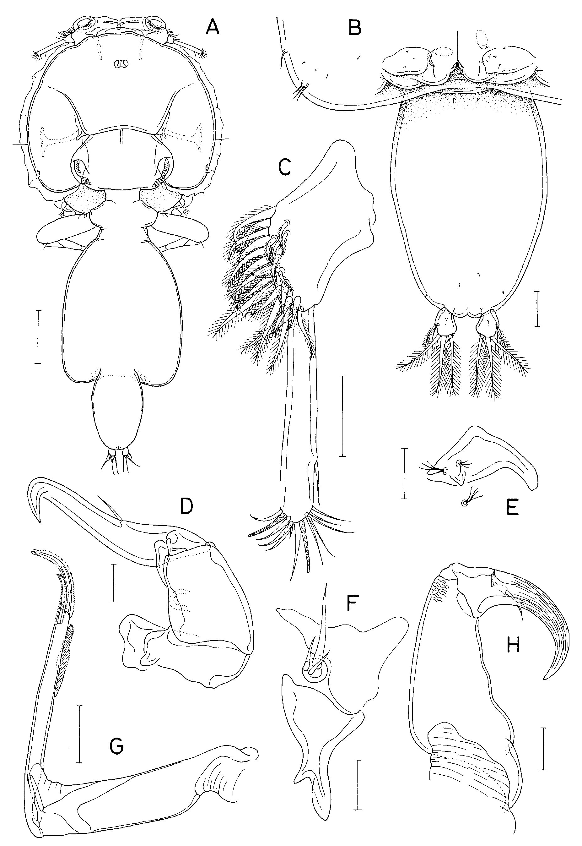
Description of female. Body ( Fig. 1 View FIGURE 1 A) 3.83 mm long. Cephalothoracic shield subcircular, 1.51 × 1.65 mm; lateral zone with T-shaped ventral rib; posterior sinus deep. Fourth pedigerous somite fused with genital complex. Genital complex gradually broadened distally, slightly truncated posteriorly, 1.35 × 1.08 mm, with rounded posterolateral corners. Abdomen ( Fig. 1 View FIGURE 1 B) 0.68 × 0.45 mm, 1-segmented, fusiform, with convex lateral margins. Caudal ramus 71 × 63 μm, slightly longer than wide, with 6 setae and 1 small dorsal setule.
Antennule ( Fig. 1 View FIGURE 1 C) 2-segmented; proximal segment with 25 pinnate and 2 naked setae; distal segment elongated, 1.3 times longer than proximal segment, with 1 naked subterminal seta on posterior margin and 11 naked setae and 2 aesthetascs distally. Antenna ( Fig. 1 View FIGURE 1 D) 3-segmented; first segment with small, tapering proximal process; second segment nearly quadrangular, with 1 adhesion pad; third segment forming long, distally strongly bent claw bearing 2 small setae. Postantennal process ( Fig. 1 View FIGURE 1 E) proximally bearing 1 small posterior subsidiary process and 2 papillae each tipped with 4 setules; another papilla located posterior to postantennal process also tipped with 4 setules.
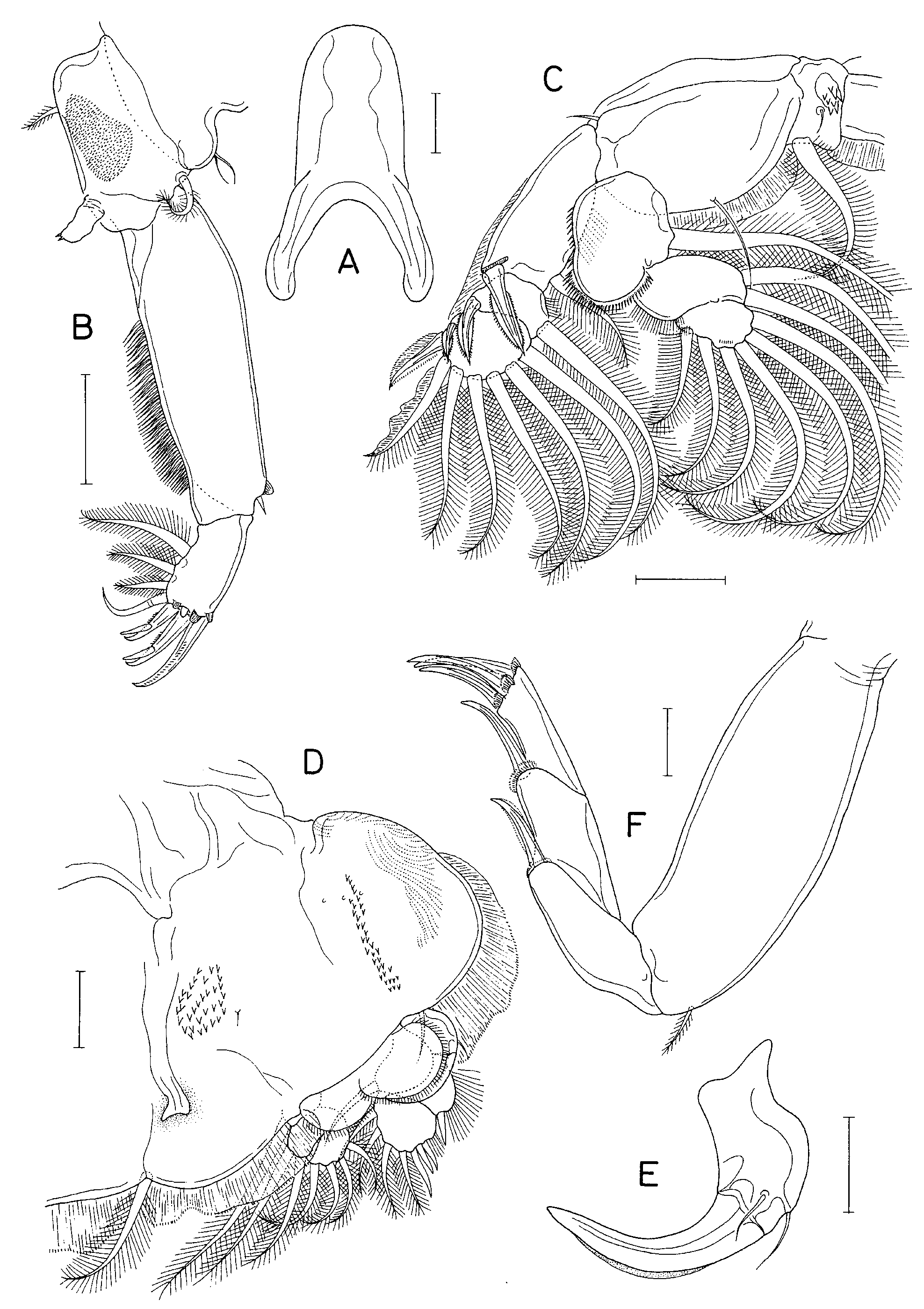
Mandible with 12 teeth distally. Maxillule ( Fig. 1 View FIGURE 1 F) consisting of anterior papilla bearing 3 unequal setae and posterior process bearing fusiform distal tine and smaller medial tine. Maxilla ( Fig. 1 View FIGURE 1 G) 2-segmented; proximal segment (lacertus) unarmed; slender distal segment (brachium) with large subdistal membrane (flabellum) on inner margin; calamus about 1.7 times longer than canna. Maxilliped ( Fig. 1 View FIGURE 1 H) 3-segmented; first segment (corpus) gradually narrowed distally, with uneven inner margin; second segment (shaft) short, with 1 distal seta; third segment almost fused with second, forming strongly curved claw with longitudinal surface striations. Sternal furca ( Fig. 2 View FIGURE 2 A) with slender, slightly incurved tines bearing blunt tips.
Armature on rami of legs 1–4 as follows:
Leg 1: exopod 1-0; III,1,3; endopod (vestigial)
Leg 2: exopod I-1; I-1; II,I,5; endopod 0-1; 0-2; 6
Leg 3: exopod I-0; I-1; III,4; endopod 0-1; 6
Leg 4: exopod I-0; I-0; III; endopod (lacking)
Leg 1 ( Fig. 2 View FIGURE 2 B) coxa with branched outer setule; basis with pinnate outer seta, smaller pinnate inner seta, and patch of numerous minute spinules on ventral surface. Proximal exopodal segment elongate, with 1 small outer distal naked seta and row of setules on inner margin; distal segment with digitiform process on distal margin; three distal spines each accompanied by flabelliform membrane; two inner distal spines bifurcating at about their midlength; distal seta longer than spines and naked; endopod flexible and tipped with 2 small processes. Leg 2 ( Fig. 2 View FIGURE 2 C) coxa with large seta on inner posterior margin, 1 patch of spinules and 1 setule on ventral surface; basis with small outer seta and 1 inner setule and membrane on inner part of posterior margin; first endopodal segment expanded posterolaterally, with spinules along outer margin; outer side of basis and first exopodal segment with broad membrane (not illustrated in Fig. 2 View FIGURE 2 C). Leg 3 ( Fig. 2 View FIGURE 2 D) protopod (apron) with adhesion pads and broad membrane on outer margin, longitudinal patch of spinules on mid-ventral surface, and patch of 25–34 spinules on inner ventral surface; spine on first exopodal segment ( Fig. 2 View FIGURE 2 E) enlarged and strongly curved; distal endopodal segment partially subdivided. Leg 4 ( Fig. 2 View FIGURE 2 F) protopod moderately expanded, with small outer distal seta; spines on first and second exopodal segments 127 and 165 μm, respectively; three spines on terminal segment 135, 156, and 146 μm from outer to inner; all spines on exopodal segments accompanied with flabelliform membranes near base. Leg 5 ( Fig. 1 View FIGURE 1 B) represented by 1 and 3 small setae on posterolateral margin of genital complex.
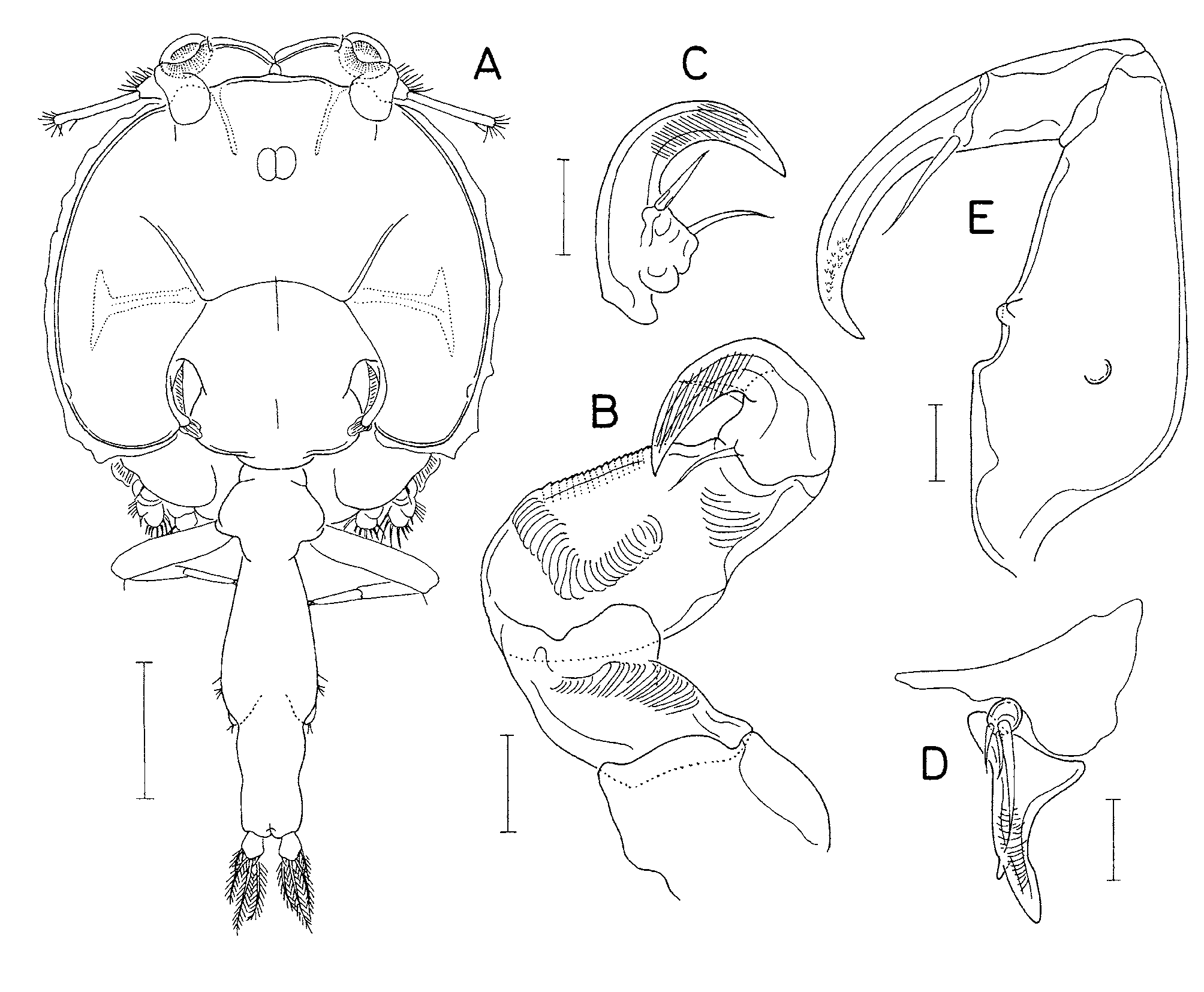
Male. Body ( Fig. 3 View FIGURE 3 A) 3.01 mm long. Cephalic shield resembles that of female. Genital complex completely fused with abdomen to form elongate genito-abdomen of 972 × 339 μm (2.87:1, excluding caudal rami). Caudal ramus 83 × 81 μm, with setules on inner margin (not figured).
Antennule armed as in female, but distal segment relatively longer than that of female. Antenna ( Fig. 3 View FIGURE 3 B) 3-segmented as in female; first segment with 1 adhesion pad; second segment with 3 adhesion pads; third segment with 2 inner proximal setae and forming strongly curved, large claw ( Fig. 3 View FIGURE 3 C). Postantennal process more slender than that of female.
Mandible and maxilla as in female. Maxillule ( Fig. 3 View FIGURE 3 D) with adhesion pad on ventral surface of posterior process. Maxilliped ( Fig. 3 View FIGURE 3 E) with 1 ventral and 3 inner small tubercles on first segment; claw with small denticles on distal part. Sternal furca with tines more slender than that of female.
Legs 1–5 as in female. Leg 6 ( Fig. 3 View FIGURE 3 A) represented by 2 small setae on each posterolateral corner of genital complex.
Hosts and distribution. Seriola quinqueradiata in Japan and Korea, and S. lalandi in Korea.
Remarks. We examined the type specimens of C. spinosus loaned from the Meguro Parasitological Museum, Tokyo. Yamaguti (1939) referred to these type specimens as “one immature and four mature specimens”, but we confirmed that they consist of four adult females and one adult male mounted on two slides. Although the specimens were mounted, they showed several characteristic features of C. spinosus : the rounded posterolateral corners of the genital double somite in the female, the absence of a lateral constriction on the abdomen, a patch of more than 20 spinules on the protopod of leg 3, and the similar sizes of the terminal spines of leg 4.
No known copyright restrictions apply. See Agosti, D., Egloff, W., 2009. Taxonomic information exchange and copyright: the Plazi approach. BMC Research Notes 2009, 2:53 for further explanation.
|
Kingdom |
|
|
Phylum |
|
|
Class |
|
|
Order |
|
|
Family |
|
|
Genus |
Caligus spinosus Yamaguti, 1939
| Choe, Mi-Kyung & Kim, Il-Hoi 2010 |
Caligus spinosus :
| Shiino 1960 |
Caligus spinosus
| Yamaguti 1939 |