
Sinodiaptomus sarsi ( Rylov, 1923 )
|
publication ID |
https://doi.org/10.15407/zoo2021.01.001 |
|
DOI |
https://doi.org/10.5281/zenodo.6422564 |
|
persistent identifier |
https://treatment.plazi.org/id/03BDDA77-8E5C-781E-8087-FE75FB4AD842 |
|
treatment provided by |
Felipe |
|
scientific name |
Sinodiaptomus sarsi ( Rylov, 1923 ) |
| status |
|
Sinodiaptomus sarsi ( Rylov, 1923)
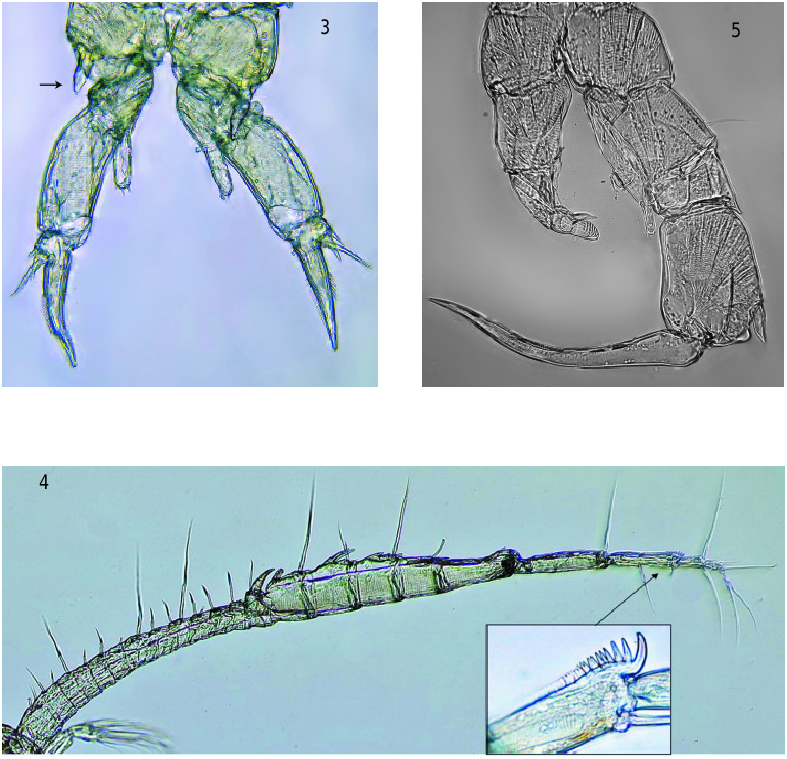
R e d e s c r i p t i o n. Female ( FIg. 1 View Figs 1–5 , A). Body length (excluding furcal setae) 2.05–2.49 mm (2.24 ± 0.13 mm, n = 15), prosome length 1.45–1.75 mm (1.58 ± 0.09 mm), prosome width 0.51 – 0.65 mm (0.58 ± 0.04 mm). The dorsal surface of last prosomal somite with a chitinous, triangular projection ( FIg. 1 View Figs 1–5 , A), which is a diagnostic character of the genus Sinodiaptomus ( Kiefer, 1932) . This somite with moderately developed wings. Left wing twolobed with inner one round. Right wing relatively pointed. Both lobes with strong hyaline spine ( FIg. 2 View Figs 1–5 ). Urosome with three somites, genital somite slightly asymmetrical, with a moderately long hyaline spine on each side, the intermediate somite short. Antennules extending beyond the caudal setae ( FIg. 1 View Figs 1–5 , A). Сoxopodite of leg 5 with long, strong, distolateral projection ( FIg. 3 View Figs 1–5 , arrow).
Male ( FIg. 1 View Figs 1–5 , B). Body length 1.9–1.95 mm (1.91 ± 0.02 mm, n = 20), prosome length 1.20–1.32 mm (1.25 ± 0.04 mm), prosome width 0.45–0.48 mm (0.46 ± 0.02 mm). Urosome has a lateral bend to the right. Antennules reach the end of caudal rami. Right antennule geniculated between segments 18 and 19; segment 15 ( FIg. 4 View Figs 1–5 ) with longer projection than that of segment 14; antepenultimate segment of right male antennule with a comb-like process ( FIg. 4 View Figs 1–5 ). Basipodite of right leg 5 with a wide round process reaching almost the distal end of the exopodite I and with a hyaline lamella on its distal inner margin ( FIg. 5 View Figs 1–5 ). Exopodite II of left leg 5 ending in a lamellate thumb-like process with short posterodistal basal projection.
No known copyright restrictions apply. See Agosti, D., Egloff, W., 2009. Taxonomic information exchange and copyright: the Plazi approach. BMC Research Notes 2009, 2:53 for further explanation.
|
Kingdom |
|
|
Phylum |
|
|
Class |
|
|
Order |
|
|
Family |
|
|
Genus |