
Fidelidiella pectinata, Jaume & Gràcia & Boxshall, 2007
|
publication ID |
https://doi.org/10.1080/00222930701228835 |
|
persistent identifier |
https://treatment.plazi.org/id/03BD87FC-706E-FFCF-FE68-FC820A10FE50 |
|
treatment provided by |
Felipe |
|
scientific name |
Fidelidiella pectinata |
| status |
sp. nov. |
Fidelidiella pectinata View in CoL sp. nov.
( Figures 1–7 View Figure 1 View Figure 2 View Figure 3 View Figure 4 View Figure 5 View Figure 6 View Figure 7 )
Material examined
Littoral cave ( sensu Stock et al. 1986) at the base of Ihnig cliffs, Tingeting Tribu, NW Lifou ( Loyalty Islands). UTM coordinates (Datum WGS84): 7706536/58 736772. Single chamber roughly 10× 3 m, entirely occupied by lake about 0.5 m deep subject to strong swell. Hyperbenthic haul with hand-held net above coral rubble after stirring up bottom with feet. Holotype: 1.76 mm, sex unknown, completely dissected and mounted on four slides (reg. no. MNHN-Am7459). Paratype: 2.11 mm, sex unknown, pereopods 6 and 7 missing; partially dissected in ethanol vial, internal tissue preserved (i.e. not treated with hot lactic acid) ( BMNH reg. no. 2006.1125). Collected by authors, 28 October 2000 .
Description of holotype
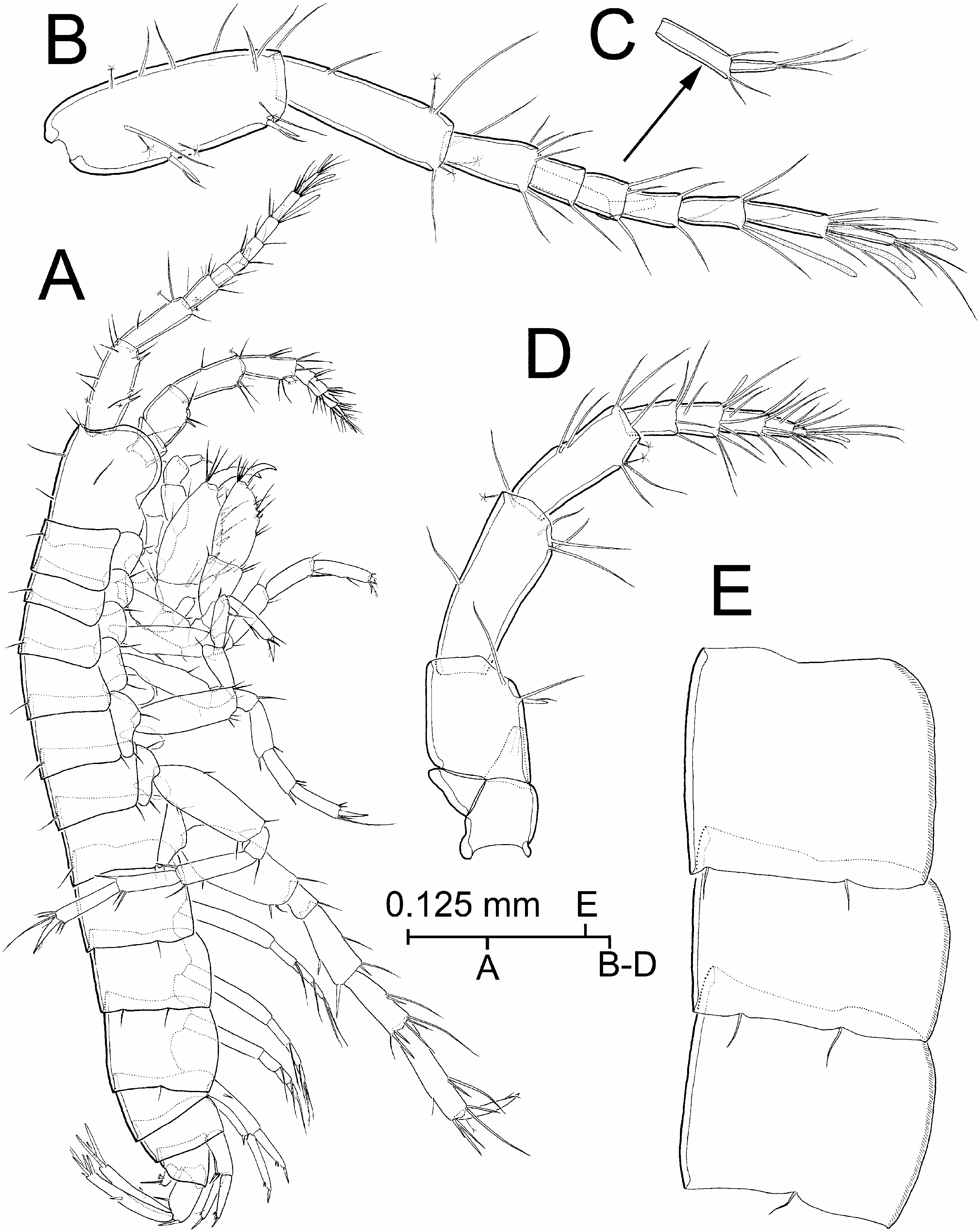
Head ( Figure 1A View Figure 1 ) longer than wide, with hardly developed rostrum, evenly rounded lateral lobe and no trace of post-antennal sinus (terms sensu Lincoln 1979, p 15); eyes absent. Body unpigmented, smooth, with sparsely set long sensillae distributed as figured over tergites. Slender seta present at posteroventral corner of each of fifth to seventh pereonites. Epimeral plates with small but distinct posterodistal corners and with 2-2-1 setae on posterior margin, respectively ( Figure 1E View Figure 1 ); ventral margin of plates fringed with microspinules.
Antennule ( Figure 1B View Figure 1 ) about 35% of body length. Proximal peduncle segment twice as long as wide, with two strong flagellate spines on posterior margin. Second peduncle segment three times longer than wide, 78% length of proximal segment. Third peduncle segment 51% length of preceding segment. Main flagellum shorter than peduncle, sixarticulate, with slender aesthetasc on each of articles 4, 5, and 6. Accessory flagellum ( Figure 1C View Figure 1 ) two-articulate, not reaching distal margin of third article of main flagellum; proximal article twice as long as distal.
Antenna ( Figure 1D View Figure 1 ) about 80% length of antennule. Fourth peduncle segment longest, 2.7 times longer than wide. Fifth peduncle segment 71% length of preceding segment, 2.5 times longer than wide. Flagellum slightly shorter than third peduncle segment, fivearticulate; tiny aesthetasc on articles 2 and 5.
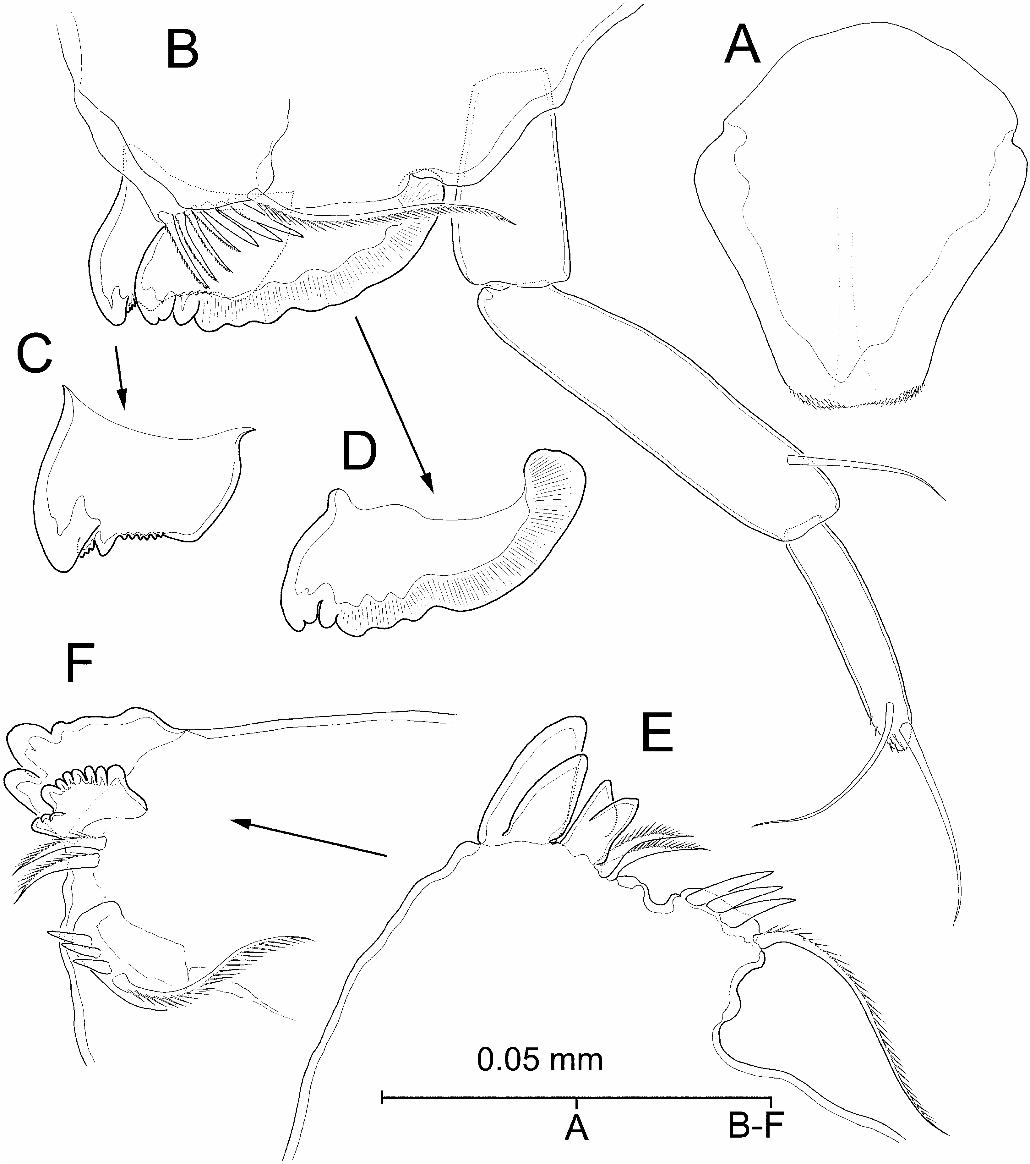
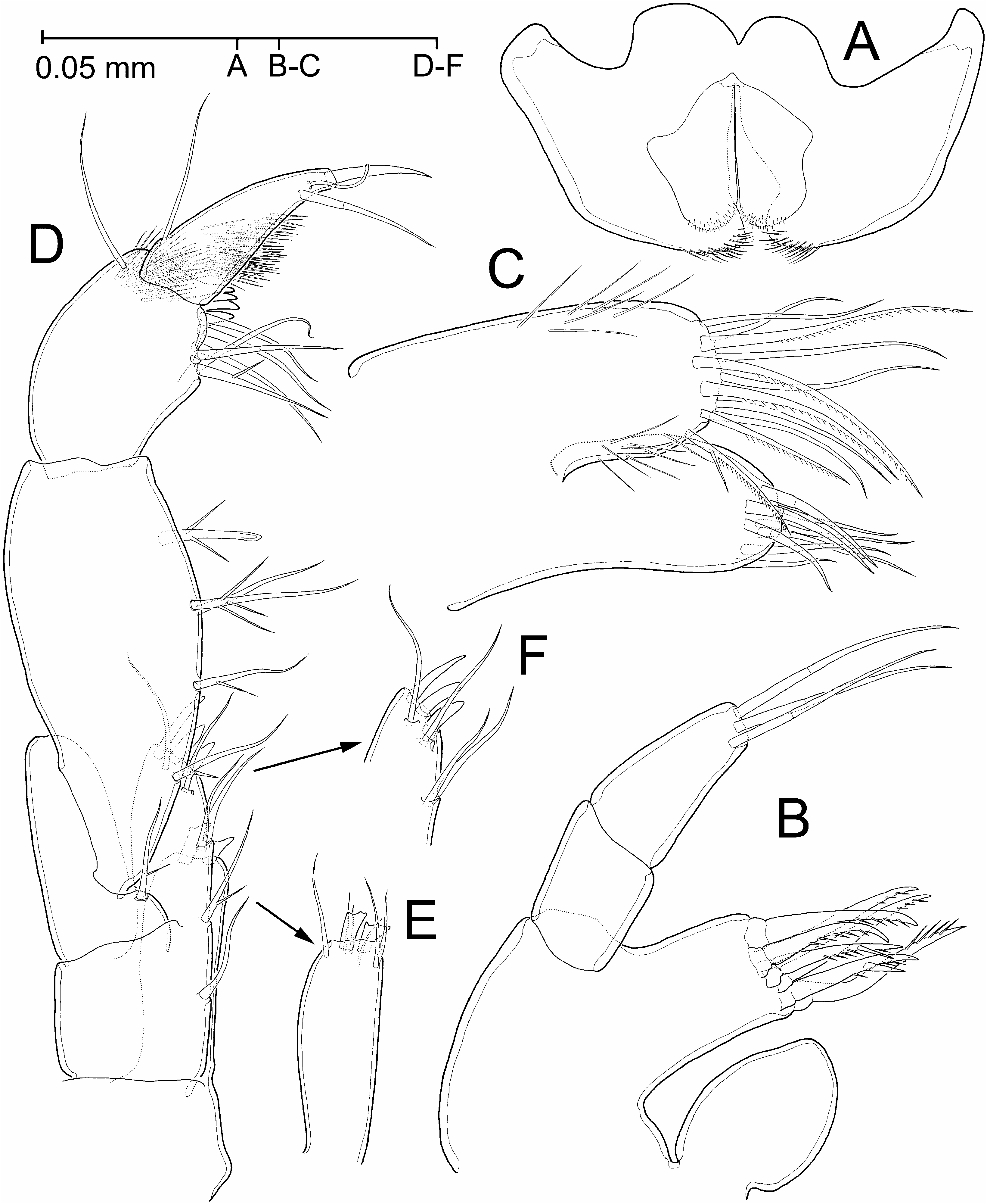
Labrum ( Figure 5A View Figure 5 ) trapezoid, slightly constricted at two-thirds of total length, with straight, hardly setulose distal margin. Paragnaths ( Figure 6A View Figure 6 ) bilobed, outer lobe with long setules distally on inner margin; inner lobe with sparsely set short setules distally.
Left mandible ( Figure 5B View Figure 5 ) with subrectangular incisor bearing three major unequal denticles, innermost largest, plus two intercalated serrate portions ( Figure 5C View Figure 5 ). Lacinia hypertrophied, wider than long, lateral half of distal margin smooth, inner half with six unequal rounded denticles ( Figure 5D View Figure 5 ). Spine row composed of four short, stiff unipinnate elements. Molar non-triturative, reduced to narrow lappet crowned with three lanceolate denticles plus long unipinnate molar seta. Palp three-segmented, second segment with subdistal seta, third segment with two (apical and subapical) setae, and patch of short spinules on tip.
Right mandible ( Figure 5E, F View Figure 5 ) differing from left counterpart in reduced spine row (comprising two spines only) and morphology of incisor and lacinia. Former indistinctly five-denticulate; latter reduced, not expanded laterally, with eight distal denticles, inner six subequal and rounded, outer two larger, outermost tricuspidate.
Maxillules ( Figure 6B View Figure 6 ) symmetrical, with two-segmented endopod (5palp), distal segment bearing two distal and one distolateral setae. Coxal endite (5inner lobe) ovoid and unarmed. Basal endite (5outer lobe) with seven unipinnate spines distally, two of them bearing hypertrophied pinnule.
Maxilla ( Figure 6C View Figure 6 ) somewhat reduced, with coxal endite (5inner lobe) slightly longer than basal endite (5outer lobe). Coxal endite with eight setae; basal endite with 11 setae distally, innermost seta with hypertrophied pinnule proximally on margin.
Maxilliped ( Figure 6D View Figure 6 ) with short endites. Distal margin of basal endite (5inner lobe) with two stout spines expanded distally, innermost bicuspidate, outermost tricuspidate, and six subdistal setae ( Figure 6E View Figure 6 ). Endite of ischium (5outer lobe) with three unequal spines distally and two setae subdistally on posterior surface, and four setae along inner margin ( Figure 6D, F View Figure 6 ); one of marginal setae with hypertrophied pinnule on medial margin. Merus (5proximal segment of four-segmented palp) strongly oblique, distomedial corner bearing seta with hypertrophied pinnule. Carpus (5palp segment 2) with five setae along medial margin, each with one or two hypertrophied pinnules proximally on margin(s), distalmost seta with expanded tip. Propodus (5palp segment 3) with six distomedial setae (one with hypertrophied pinnule) and single distolateral seta; four strong rounded processes on anterior side of distomedial corner; tuft of long spinules on anterior side of distolateral corner. Dactylus with one proximolateral seta; two unequal setae, one long and stiff, other slender, subdistally on medial margin; tuft of long spinules on anterior surface of segment. Unguis slender, about half length of preceding segment.
Coxal plates ( Figure 1A View Figure 1 ) wider than long, plates 2–5 each with anterior margin overlapping one in front, plates 5–6 with posterior margin overlapping one to rear. Coxal gills apparently unstalked, on pereopods 4–6 ( Figures 1A View Figure 1 , 7B–D View Figure 7 ).
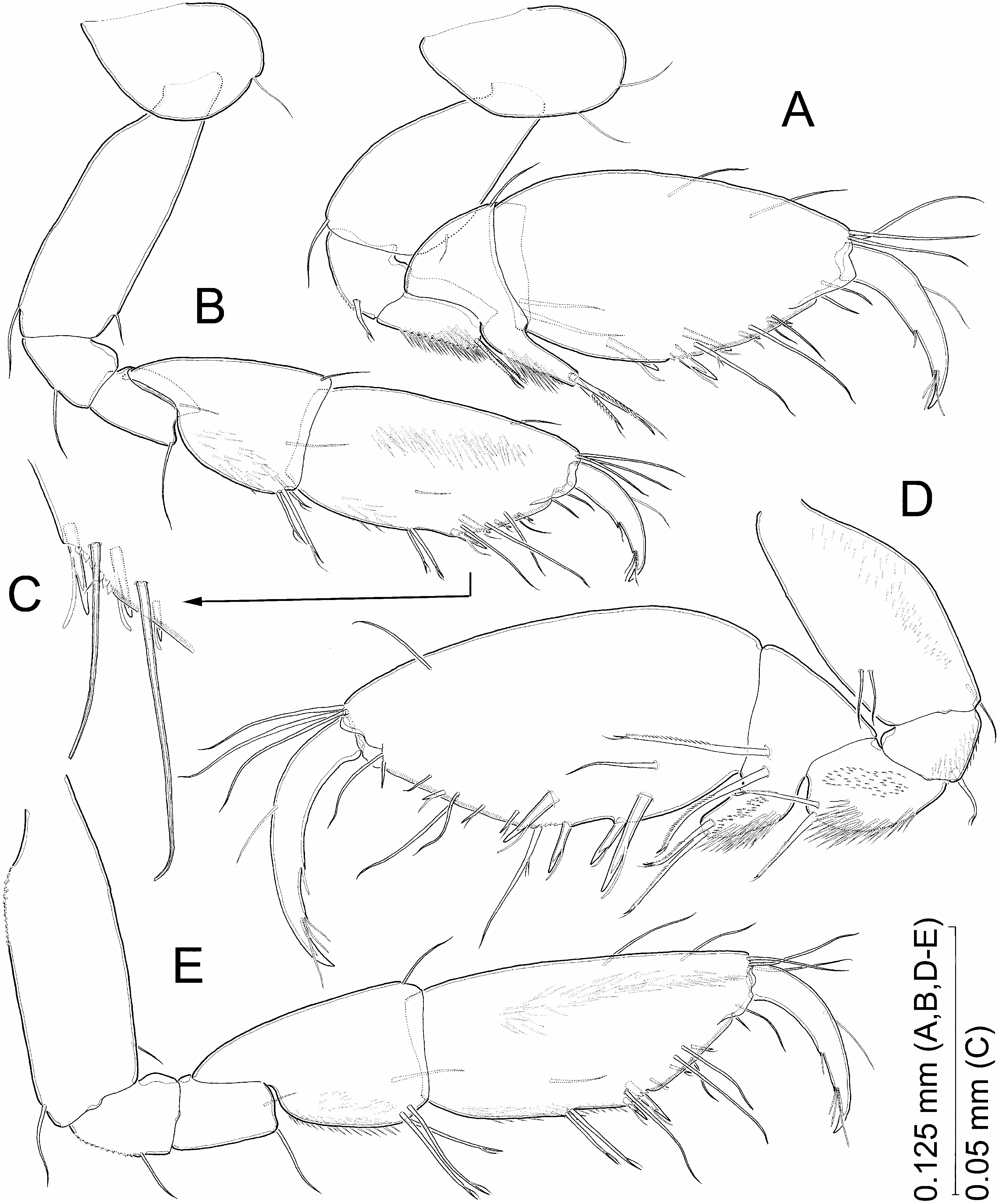
Coxa of first gnathopod ( Figure 3A View Figure 3 ) roughly trapezoid, about 1.7 times wider than long, with evenly rounded anterior margin and two setae close to anterodistal corner. Basis short and stout, about 1.8 times longer than wide. Merus with posteromedial surface densely spinulose. Carpus posterodistal corner produced into digitiform process crowned with two pinnate flagellate spines; posteromedial surface of segment with patch of long spinules. Propodus larger than second gnathopod counterpart, ellipsoidal, about 1.9 times longer than wide, with palm angle placed at about 54% of maximum length of segment. Two stout flagellate spines sparsely set around palm angle on medial surface of segment, plus submarginal spine about midway along posterior margin, also on medial surface of segment. Palm margin convex, hyaline, smooth except for short row of tiny serrations near palm angle; two short flagellate spines on medial side of margin, and seta with two minute pinnae on lateral side. Nail short, not reaching palm angle, with two indentations along posterior margin harbouring one and three short setae, respectively.
Coxa of second gnathopod ( Figure 3B View Figure 3 ) roughly ovoid, about 1.5 times wider than long, with seta on anterodistal corner. Basis longer and proportionally more slender than in gnathopod 1 (about 2.9 times longer than wide and 1.4 times longer than basis of first gnathopod). Merus with hardly developed posterodistal lobe. Carpus about 1.6 times longer than wide, with subparallel anterior and posterior margins, and with patch of long spinules on posteromedial surface. Propodus 1.9 times longer than wide, with subparallel anterior and posterior margins, 1.3 times longer than carpus; posterior margin with two slender bifid spines at about two-thirds of length of margin. Palm angle placed at 61% maximum length of segment, with two strong bifid spines submarginally on medial surface of segment (see Figure 3C View Figure 3 ). Palm margin hyaline, smooth except for proximal serrate portion (see Figure 3C View Figure 3 ); armature comprising three short flagellate spines along medial side. Medial surface of propodus with longitudinal patch of long spinules close to anterior margin, and with patch of sparsely set spinules near posterior margin. Nail as in gnathopod 1.
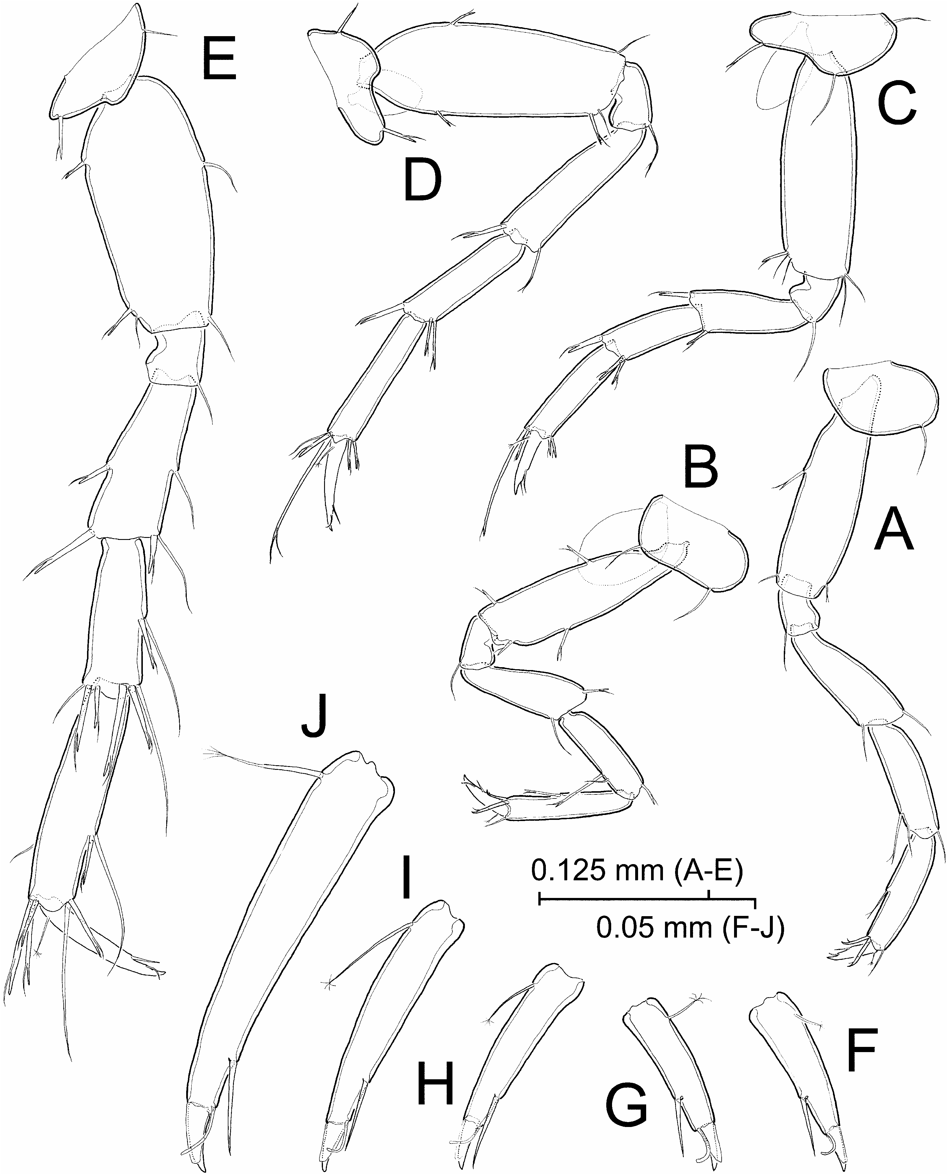
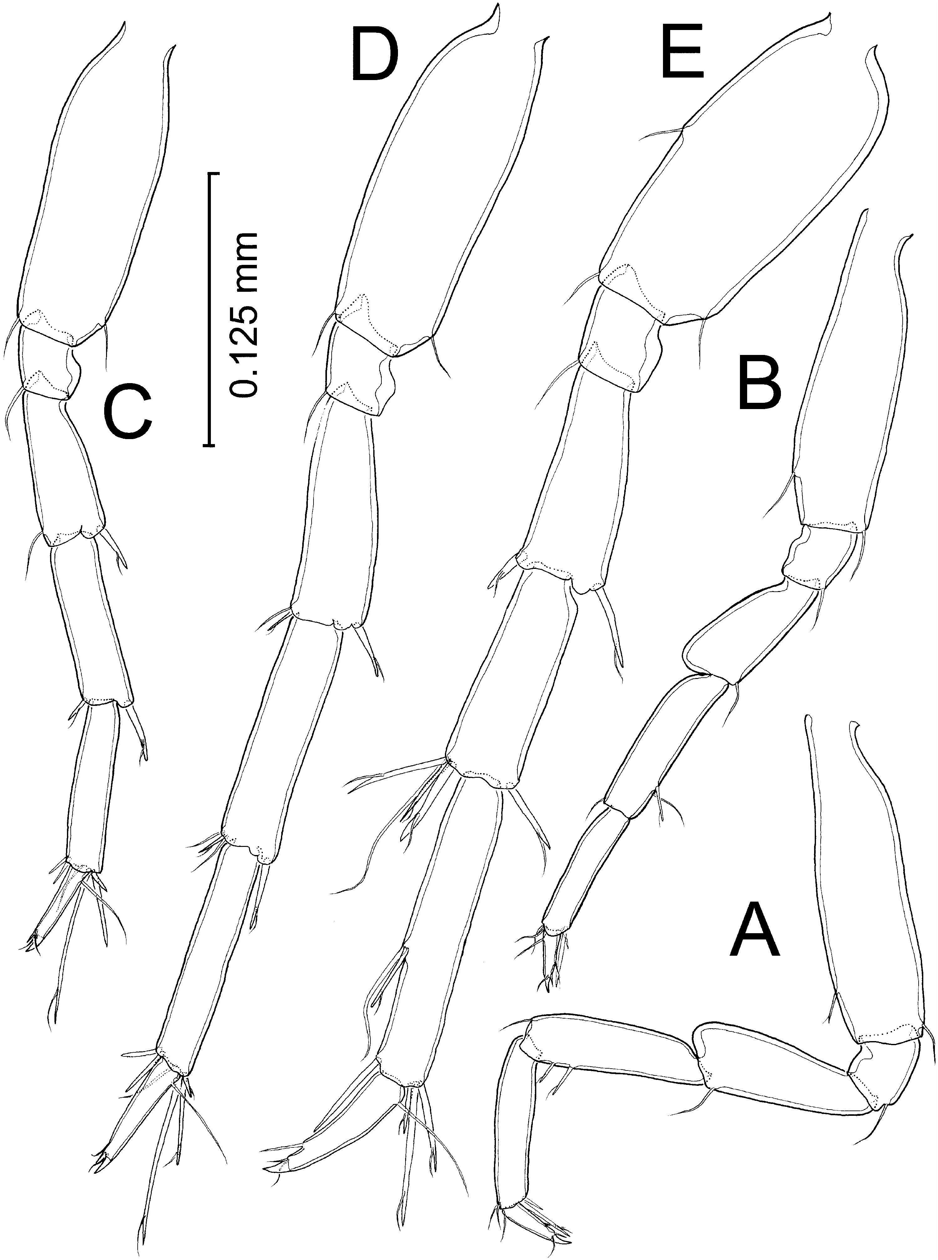
Pereopods 3–5 with narrow bases, those of pereopods 6–7 slightly expanded proximally. Pereopod 3 ( Figure 7A View Figure 7 ) with coxa roughly ovoid, about twice as wide as long, with evenly rounded anterior margin and slightly convex distal margin; seta placed on anterodistal corner of plate. Two stout recurved flagellate spines on posterodistal corner of propodus.
Coxa of pereopod 4 ( Figure 7B View Figure 7 ) roughly rectangular, twice as wide as long, with evenly rounded anterior margin and convex posterior margin; seta present at antero- and posterodistal corner of plate. Rest of limb about similar to preceding pereopod except for shorter length (attaining only 92.4% of third pereopod length, mainly due to shorter basis, merus, and carpus).
Pereopod 5 ( Figure 7C View Figure 7 ) as long as preceding limb. Coxa with proximal seta implanted submarginally near anterior margin on medial surface, distal seta on anterior lobe, and flagellate spine on posterodistal corner. Stout flagellate spine(s) at antero- and posterodistal corner of merus, carpus, and propodus; one of spines on anterodistal corner of propodus elongate, widely surpassing nail tip.
Pereopod 6 ( Figure 7D View Figure 7 ) about 1.3 times longer than preceding limb. Coxa with slender flagellate spine on anterior margin and at distolateral corner. Armature of rest of pereopod similar to preceding limb.
Pereopod 7 ( Figure 7E View Figure 7 ) elongated, about 1.2 times longer, and bearing more spines than preceding pereopod.
Relative length of nail (dactylus + unguis) of pereopods 3–7 as follows: 43:41:51:67:100 (see Figure 7F–J View Figure 7 ). Each with part corresponding to dactylus bearing stiff seta subdistally and slender seta distally on posterior margin, and with penicillate seta proximally on anterior margin. Unguis part almost completely covered anteriorly by hyaline scar. Pereopods lacking any trace of lenticular (5Hertzog’s) organs.
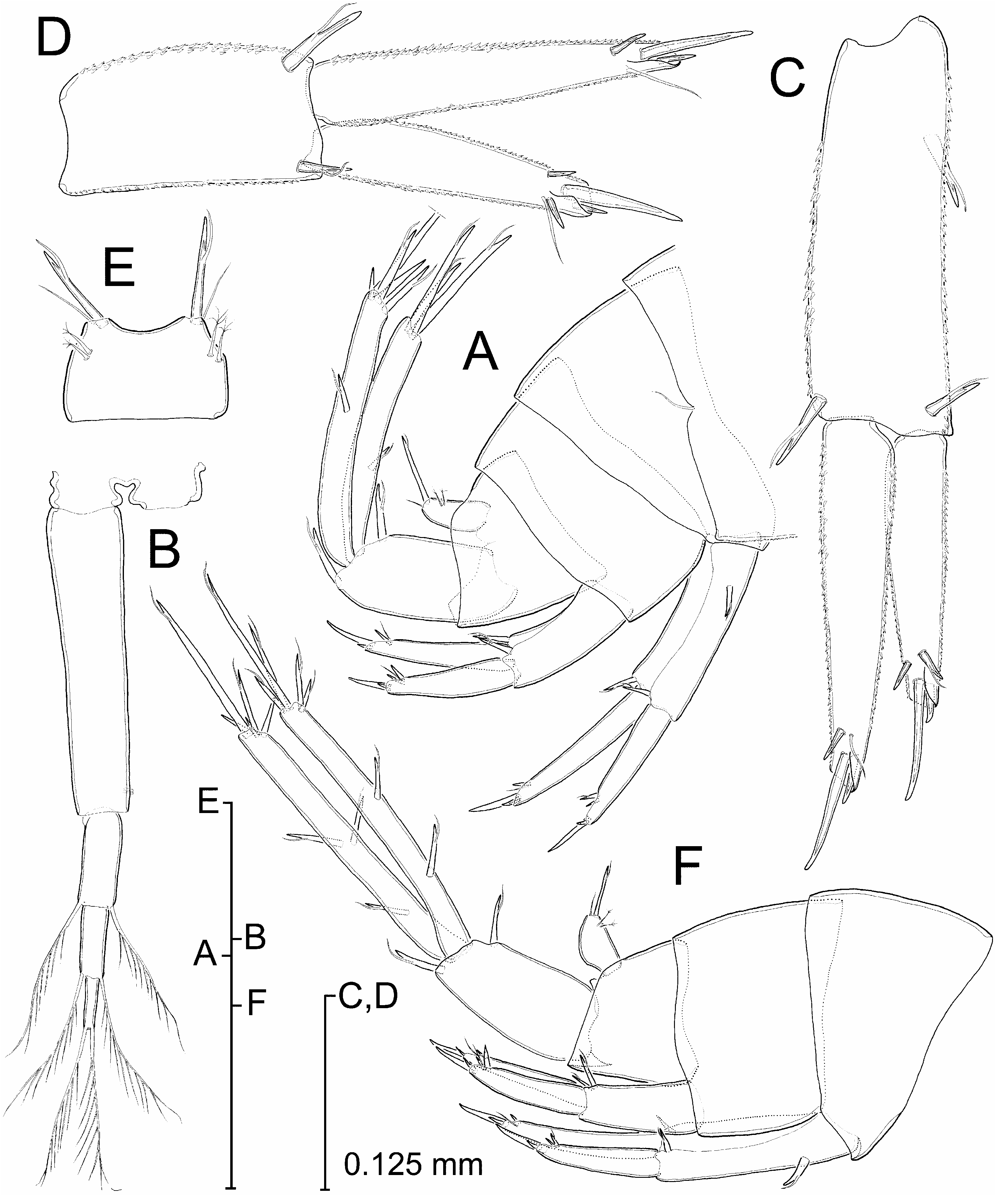
Pleopods 1–3 ( Figures 1A View Figure 1 , 4B View Figure 4 ) sub-similar, uniramous, lacking secondary sexual characters. Protopod with two retinacles subdistally on medial margin. Presumed exopod three-articulate, with two plumose setae per article. Third pleopod with exopodal articles comparatively shorter and thicker than counterparts of preceding pleopods.
Uropod 1 ( Figure 4A, C View Figure 4 ) biramous, exopod shorter than endopod, both shorter than protopod (67 and 90% of protopod length, respectively). Protopod elongate, about three times longer than wide, with basofacial (5proximolateral) spine, and with flagellate spine on dorsolateral (5posterolateral) and dorsomedial (5posteromedial) corner. Exopod with two distal spines (inner elongate) and one subdistal spine dorsomarginally on each side, outer flagellate, inner simple. Endopod with same armature as exopod except for outer subdistal spine substituted here by slender simple seta. Lateral and medial margins of protopod and rami covered with short denticles.
Uropod 2 ( Figure 4A, D View Figure 4 ) protopod with flagellate spine on dorsolateral and dorsomedial corner. Exopod attaining 80% of endopod length, being slightly longer than protopod. Two distal spines (inner elongate) and one subdistal spine arrayed dorsomarginally at each side; slender simple seta near insertion of outer subdistal spine. Endopod with same armature as exopod except for lack of outer subdistal spine. Lateral and medial margins of protopod and rami covered with short denticles.
Uropod 3 ( Figure 4A View Figure 4 ) long, with unisegmented rami, endopod 89% length of exopod. Protopod 65% length of exopod, with stout flagellate spine on dorsolateral and dorsomedial corner. Exopod with flagellate spine at about 57% of distance along lateral margin and with five apical spines, three of them flagellate, other two simple. Endopod with flagellate spine at 39% of distance along medial margin and with three flagellate spines on tip.
Telson ( Figure 4E View Figure 4 ) wider than long, with distal margin shallowly excavate. Long bifid spine and slender simple seta at each distal corner, plus two tiny penicillate setae at each lateral margin.
Description of paratype
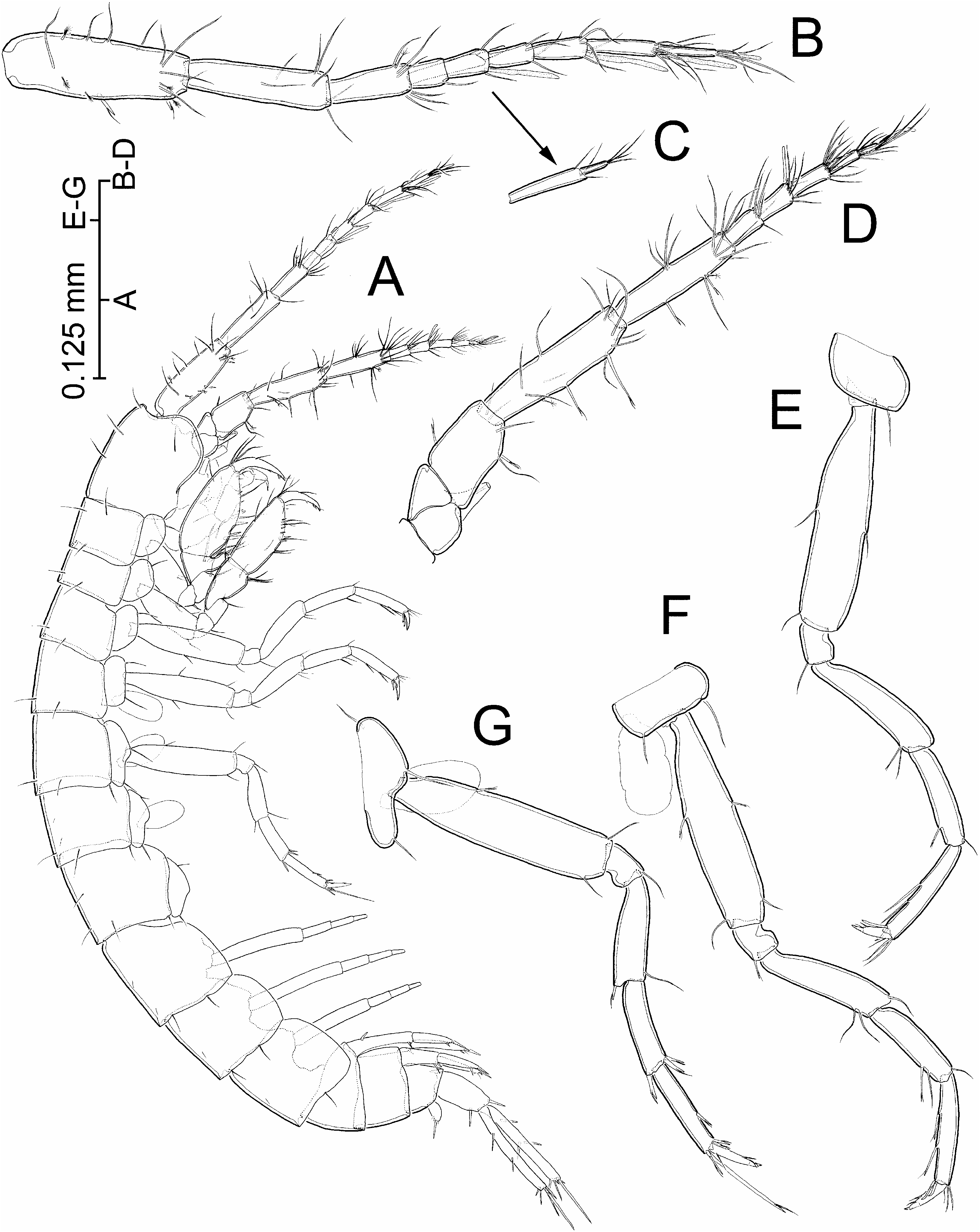
Body about 20% larger than holotype, with pereopods 6 and 7 missing ( Figure 2A View Figure 2 ). Differing from holotype as follows: (1) aesthetasc present on second article of main flagellum of antennule ( Figure 2B View Figure 2 ; aesthetasc apparently absent in holotype); (2) fourth and fifth peduncle segments of antenna proportionally more slender and elongate (4.2 and 4.4 times longer than wide, respectively; fifth segment 84% length of fourth segment; see Figure 2C View Figure 2 ; corresponding values for holotype 2.7, 2.5, and 71%, respectively); (3) first gnathopod with one additional spine on posterior margin of propodus, and additional short flagellate spine on palm margin (compare Figure 3D and 3A View Figure 3 ); (4) propodus of second gnathopod ( Figure 3E View Figure 3 ) proportionally longer (2.3 times longer than wide versus 1.9 in holotype), with slightly more spinulose posterior margin, and with one additional short flagellate spine on palm margin; (5) propodus of third and fourth pereopods with two spines on posterior margin ( Figure 2E, F View Figure 2 ; only one spine present in holotype); (6) rami of third uropods ( Figure 4F View Figure 4 ) differing in number of armature elements: two flagellate spines on outer margin of exopod (only one in holotype); three flagellate spines on inner margin of endopod, two of them inserted close together (versus only one in holotype); and five terminal spines on endopod (only three in holotype).
Etymology
Species name derived from the Latin pecten (5comb), and referring to the peculiar row of stout rounded processes that the new taxon displays anteriorly on the distomedial corner of the fourth segment of the maxillipedal palp.
Variability
The paratype of Fidelidiella pectinata gen. et sp. nov. differs from the holotype in several respects, most probably linked to differences in body size (namely the marginal armature of rami of the third uropods; see Hovenkamp et al. 1983 for a similar situation described in Bogidiella cyrnensis Hovenkamp, Hovenkamp and van der Heide, 1983 ). Since both specimens share remarkable features, such as the peculiar mouthparts mentioned in the generic diagnosis, plus other characters such as the presence of a seta anteriorly on the inner surface of the coxa of the fifth pereopod, an elongate slender flagellate spine on the propodus of the same pereopod, and a seta with two minute pinnae on the palm margin of the first gnathopod (see Figure 3A, D View Figure 3 ), we tentatively consider both specimens to be conspecific.
Remarks
The new taxon from Lifou conforms precisely to the restricted concept of the Bogidiellidae as introduced by Koenemann and Holsinger (1999, p 784). With regard to its generic assignment, neither of the two specimens collected displays penile papillae, oostegites, or any secondary sexual characters on the pleopods and/or uropods permitting their unequivocal sexing. This could represent a taxonomic handicap since many bogidiellid genera are defined on the basis of secondary sexually dimorphic characters only ( Stock 1981; Koenemann and Holsinger 1999). Nevertheless, the new taxon shows a combination of non-sexually dimorphic characters shared only by members of three out of the 36 genera comprising the family (see Koenemann and Holsinger 1999, Table I, Appendix B): (1) Actogidiella Stock, 1981 , a monotypic genus from the marine interstitial medium of the British Virgin Islands ( Stock 1981); (2) Bogidiella Hertzog, 1933 of the so-called ‘‘ albertimagni -group’’, or ‘‘group A’’ ( sensu Koenemann and Holsinger 1999), a cluster of 14 Palaearctic species with a single outlier in South America; and (3) Bogidomma Bradbury and Williams, 1996 , a monotypic genus from inland groundwaters of Barrow Island, NW Australia ( Bradbury and Williams 1996). This shared set of characters comprises: (1) twoarticulate accessory flagellum of antennule; (2) two-segmented endopod of maxillule; (3) endopod of pleopods 1–3 wanting; (4) third uropod with aequiramous rami; (5) coxal gills present on pereonites 4–6 only; (6) coxal plates wider than long; and (7) telson lacking subapical spines.
Despite these shared similarities, the new taxon from Lifou differs from any bogidiellid known thus far in the possession of a modified lacinia on the left mandible, hypertrophied and expanded laterally, and of a transverse row of strong rounded processes on the anterior side of the distomedial corner of the fourth segment of the maxillipedal palp (latter character shared only with the monotypic Cabogidiella Stock and Vonk, 1992 ). Since the taxonomy of the family at the genus level is rather confused, with many genera and species showing numerous parallelisms and mosaic patterns of character expression, we consider that the erection of a new genus is fully justified by the apomorphies exhibited by the mouthparts of the new taxon.
Additional differences between the new taxon and the species of the Bogidiella albertimagni -group include the morphology of the molar process of the mandibles, reduced to a narrow lappet instead of being columnar and triturative, and the unarmed condition of the coxal endite of the maxillule. The new taxon differs from Actogidiella cultrifera Stock, 1981 also in the unarmed condition of the coxal endite of the maxillule, as well as in the possession of an unmodified, instead of an inflated, second segment of the mandibular palp (see Stock 1981). Apart from the modified left lacinia mobilis and the row of rounded processes present on the fourth segment of the maxillipedal palp, Fidelidiella differs from Bogidomma australis Bradbury and Williams, 1996 in the lack of eyes and in the unmodified condition of the accessory flagellum of the antennule (versus eyes well developed and flagellum with posterior distal corner of proximal article extending to mid-length of the tiny distal article in Bogidomma ). In addition, the propodus of the first and second gnathopods of Bogidomma is comparatively more elongated and has a more oblique palm margin than in the new taxon. Nevertheless, the condition of the posterior margin of the propodus of the second gnathopod of the paratype ( Figure 3D View Figure 3 ) is reminiscent of the condition displayed in Bogidomma , where this margin is produced about midway into a pointed process with two huge spines.
As stated above, Cabogidiella littoralis Stock and Vonk, 1992 , from the shallow marine interstitial of the Cape Verde Islands, displays a row of stout processes on the fourth segment of the maxillipedal palp, a feature shared only with the new taxon, although Bogidiella balearica Dancau, 1973 (a member of the albertimagni -group), and perhaps also the monotypic Actogidiella Stock, 1981 display a row of short, stout spines in a position homologous to that occupied by the integumentary processes mentioned above (see Stock 1981, Figure 11h View Figure 11 ; Jaume 1990, Figure 1g View Figure 1 ; Stock and Vonk 1992). However, Cabogidiella differs from the new taxon in: (1) the presence of coxal gills on gnathopod 2 and pereopods 3–6 (gills present only on pereopods 4–6 in Fidelidiella gen. nov.); (2) uropod 1 of both sexes with both rami styliform (versus rami unmodified in known, unsexed specimens of Fidelidiella ); (3) protopod of uropod 1 lacking basofacial spine (versus spine present in Fidelidiella ); (4) telson non-excavate (shallowly excavate in Fidelidiella ); (5) coxal endite of maxillule with two setae (unarmed in Fidelidiella ); and (6) segment 2 of mandibular palp swollen distally (not swollen in Fidelidiella ), among other features.
This is the third and most easterly record of bogidiellid amphipods from the SW Pacific: other records from the region are Xystriogidiella capricornea ( Stock 1984) from Heron Island, NE Australia (see Stock 1984), and unidentified putative bogidiellids reported from interstitial waters of river alluvia in New Caledonia ( Mary and Marmonier 2000).
No known copyright restrictions apply. See Agosti, D., Egloff, W., 2009. Taxonomic information exchange and copyright: the Plazi approach. BMC Research Notes 2009, 2:53 for further explanation.