
Prorates ballmeri Nagatomi and Liu
|
publication ID |
https://doi.org/ 10.11646/zootaxa.76.1.1 |
|
persistent identifier |
https://treatment.plazi.org/id/03BD8789-FF91-8B66-FEA9-FBEF69FEDECA |
|
treatment provided by |
Plazi |
|
scientific name |
Prorates ballmeri Nagatomi and Liu |
| status |
|
Prorates ballmeri Nagatomi and Liu View in CoL
( Figs. 15 View FIGURES 1 5 )
Redescription and variation of males
Head. Dark brown; gray pruinose. Holoptic, eyes touching above frontal triangle. Inner eye margin curved around base of antenna, but not sharply indented; shiny black with an expanded portion of shiny black adjacent to antenna. Dorsal ommatidia larger than ventral with a clear horizontal line at level of antenna demarcating upper and lower ommatidia. Postocellar setae short, filiform, pale yellow. Gena narrow, pale yellow setose. Postgena pale yellow setose. Occiput lacking setae. Antenna dark brown, some specimens with pedicel light brown. Scape and pedicel gray pruinose; with a few fine, brown setae dorsally. Flagellomeres with short, fine, golden setae that are bent towards the apex. First flagellomere wider at base and slowly tapering towards apex. Second flagellomere cylindrical, slightly narrower apically. Style conical, terminal. Maxillary palpus one segmented; longer than antennae; cylindrical; yellow for basal 2/3 and brown on apical 1/3 – 1/2; with long, filiform, pale yellow setae. Mouth parts longer than head height; dark brown; sparsely yellow setose; labellar surface with few long, goldenbrown setae.
Thorax. Dark brown, except postalar callous lighter; gray pruinose throughout. Scutum and scutellum with pale yellow, fine filiform setae. Scutum with sublateral and dorsocentral brown pruinose vittae. Scutal and scutellar macrosetae pale, goldenyellow to light brown; sometimes pale at base and dark at apices. Notum with one or two notopleural, one postalar, and one scutellar pairs of macrosetae. Prosternum, proepisternum, proepimeron, posterior anepisternum, and ventral katepisternum short, filiform, pale golden setose; amount of setae varies considerable among specimens. Legs. Dark brown, apices of forecoxa and femora light brown to yellow; sparsely gray pruinose; short, filiform, pale golden setose. Fore and midcoxae with long, filiform, pale golden setae anteriorly; hindcoxa with long, filiform, pale golden setae anterolaterally to posterolaterally; hind coxal knob extremely reduced, present as a low rounded point. Wing. Membrane hyaline, microtrichiose throughout; pterostigma pale brown; costa reaching R5; vein M2 arising from M1 or separately from discal cell; veins M1, M2, and M3 not reaching wing margin. Halter knob off white; base of stalk light brown.
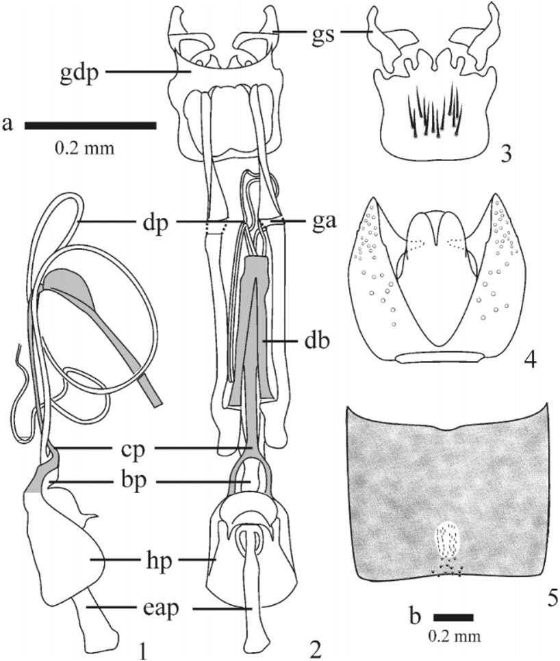
Abdomen. Tergites and sternites dark brown, sparsely gray pruinose; sparsely short, fine filiform pale golden setose, with lateral setae and setae on basal segments longer. Modified setae on tergite 2 composed of an anterior pale, spiculate patch arranged in an ovoid pattern, longer than wide, and a posterior dark brown, broad, truncate patch arranged in a triangular pattern, spread out wider posteriorly ( Fig. 5 View FIGURES 1 5 ).
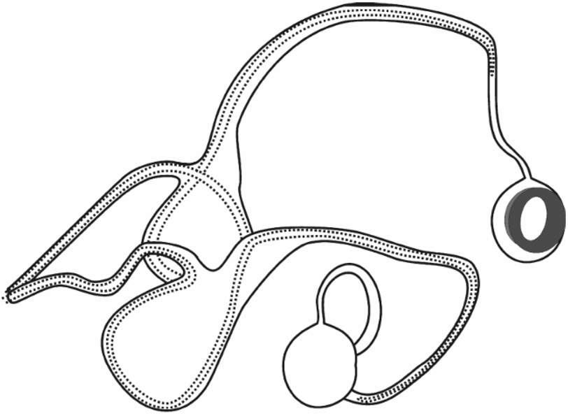
Terminalia. Tergite 8 only slightly modified, similar to previous segments; approximately half the length of tergite 7; anterior margin slightly acuminate medially, posterior margin emarginate medially; with one pair of sensory setae; dark brown; sparsely gray pruinose; sparsely short, fine filiform pale golden setose. Sternite 8 approximately half the length of sternite 7; quadrate, but wider posteriorly; brown; sparsely gray pruinose; sparsely short, fine filiform pale golden setose. Tergite 9 ( Fig. 4 View FIGURES 1 5 ); brown, light brown along margins; anterior connection between right and left halves dark brown; sparsely gray pruinose; sparsely short, fine filiform pale golden setose. Cerci ( Fig. 4 View FIGURES 1 5 ) bifurcate, subequal to extension of hypoproct; minutely setose apically. Hypoproct ( Fig. 4 View FIGURES 1 5 ) bluntly pointed posteriorly; minutely setose ventrally and apically. Subepandrial sclerite partially sclerotized. Gonocoxites ( Fig. 2 & 3 View FIGURES 1 5 ); brown; long, filiform, light brown setose ventromedially; gonocoxal ventral process projecting posteriorly and bifurcate apically. Gonocoxal apodeme ( Fig. 2 View FIGURES 1 5 ) brown, with an area just anterior to anterior gonocoxite margin dorsoventrally flattened and light brown, connection with dorsal bridge light brown. Gonostylus ( Fig. 3 View FIGURES 1 5 ) curved dorsomedially at apex; apex dorsoventrally flattened, scooplike; “crossshaped” when viewed dorsally or ventrally; brown; glabrous. Dorsal bridge and cordlike phallus brown ( Figs. 1 & 2 View FIGURES 1 5 ). Hanging bell phallus ( Figs. 1 & 2 View FIGURES 1 5 ) short and broad; posterior ejaculatory bulb rounded with a dorsal, pointed extension; brown. Distiphallus ( Fig. 2 View FIGURES 1 5 ) light brown, bifurcate at base, distal coils longer than gonocoxal apodeme. Aedeagal apodeme ( Figs. 1 & 2 View FIGURES 1 5 ) robust, cylindrical; posterior end broadened laterally and ventrally creating a basketshaped posterior face.
Description and variation of females
Generally lighter in color, but similar to males except as follows.
Head. Dichoptic, frons at its narrowest width as wide as or wider than ocellar tubercle. Inner eye margin with shiny black edge less pronounced, but expanded portion of shiny black adjacent to antenna more pronounced. Ommatidia of uniform size. Scape and pedicel dark to light brown.
Thorax. Legs yellow to light brown.
Terminalia ( Fig. 6 View FIGURE 6 ). Tergite 8 longer than wide; anterior margin straight; posterior margin with a narrow strip of cuticle connecting to tergite 9; dark brown; sparsely gray pruinose; sparsely short, fine filiform, pale golden setose. Membrane between tergite 8 and tergite 9 short, fine filiform, pale golden setose. Sternite 8 ( Fig. 6 View FIGURE 6 ) longer than wide; anterior margin straight; posterior lobe membranous, short, filiform, pale golden setose. Acanthophorites ( Fig. 6 View FIGURE 6 ) joined narrowly at dorsum and joined anterolaterally to sternite 10, with 5 pairs of robust, acuminate spines. Sternite 10 ( Fig. 6 View FIGURE 6 ) broader posteriorly; anterior margin with three anteriorly projecting points; ventrally brown; short, fine filiform, pale golden setose with a posterolateral to medial fringe of much longer setae. Cerci ( Fig. 6 View FIGURE 6 ) and hypoproct bulbous and membranous; minutely setose. Furca ( Fig. 6 View FIGURE 6 ) pearshaped, narrower anteriorly; posterolateral corners expanded laterally into hornshaped extensions that curve anteriorly. Spermathecal ducts and spermathecal sac duct ( Fig. 6 View FIGURE 6 ) arising from a common gonopore on the membrane of the furca. Spermathecal duct wider basally with a membranous expansion ( Fig. 6 View FIGURE 6 , me) at approximately half of its length then narrowing before terminating at the spermatheca; total length approximately ten furcal lengths. Spermatheca ( Fig. 6 View FIGURE 6 ) doughnut shaped with a sclerotized collar and neck on one side. Spermathecal sac duct subequal in width to the base of the spermathecal duct, slowly expanding towards sac; total length approximately four furcal lengths. Sac spherical, its length, width, and height subequal.
Distribution. Disjunct within the Great Basin Desert of the United States. It has been collected in Mono and Inyo Counties in California and Emery, Garfield, and Wayne Counties in Utah.
Ecology. Specimens have been taken by hand netting and are readily collected in numbers in Malaise traps. They are known to occur in dry washes and creek beds that range in habitats from Artemisia zones at 1375 m through the transition zone and into Juniperus / Pinus zones at 2100m.
Specimens Examined. USA, California, Mono County: 2 male paratypes (MEI 1418956, UCR), S[c]herwin Summit, 6500 [ft], 24.V.1985, G.R. Ballmer. Inyo County : 2 males (MEI 1418978), 6 females (MEI 141899904) 17 mi E Big Pine, Death Valley Road, 6925 [ft], pinonjuniper zone, 2124.VI.1992, Malaise, ME Irwin, DK Yeates. Utah, Emery County: 1 male (MEI 141842) and 1 female (MEI 141843) in copula, 2 km S Hatt’s Ranch, 27 km SW Green River, Malaise in dry wash, 2126.V.2002, ME Irwin, FD Parker, 1375 m, 38°50.5’N, 110°22.9’W; 1 female (MEI 141845), 2 males (MEI 141846 847), same data; 1 female (MEI 141844) same locality, 1921.V.2002. Garfield County: 1 male (MEI 141848), Burr Trail, 45 km SE Boulder, Malaise in dry canyon wash, juniper woodlands, 2327.V.2002, ME Irwin, FD Parker, 2190 m, 37°52.9’N, 111°06.7’W; 4 males (MEI 1419058), 2 females (MEI 14190910) Calf Crk., 10 km S Boulder, Malaise in side canyon, riparian vegetation, 2325.V.2002, ME Irwin, FD Parker, 1750 m, 37°47.6’N, 111°24.9’W; 1 male (MEI 141853) same locality 2627.V.2002; 2 males (MEI 141863, 141866), 6 females (MEI 14185051, 14187780), Hall’s Creek Overlook Road, nr main rd., Malaise in rugged rock wash, 2327.V.2002, ME Irwin, FD Parker, 1670 m, 37°44.6’N, 110°55.2’W; 8 males (MEI 14185461), 10 females (MEI 141849, 1418819), Trachyte Ck at Hwy 276, Malaise on sandy bank, 2227.V. 2002, 1560 m, ME Irwin, FD Parker, 37°57.4’N, 110°34.3’W. Wayne County: 10 males (MEI 141852, 141862, 141864 5, 14186772), 6 females (MEI 1418738), Bull Mountain Road, 18 km S Hanksville, 22 27.V.2002. Malaise in dry wash, ME Irwin, FD Parker, 1620 m, 38°13.8’N, 110°40.6’W. Deposition of specimens is as follows: BYU (MEI 14190510); EMUS (MEI 1418545, 1418623, 1418778, 1418823); INHS (MEI 1418567, 141861, 1418846); LACM (MEI 1418668, 14187981); UCR (MEI 14185860, 1418879); USNM (MEI 1418523, 1418645, 14186971, 1418746). The remaining specimens are deposited in MEI.
The description of the associated male and female genitalia ( Fig. 7 View FIGURE 7 )
The terminalia are in opposite orientation to each other so that the ventral surface of the female’s sternite 8 is facing ventrally while the ventral surface of the male’s sternite 8 and gonocoxites are facing dorsally. The external terminalia are in close association, but the sclerites of the two sexes are not touching one another. Neither the male’s gonostyli, which are articulated and musculated ( Ovtshinnikova & Yeates 1998) and seemingly would serve a copulatory function, nor tergite 9, which has been suggested to possibly serve a clasping function in Asiloidea ( Sinclair et al. 1994) is in contact with any female sclerites. The possibility exists that the specimens were once in closer proximity, but in death the genital sclerites retracted from one another.
The male’s distiphallus is completely inserted into and partially uncoiled in the female’s spermathecal duct ( Fig. 7 View FIGURE 7 ). Each of the separate bifurcations of the distiphallus enters into a different spermathecal duct, completes a full 360degree turn in a membranous sac located approximately halfway along the spermathecal duct, then continues further into the duct, finally reaching a point approximately 3/4 down the length of the spermathecal duct ( Fig. 7 View FIGURE 7 ). The tips of the distiphallus do not reach the spermathecae. The uncoiling of the distiphallus occurs entirely within the female abdominal cavity such that the basiphallus of the aedeagus is located at the entrance of the female gonopore ( Fig. 7 View FIGURE 7 ). The spermathecal sac is independent of any association with the male structures and “floats” freely within the abdominal cavity of the female.

No known copyright restrictions apply. See Agosti, D., Egloff, W., 2009. Taxonomic information exchange and copyright: the Plazi approach. BMC Research Notes 2009, 2:53 for further explanation.
|
Kingdom |
|
|
Phylum |
|
|
Class |
|
|
Order |
|
|
Family |
|
|
Genus |